
 E-submission
 KOTA
 TOTA
 TOTS

Current issue
- Page Path
- HOME > Browse articles > Current issue
- Volume 39(4); October 2026
-


Review Articles
- Minimally invasive plate osteosynthesis of the clavicle and humerus: indications, surgical techniques, and clinical outcomes
- Hoon-Sang Sohn, Yunjong Choi, Sang-Jin Shin
- J Musculoskelet Trauma 2026;39(4):275-288. Published online August 21, 2026
- DOI: https://doi.org/10.12671/jmt.2026.00220

-
 Abstract
 PDF
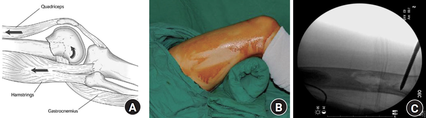
- Minimally invasive plate osteosynthesis (MIPO) is an established strategy for lower-extremity fractures because it preserves periosteal vascularity, maintains fracture hematoma, and promotes secondary bone healing through relative stability. Its use has expanded to fractures around the shoulder girdle, including fractures of the clavicle, proximal humerus, humeral shaft, and distal humerus. However, adoption in the upper extremity has been more cautious because of complex regional anatomy, narrow soft-tissue corridors, and the proximity of major neurovascular structures. This review summarizes current evidence on the indications, surgical techniques, clinical outcomes, and complications of MIPO for clavicle and humerus fractures. For clavicle fractures, MIPO is most commonly indicated for displaced multifragmentary midshaft fractures and provides union rates and functional outcomes comparable to those of conventional open reduction and internal fixation, with potential advantages in soft-tissue preservation and cosmesis. In proximal humerus fractures, MIPO performed through a deltoid-splitting approach has yielded favorable outcomes in selected two- and three-part fractures, whereas four-part fractures have less predictable outcomes and higher complication rates. In humeral shaft fractures, anterior MIPO provides stable fixation without routine radial nerve exploration and has shown reliable union with fewer complications in selected fracture patterns. For extra-articular distal humerus fractures, minimally invasive anterior and posterior plating techniques, including the use of an inverted proximal humeral internal locking system plate, have shown favorable clinical and radiographic outcomes despite technical challenges related to limited distal fixation. Overall, MIPO can achieve high union rates and satisfactory functional outcomes in clavicular and humeral fractures when indications are appropriate, safe anatomical corridors are respected, and surgical technique is meticulous. Level of evidence: Not applicable.
- 571 View
- 11 Download

- Locked plating in older patients with distal femur fractures: strategies to avoid complications
- Je-Hyun Yoo
- J Musculoskelet Trauma 2026;39(4):289-298. Published online August 24, 2026
- DOI: https://doi.org/10.12671/jmt.2026.00234
-
Abstract
PDF
- Distal femur fractures in older patients with osteoporosis are difficult to treat because poor bone quality reduces screw purchase and fixation stability. Minimally invasive locked plating with an angularly stable locking compression plate is widely used to obtain secure fixation in osteoporotic distal femur fractures and offers improved biomechanical performance. However, complications such as implant failure and peri-implant fracture continue to occur. This narrative review summarizes current principles of minimally invasive lateral locked plating for osteoporotic distal femur fractures in older patients and discusses practical strategies to reduce fixation-related complications based on the available literature and the author’s clinical experience. Level of evidence: Not applicable.
- 446 View
- 4 Download
- Current concepts in distal clavicle fractures: an evidence-based update
- Du-Han Kim, Hyuk-Joon Sohn, Byung-Chan Choi, Chul-Hyun Cho
- J Musculoskelet Trauma 2026;39(4):299-309. Published online September 4, 2026
- DOI: https://doi.org/10.12671/jmt.2026.00241
-
Abstract
PDF
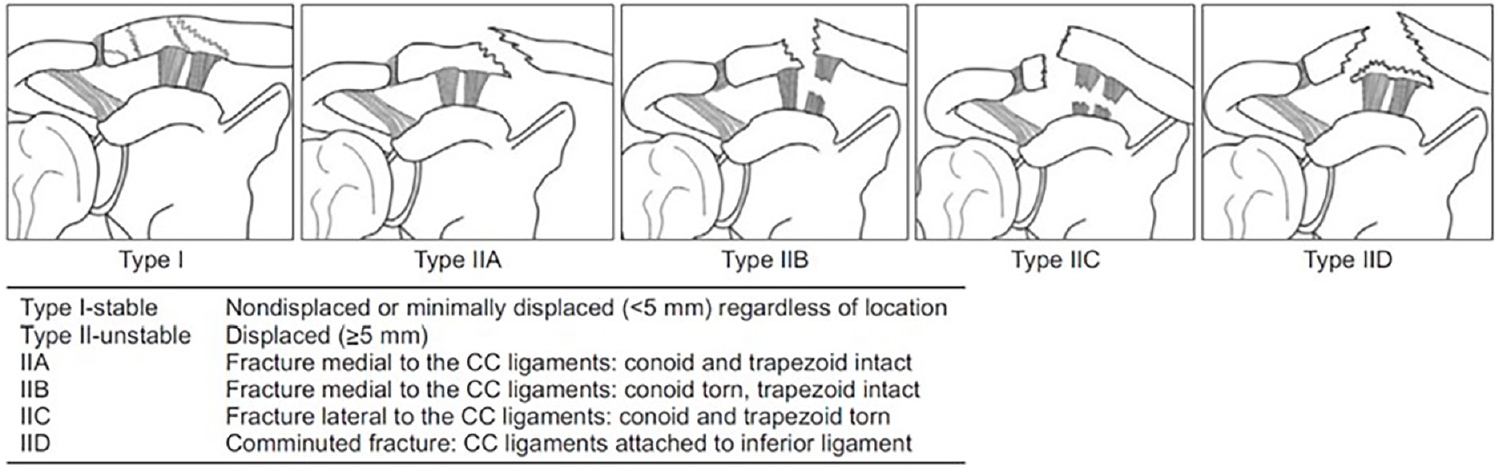
- Distal clavicle fractures (DCFs) account for 10%–30% of all clavicle fractures and remain difficult to manage because fracture stability depends heavily on coracoclavicular ligament integrity, plain radiographic classification has limited reliability, and distal fragment size varies substantially, complicating fixation planning. This review provides an evidence- based update on the anatomy, classification, imaging, and management of DCFs, incorporating recent literature, including population-specific cadaveric data and three-dimensional fracture mapping. The conoid and trapezoid ligaments have distinct osseous footprints that influence fracture stability in both the Neer/Craig and Cho classification systems; however, interobserver reliability remains only fair-to-moderate for both systems, particularly when conoid ligament integrity must be inferred. Three-dimensional computed tomography mapping has shown that posterior displacement and angulation are nearly universal but often underrecognized on standard radiographs, supporting selective computed tomography use when radiographs do not adequately characterize fracture morphology, comminution, or multiplanar displacement. Magnetic resonance imaging should be reserved for cases in which ligament integrity remains diagnostically ambiguous. Stable fractures generally have excellent outcomes with conservative treatment using a simple arm sling. For unstable fractures, operative and conservative treatment yield similar 1-year functional outcomes despite different union rates (96% vs. 63%), with conservative treatment carrying a nonunion risk of approximately one-third and operative treatment carrying a technique-dependent reoperation burden, ranging from planned implant removal in most hook plate cases to less than 10% with coracoclavicular stabilization constructs. When surgery is indicated, technique selection should be guided by fracture morphology and surgeon experience: anatomic locking plate fixation with or without coracoclavicular augmentation for Neer IIA/Cho IIA fractures, dedicated coracoclavicular stabilization for Neer IIB fractures (Cho IIB/IIC), and hook plate fixation as a salvage construct for Neer V/Cho IID fractures. Management should integrate classification, imaging findings, union risk, reoperation risk, expected recovery, and patient-specific factors through shared decision-making. Level of evidence: Not applicable.
- 699 View
- 16 Download
Systematic Reviews
- Impact of posterior wall involvement and fixation on clinical and radiographic outcomes of associated both-column acetabular fractures: a systematic review and meta-analysis
- Chul-Ho Kim, Eic Ju Lim
- J Musculoskelet Trauma 2026;39(4):310-322. Published online October 19, 2026
- DOI: https://doi.org/10.12671/jmt.2026.00248
-
Abstract
PDF
 Supplementary Material
- Background
Posterior wall (PW) fractures are frequently associated with both-column (ABC) acetabular fractures. However, whether the presence of a PW fragment or the need for internal fixation affects prognosis remains a topic of debate. This study compared postoperative outcomes, including reduction quality and clinical results, according to the presence of an associated PW fracture and whether PW fixation was performed.
Methods
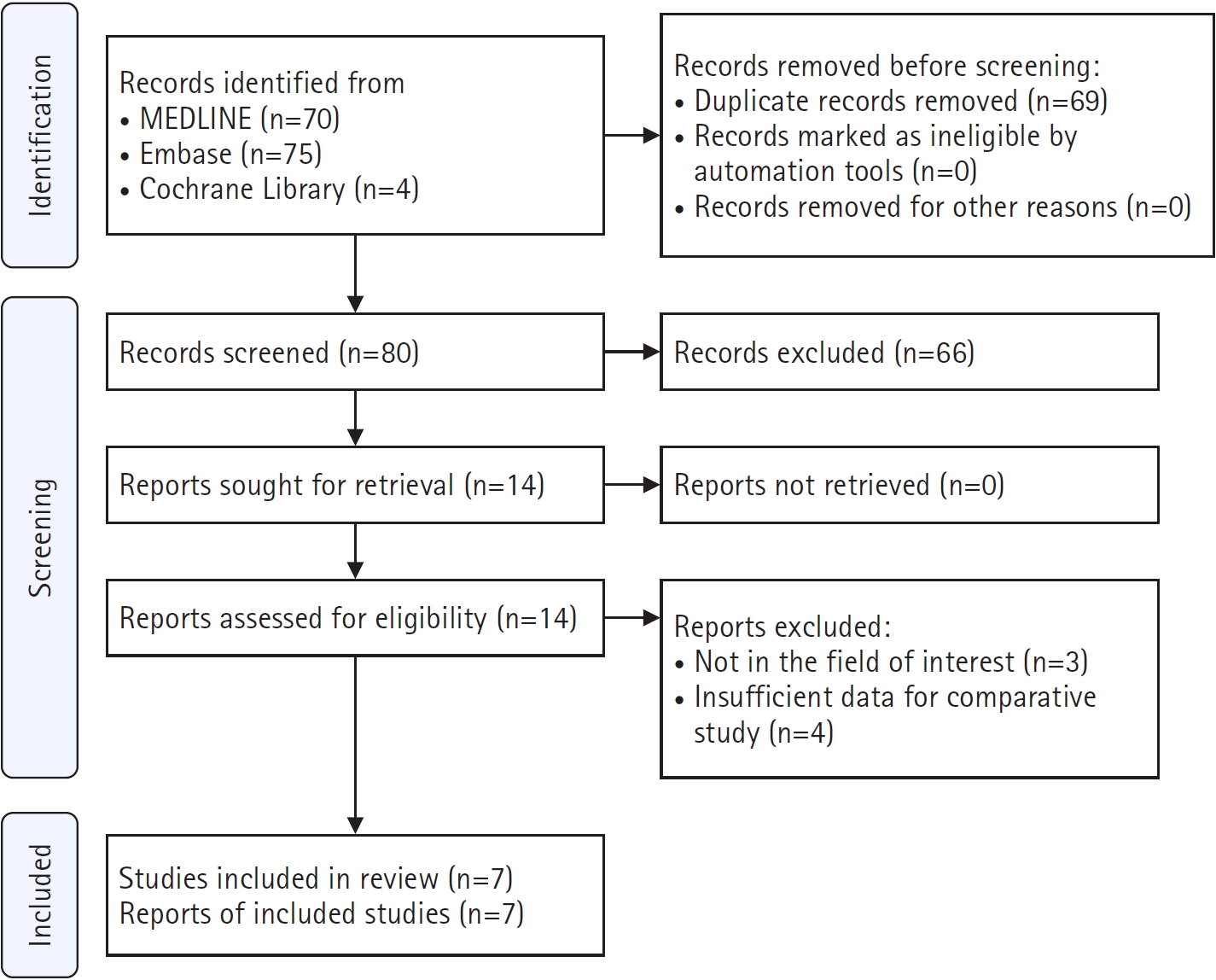
This systematic review and meta-analysis followed the Preferred Reporting Items for Systematic Reviews and Meta-Analyses (PRISMA) 2020 guidelines. MEDLINE, Embase, and the Cochrane Library were searched for articles published up to December 10, 2025. Comparative studies of ABC fractures were included if they evaluated the presence or absence of PW involvement or compared internal fixation with nonfixation of the PW component. Clinical and radiographic outcome data were extracted.
Results
Seven retrospective studies including 434 analyzable patients were included. No clear differences were found in the odds of poor reduction (odds ratio [OR], 0.91; 95% confidence interval [CI], 0.43–1.94; P=0.80), osteoarthritis (OR, 1.04; 95% CI, 0.37–2.92; P=0.94), or conversion to total hip arthroplasty (OR, 0.63; 95% CI, 0.23–1.71; P=0.36) according to the presence of a PW fragment in ABC fractures. Among ABC fractures with PW involvement, nonfixation was associated with numerically higher odds of poor reduction than fixation, although the difference was not statistically significant (OR, 1.47; 95% CI, 0.46–4.76; P=0.52). In unadjusted comparisons of non-randomized cohorts, PW fixation was associated with significantly longer operative time (mean difference [MD], 70.38 min; 95% CI, 24.46–116.29; P=0.003) and greater intraoperative blood loss (MD, 351.36 mL; 95% CI, 244.97–457.74; P<0.00001). The final modified Merle d'Aubigné-Postel score did not differ significantly between the fixation and nonfixation groups (MD, 0.58 points; 95% CI, −0.57 to 1.74; P=0.32).
Conclusions
Current evidence does not clearly support routine fixation of all PW fragments. In unadjusted comparisons of non-randomized cohorts, patients who underwent PW fixation had longer operative time and greater blood loss. These differences cannot be attributed to fixation itself because of confounding by indication and the frequent use of an additional posterior approach. Selective fixation based on residual displacement, joint congruency, and hip stability appears reasonable, although the certainty of evidence remains limited. Level of evidence: Not applicable.
- 101 View
- 2 Download
- Definitions and assessment of residual distal radioulnar joint instability after operative fixation of distal radius fractures: a methodological systematic review
- Jae Ho Cho, Seungyeob Sakong, Sumin Lim
- J Musculoskelet Trauma 2026;39(4):323-340. Published online October 19, 2026
- DOI: https://doi.org/10.12671/jmt.2026.00290
-
Abstract
PDFSupplementary Material
- Background
Residual distal radioulnar joint (DRUJ) instability after operative fixation of distal radius fractures serves as an eligibility criterion, treatment indication, and outcome, but previous syntheses report inconsistent definitions. This methodological systematic review examined how clinical reports define, assess, and report it, and whether they distinguish it from structural triangular fibrocartilage complex (TFCC) lesions.
Methods
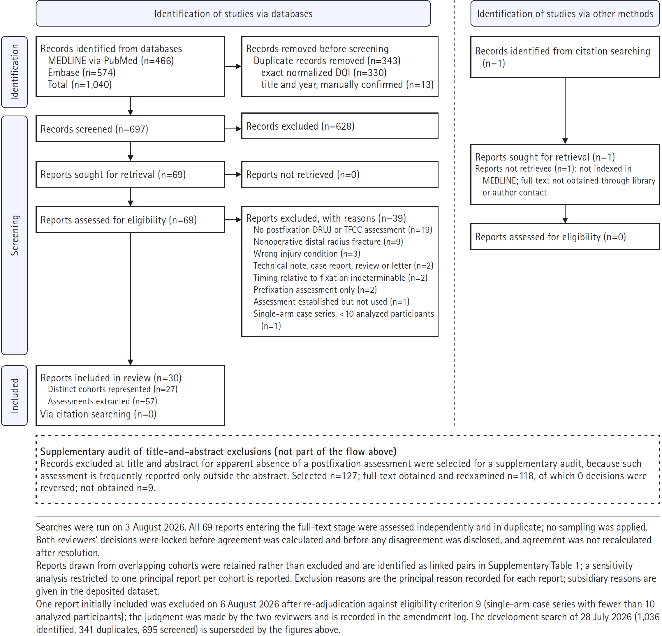
MEDLINE (PubMed) and Embase were searched from 2015 to 3 August 2026 for adult clinical reports in which residual DRUJ instability was assessed after fixation, or inferred from TFCC lesion characteristics, and used for a treatment decision or as an outcome. Every report entering full-text eligibility was assessed independently in duplicate; data were locked before agreement was calculated and disagreements disclosed. No riskof- bias tool was applied: neither treatment effects nor diagnostic accuracy were estimated. Findings are counts and proportions; no meta-analysis was performed.
Results
Of 1,040 records, 697 were screened and 69 assessed as full texts; 30 reports of 27 cohorts were included, contributing 57 assessments. In the treatment-decision context, 12 reports stated an operational definition (40.0% of all 30; 50.0% of the 24 containing such an assessment), four stated neither required element (16.7% of 24), and six contained no such assessment and were classified as not applicable. At the assessment level, the positive criterion was vague in 43.9% and absent in 17.5%, and explicit for 48.0% of intraoperative but 25.9% of final follow-up assessments. No report inferred instability from a TFCC lesion instead of testing the joint; one used the two constructs interchangeably, 14 (46.7%) distinguished them, and 15 (50.0%) addressed instability without structural verification. A comparable prevalence estimate was extractable from only 9 of 57 assessments (15.8%).
Conclusions
Residual DRUJ instability is therefore assessed frequently but is often incompletely defined, and until these elements are stated consistently, frequency estimates cannot be compared across studies. Level of evidence: Not applicable.
- 112 View
- 5 Download
Original Articles
- Multiple blocking screws reduce peak stress in a smaller-diameter tibial nail to a level similar to that in a canal-filling nail: a comparative finite element analysis
- Jungtae Ahn, Se Lin Jeong, Gu-Hee Jung
- J Musculoskelet Trauma 2026;39(4):341-353. Published online October 13, 2026
- DOI: https://doi.org/10.12671/jmt.2026.00255
-
Abstract
PDF
- Background
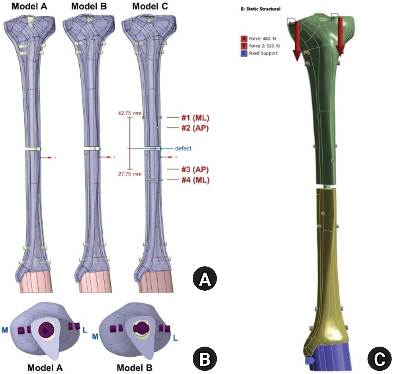
Cortical contact at the medullary isthmus permits cortical load sharing and constrains nail motion, and is considered important for stable intramedullary nailing. Blocking screws may provide the latter function. This finite element analysis examined whether adding multiple blocking screws to a smaller-diameter nail would reduce peak nail stress to a level similar to that of a canal-filling construct.
Methods
Three configurations were modeled in a tibia with a 5-mm diaphyseal defect at the isthmus: an 11-mm canal-filling nail (model A), an 8-mm nail alone (model B), and an 8-mm nail with four 3.5-mm blocking screws positioned circumferentially (model C). An 800-N static axial load was applied at a medial-to-lateral ratio of 3:2 with the distal end fixed. Total deformation and von Mises stresses in bone, nail, and screws were analyzed using a linear elastic, small-deformation formulation.
Results
Maximum total deformation was 5.60, 33.33, and 18.82 mm in models A, B, and C, respectively, occurring at the proximal tibia in all models. Peak nail stress in model B was 891.9 MPa, slightly exceeding the assumed 880-MPa yield threshold, whereas peak stresses were similar in models A (272.9 MPa) and C (265.8 MPa). Screw stress was asymmetric: 57.9 and 58.2 MPa in the posterior and medial screws versus 3.2 and 6.8 MPa in the lateral and anterior screws, indicating differential mechanical engagement of the blocking screws.
Conclusions
Under the single static loading condition evaluated, four circumferential blocking screws reduced the peak nail stress of a smaller-diameter nail to a level similar to that of a canal-filling nail. Total deformation nevertheless remained approximately 3.4 times that of the canal-filling construct, and peak bone stress differed between constructs. The asymmetric screw-stress distribution was consistent with differential mechanical engagement of the blocking screws. Mechanical equivalence was not established for outcomes other than peak nail stress. Level of evidence: Not applicable.
- 208 View
- 4 Download
- Percutaneous Kirschner-wire fixation followed by an opencast to permit protected daily and sports activities in patients with tendinous mallet finger: a case series at a tertiary hospital in Korea
- Hyeon Jun Kim, Ji Woong Kim, Min Kyun Cho, Sung Yoon Jung
- J Musculoskelet Trauma 2026;39(4):354-364. Published online October 20, 2026
- DOI: https://doi.org/10.12671/jmt.2026.00269
-
Abstract
PDF
- Background
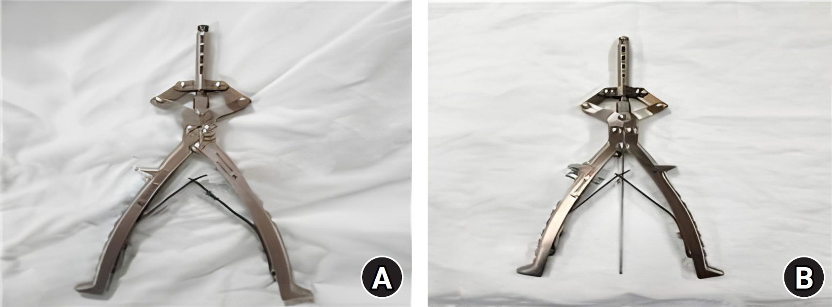
Dong-A University Hospital’s protocol for tendinous mallet finger requires 12 weeks of continuous full-time distal interphalangeal (DIP) joint splinting, which may restrict activities and complicate hygiene. This study evaluated the feasibility, short-term safety, and 6-month clinical outcomes of buried Kirschner-wire fixation followed by opencast application.
Methods
This retrospective case series screened 36 patients with mallet finger between January 2024 and October 2025. Twelve met the exclusion criteria, leaving 24 eligible patients; eight elected nonoperative treatment, and 16 chose surgery for acute closed tendinous mallet finger and constituted the study cohort. The DIP joint was fixed in approximately 15° of hyperextension using one or two buried 1.1-mm Kirschner wires. An opencast was applied at 1 week. The wires were routinely removed and active motion was initiated at 8 weeks, followed by a removable brace during outdoor or sports activities for 4 weeks. Outcomes were evaluated at 6 months.
Results
The mean preoperative extension lag was 30.0°. At 6 months, the mean residual extension lag was 1.9°, no flexion loss was observed, and Crawford outcomes were excellent in 62.5% and good in 37.5%. Two superficial pin-site infections resolved with oral antibiotics and local wound care. A separate case of wire loosening with painful subcutaneous prominence was managed with an aluminum splint. All patients resumed daily activities and showering at 1 week, when protected sports were also permitted; exact return dates and activity intensity were not recorded.
Conclusions
Buried Kirschner-wire fixation followed by opencast application may be an option for selected patients who wish to avoid prolonged full-time splinting. The protocol permitted protected activities from week 1 and active DIP motion from week 8; these findings support its feasibility but do not establish earlier return to unrestricted activity. Level of evidence: IV.
- 64 View
- 2 Download
- Proximal screw number and neck-shaft angle after locking plate fixation of proximal humerus fractures at a single center in Korea: a retrospective cohort study
- Dong Uk Jin, Se Ho Kang, Yeong Chan Kim, Kyu-Hak Jung, Sang-Pil So
- J Musculoskelet Trauma 2026;39(4):365-373. Published online October 20, 2026
- DOI: https://doi.org/10.12671/jmt.2026.00276
-
Abstract
PDF
- Background
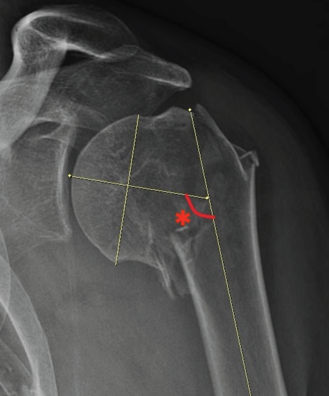
The neck-shaft angle (NSA) may decrease between the immediate postoperative assessment and radiographic union after locking plate fixation of proximal humerus fractures. However, the relationship between the number of proximal humeral head screws and postoperative NSA remains unclear. This study compared postoperative NSA and the change in NSA (ΔNSA) among groups defined by the number of proximal screws.
Methods
This retrospective cohort study included consecutive patients who underwent locking plate fixation of a proximal humerus fracture between August 2025 and June 2026. Patients were classified into three groups according to the number of proximal screws: ≤7, 8, or 9. NSA was measured preoperatively, immediately postoperatively, and at radiographic union, hereafter referred to as the final assessment. Final NSA, ΔNSA, the proportions of patients with varus malalignment (final NSA <120°) and angular loss of reduction (ΔNSA ≤−10°), and complication rates were compared among groups. Trends across increasing screw numbers were also assessed.
Results
Nineteen patients were evaluated: nine in the ≤7-screw group, four in the 8-screw group, and six in the 9-screw group. Immediate postoperative NSA was 140.1°±5.7°, 136.2°±11.8°, and 134.0°±6.2° in the three groups, respectively; final NSA was 131.6°±9.2°, 129.5°±16.1°, and 130.2°±8.3°; and ΔNSA was −8.6°±8.8°, −6.8°±4.6°, and −3.8°±5.2°. None of these outcomes differed significantly among groups (all P>0.05). Varus malalignment occurred in 22.2%, 25.0%, and 16.7% of patients, respectively (P=0.944). Angular loss of reduction occurred in 44.4%, 25.0%, and 0%, respectively; neither the overall group difference (P=0.159) nor the trend across groups (P=0.056) reached statistical significance. Complication rates also did not differ significantly among groups.
Conclusions
In this small retrospective cohort, the number of proximal screws was not significantly associated with final NSA or ΔNSA. Although the proportion of patients with angular loss of reduction decreased numerically as the screw number increased, this trend was not statistically significant and should be regarded as exploratory. Level of evidence: III.
- 95 View
- 4 Download
Case Reports
- Progressive forearm instability after anatomical union of a radial head fracture: a case report of a challenging Essex-Lopresti injury
- SeongJu Choi
- J Musculoskelet Trauma 2026;39(4):374-381. Published online July 20, 2026
- DOI: https://doi.org/10.12671/jmt.2026.00157
-
Abstract
PDF
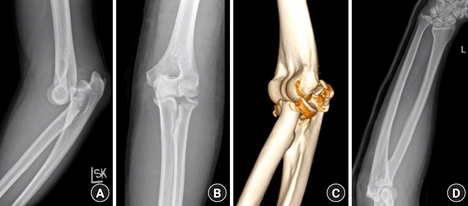
- Essex-Lopresti injuries remain challenging because anatomical restoration of bony structures does not always re-establish functional forearm stability. We report the case of a 51-year-old male patient who sustained an elbow dislocation, comminuted radial head and neck fracture, and distal radioulnar joint (DRUJ) dissociation after falling from a bicycle. He underwent open reduction and internal fixation of the radial head, interosseous membrane augmentation using a suture-button construct, and temporary DRUJ pinning. Immediate postoperative radiographs showed apparent restoration of radial length, and radiographic union of the radial head was achieved. However, ulnar-sided wrist pain initially improved but progressively recurred by 6 months, with increasing ulnar-positive variance and DRUJ widening. Elbow motion was preserved, but forearm rotation was limited, and computed tomography demonstrated heterotopic ossification around the radial head. Ulnar shortening osteotomy and radial head arthrolysis were performed as a second procedure, after which mechanical instability developed, with irreducible anterior dislocation of the radial head. A third operation, consisting of annular ligament reconstruction using a palmaris longus autograft, restored proximal radioulnar joint stability, although substantial limitation of forearm rotation persisted. This case highlights that even radiographic union and multistructural initial management may fail to restore balanced forearm mechanics in Essex-Lopresti injury. Level of evidence: V.
- 533 View
- 8 Download
- Reconstruction of a 20-cm tibial bone defect after recurrent fracture-related infection using the induced membrane technique and rhBMP-2 augmentation with limited autogenous bone availability: a case report
- Whee Sung Son
- J Musculoskelet Trauma 2026;39(4):382-392. Published online August 21, 2026
- DOI: https://doi.org/10.12671/jmt.2026.00143

-
Abstract
PDF
- Fracture-related infection (FRI) poses the combined challenges of infection control and reconstruction of critical-sized bone defects. These challenges are amplified in recurrent cases with severely limited autogenous bone availability. We report a 61-year-old male patient with recurrent right tibial FRI after multiple operations and a 20-cm segmental defect. Suspected residual infectious foci were mapped by comparing preoperative fluorodeoxyglucose positron emission tomography/computed tomography (PET/CT) with post-debridement CT and were evaluated using site-specific pre- and post-debridement cultures. After a second targeted debridement, all post-debridement cultures were negative, and no clinical or laboratory evidence of active infection was observed during a 2-week antibiotic-free interval. The defect was subsequently reconstructed using the induced membrane technique with 30 cm³ of autogenous cancellous bone, 60 cm³ of demineralized bone matrix, and 9 mg of recombinant human bone morphogenetic protein-2 (rhBMP-2) delivered on a 20 cm³ hydroxyapatite carrier. Radiographic union was achieved, and no clinical evidence of infection recurrence was observed at 1 year. This case suggests the feasibility of combining PET/CT- and culture-guided anatomically targeted debridement with rhBMP-2-augmented induced membrane reconstruction when autogenous graft volume is severely limited. Level of evidence: V.
- 386 View
- 10 Download
 First
 Prev



