
 E-submission
 KOTA
KOTA TOTA
TOTA TOTS
TOTS
Articles
- Page Path
- HOME > > Ahead-of print articles > Article
-
Review Article
- Current concepts in distal clavicle fractures: an evidence-based update
-
Du-Han Kim
 , Hyuk-Joon Sohn, Byung-Chan Choi, Chul-Hyun Cho
 -
DOI: https://doi.org/10.12671/jmt.2026.00241
Published online: September 4, 2026
Department of Orthopedic Surgery, Keimyung University Dongsan Hospital, Keimyung University School of Medicine, Daegu, Korea
- Correspondence to: Chul-Hyun Cho Department of Orthopedic Surgery, Keimyung University Dongsan Hospital, Keimyung University School of Medicine, 1035 Dalgubeol-daero, Dalseo-gu, Daegu 42601, Korea Tel: +82-53-258-4771 Email: oscho5362@dsmc.or.kr
• Received: June 24, 2026 • Revised: July 17, 2026 • Accepted: July 20, 2026
© 2026 The Korean Orthopaedic Trauma Association
This is an Open Access article distributed under the terms of the Creative Commons Attribution Non-Commercial License (http://creativecommons.org/licenses/by-nc/4.0/) which permits unrestricted noncommercial use, distribution, and reproduction in any medium, provided the original work is properly cited.
- 462 Views
- 7 Download


Abstract
- Distal clavicle fractures (DCFs) account for 10%–30% of all clavicle fractures and remain difficult to manage because fracture stability depends heavily on coracoclavicular ligament integrity, plain radiographic classification has limited reliability, and distal fragment size varies substantially, complicating fixation planning. This review provides an evidence-based update on the anatomy, classification, imaging, and management of DCFs, incorporating recent literature, including population-specific cadaveric data and three-dimensional fracture mapping. The conoid and trapezoid ligaments have distinct osseous footprints that influence fracture stability in both the Neer/Craig and Cho classification systems; however, interobserver reliability remains only fair-to-moderate for both systems, particularly when conoid ligament integrity must be inferred. Three-dimensional computed tomography mapping has shown that posterior displacement and angulation are nearly universal but often underrecognized on standard radiographs, supporting selective computed tomography use when radiographs do not adequately characterize fracture morphology, comminution, or multiplanar displacement. Magnetic resonance imaging should be reserved for cases in which ligament integrity remains diagnostically ambiguous. Stable fractures generally have excellent outcomes with conservative treatment using a simple arm sling. For unstable fractures, operative and conservative treatment yield similar 1-year functional outcomes despite different union rates (96% vs. 63%), with conservative treatment carrying a nonunion risk of approximately one-third and operative treatment carrying a technique-dependent reoperation burden, ranging from planned implant removal in most hook plate cases to less than 10% with coracoclavicular stabilization constructs. When surgery is indicated, technique selection should be guided by fracture morphology and surgeon experience: anatomic locking plate fixation with or without coracoclavicular augmentation for Neer IIA/Cho IIA fractures, dedicated coracoclavicular stabilization for Neer IIB fractures (Cho IIB/IIC), and hook plate fixation as a salvage construct for Neer V/Cho IID fractures. Management should integrate classification, imaging findings, union risk, reoperation risk, expected recovery, and patient-specific factors through shared decision-making.
Introduction
Distal clavicle fractures (DCFs) account for 10%–30% of all clavicle fractures and remain a challenging clinical problem due to their unique anatomical complexity and the lack of consensus on optimal management [1-6].
Fracture instability in DCFs is largely determined by the integrity of the coracoclavicular (CC) ligament complex: ligamentous integrity—not fracture morphology alone—determines stability and guides treatment, yet available classification systems demonstrate only fair-to-moderate interobserver reliability on standard radiographs [4,5,7], leaving surgeons with inconsistent classification of the same fracture pattern. Additionally, the small and often comminuted distal fragment makes surgical fixation technically demanding, and no single operative technique has demonstrated clear superiority. Nonunion rates approaching 31% with conservative treatment have prompted surgical intervention for unstable fractures [8], yet operative cohorts report aggregate complication rates exceeding 40% (implant-related pain, hardware failure, infection, scar pain, wound issue, and nonunion) and similar reoperation rates, creating an unresolved risk-benefit dilemma [8-10]. The paucity of high-quality randomized evidence comparing fixation strategies further limits the strength of current recommendations. This narrative review of the literature was conducted using PubMed and Google Scholar. Studies published between 2015 and 2025 were preferentially included, with earlier seminal works retained where appropriate. Priority was given to systematic reviews, meta-analyses, and randomized controlled trials. In this context, the present review aims to provide an evidence-based update on the classification, imaging, and surgical management of DCF, incorporating the most recent literature to guide contemporary clinical decision-making.
Written informed consent was obtained from all patients whose clinical images appear in Figs. 2–5 for publication of their imaging data.
Surgical anatomy of the CC ligament complex
A precise understanding of CC ligament anatomy is fundamental to interpreting fracture instability and selecting fixation strategies. The CC complex comprises two components—the conoid ligament (CL) and the trapezoid ligament (TL)—each with defined osseous footprints that directly influence implant placement.
The CL originates from the posteromedial coracoid dorsum in a C-shaped configuration and ascends posterolaterally to insert on the conoid tubercle of the posteroinferior clavicle, forming an inverted cone with a tapered inferior apex [11]. Pooled data from 13 cadaveric studies (n=614 shoulders) place the center of the CL clavicular footprint at a weighted mean of 34.9 mm (range, 23.2–46.4 mm) from the lateral clavicular margin, with a footprint width of 18.0 mm and length of 5.9 mm [11]. At the coracoid, the attachment center lies approximately 35.4 mm from the coracoid tip, with a footprint of 5.4 mm width and 11.0 mm length [11]. The TL arises from the anterosuperior coracoid, anterior to the CL footprint, and courses in a posteromedial-to-anterolateral direction to attach along the trapezoid line on the anteroinferior clavicle, anterolateral to the conoid tubercle [11]. It is 2–3 times thicker at its clavicular insertion than at its coracoid origin. The center of the TL clavicular footprint lies a weighted mean of 25.3 mm from the lateral clavicle margin, with a width of 14.6 mm and length of 9.6 mm [11].
Implant design parameters for CC fixation—including TightRope tunnel intervals and suture anchor footprint targets—are largely derived from Western cadaveric datasets, raising concerns about direct extrapolation to Asian patients. A cadaveric study by Chung et al. [12] analyzing 102 Korean shoulders reported that the conoid footprint center lies 42.8±3.4 mm (men) and 39.9±3.3 mm (women) from the lateral clavicle margin—approximately 8 mm more medial than the Western pooled mean of 34.9 mm [11]. Trapezoid distances were comparable between populations (21.7±3.2 mm vs. 25.3 mm pooled) [11,12]. The interligament angle between CL and TL measured 25°±8° (male) and 28°±5° (female) on the anteroposterior (AP) view and 19°±3°/17°±3° on the lateral view [12], suggesting that purely vertical CC reconstruction may inadequately restore the oblique native trajectory of the CL. These findings indicate that population-adjusted landmarks warrant consideration in preoperative planning for Korean and other Asian patients.
Classification systems
The Craig modification of the Neer system classifies DCF into five types based on CC ligament involvement. Types I and III are stable and managed conservatively; types II and V are unstable, carrying nonunion rates of 31% with conservative treatment [8]. The clinically critical type II subclassification—IIA (the fracture line is medial to the CC ligaments) versus IIB (the fracture line is between the CL and TL) directly governs operative strategy but is the distinction most vulnerable to misclassification on plain radiographs. Interobserver agreement for the Neer system consistently falls in the fair-to-moderate range (κ=0.11–0.40) [6], and a multicenter study confirmed it carried the lowest agreement among four systems evaluated (mean κ=0.368) regardless of surgeon subspecialty [13]. Magnetic resonance imaging (MRI) reclassification changed treatment recommendations in 5% of initially radiograph-typed cases, predominantly upgrading stable fractures to operative indications [14].
The Cho classification divides fractures into type I (stable, displacement <5 mm) and type II (unstable, ≥5 mm), with type II further subclassified into four subtypes: IIA (fracture medial to the CC ligament: conoid and trapezoid intact); IIB (fracture medial to the CC ligament: conoid torn, trapezoid intact); IIC (fracture lateral to the CC ligament: both ligaments torn); and IID (comminuted fracture, CC ligaments attached to inferior fragment) (Fig. 1) [15].
Each subtype is paired with a recommended fixation construct—locking plate for IIA and IID, CC fixation for IIB and IIC—providing treatment guidance absent from the Neer system [15]. Reliability was moderate interobserver (κ=0.434) and substantial intraobserver (κ=0.644), with no significant difference between shoulder specialists and orthopedic fellows [15].
Both systems share a common vulnerability: the Neer IIA/IIB and Cho IIA/IIB distinction depends on CL integrity, which plain radiographs cannot directly demonstrate. Routine measurement of the CC distance and fracture angle supplements classification accuracy; MRI should be considered when radiographic classification is ambiguous and the consequence of undertreating an unstable fracture is high [14].
Imaging and diagnosis
Plain radiography remains the primary imaging modality for DCF. The standard AP shoulder view supplemented by the Zanca view (10°–15° cephalad tilt, 50% reduced penetration) provides optimal visualization of the acromioclavicular (AC) joint and distal clavicle [16]. The axillary lateral view is essential to assess posterior displacement. Bilateral comparative views enable measurement of the CC distance (CCD); a contralateral difference of ≥5 mm or absolute CCD ≥15 mm suggests CC ligament disruption [16].
Computed tomography (CT) provides superior delineation of fracture line morphology, comminution extent, fragment number, and intra-articular involvement compared with plain radiography—information directly relevant to fixation planning when the distal fragment is small or the fracture pattern is complex [17]. A critical limitation of existing classification systems is their exclusive focus on the coronal plane, which systematically underestimates the three-dimensional (3D) complexity of DCFs. Liu et al. [17] applied 3D fracture mapping to 81 consecutive DCFs—the largest such series reported to date—using CT-based digital reconstruction and virtual repositioning to quantify multiplanar displacement. Key findings included: 92.59% of cases showed posterior displacement (mean, 6.30 mm) and posterior angulation (mean, 9.24°); 75.31% exhibited superior displacement (mean, 5.10 mm); and 91.36% demonstrated shortening at the fracture site (mean, 3.93 mm). Fracture lines were predominantly concentrated in the distal one-third of the clavicle, with highest density at the acromion end. The study identified posterior displacement and posterior angulation as the two key biomechanical predictors of therapeutic failure, consistent with the high nonunion rate in conservatively managed Neer type IIB fractures [17]. These findings demonstrate that standard radiographic classification may miss clinically consequential sagittal-plane and horizontal instability. CT should be considered when plain radiographic classification is uncertain or horizontal displacement cannot be confidently excluded (Fig. 2) [17].
MRI is the only modality capable of directly visualizing CC ligament integrity and is uniquely valuable when fracture stability classification is uncertain on radiograph. A prospective study demonstrated that MRI reclassified 8.5% of cases initially typed on plain radiographs, with treatment changes in 5%—predominantly upgrading stable fractures to operative indications due to occult CL rupture [14]. MRI additionally identifies concurrent soft tissue injuries including rotator cuff tears and AC capsular disruption. Selective use is recommended when the Neer IIA/IIB or Cho IIA/IIB distinction cannot be confidently established on radiograph and the clinical consequence of misclassification is high (Fig. 3).
Based on current evidence, the following stepwise approach is recommended: (1) bilateral AP and Zanca views with CCD measurement as the initial standard; (2) axillary lateral view to assess posterior displacement in all cases; (3) CT when comminution, fragment size, or sagittal/horizontal displacement cannot be assessed on standard views; (4) MRI when CC ligament integrity cannot be determined radiographically and the stability classification would change management [14,17].
Conservative treatment
Conservative management remains the standard of care for stable DCFs—Neer type I and III, or Cho type I—where the CC ligament complex is intact and displacement is minimal (<5 mm) (Fig. 4).
For unstable displaced fractures (Neer type II, Cho type II), conservative treatment may be appropriate in elderly or low-demand patients, those with significant medical comorbidities precluding surgery, or patients who explicitly prefer to avoid operative intervention after informed counseling [9,18]. A simple arm sling is the preferred immobilization device and has largely supplanted the figure-of-eight bandage, which confers no demonstrated advantage in union rate or functional outcome and is associated with neurovascular complications and patient discomfort [19].
The systematic review by Thurston et al. [9]—the largest to date, encompassing 779 patients across 11 studies—reported an overall union rate of 63.2% (range, 22.2%–94.4%) with conservative treatment, compared with 96.3% (range, 87.9%–100%) in operatively managed cohorts. Despite this, none of the seven comparative studies demonstrated any statistically significant difference in patient-reported outcomes. Mean Constant-Murley scores were 91.8 (nonoperative) versus 87.4 (operative), and mean Disabilities of the Arm, Shoulder, and Hand (DASH) scores were 7.8 versus 6.4, respectively [9]. The multicenter study by Hall et al. [18], conducted across seven Canadian level I trauma centers, randomized 57 patients with completely displaced Neer type II fractures to operative plate fixation or conservative sling management. At 1 year, there was no significant difference in DASH scores (operative 6.7 vs. nonoperative 10.8, P=0.31) or Constant-Murley scores (89.9 vs. 90.0, P=0.88). Operative management achieved a significantly higher union rate at 12 months (96% vs. 63%, P=0.01) and the surgical group showed earlier return to sports at 6 months (78% vs. 44%, P=0.015) [18].
The overall complication rate in nonoperatively managed displaced DCF was 45.1%, with nonunion accounting for 31.3%, CC ligament calcification for 7.4%, AC joint arthrosis for 1.6%, and impingement for 1.6% [9]. Only 11.1% of nonoperatively managed patients required delayed surgery, predominantly for symptomatic nonunion (88.1% of delayed surgical cases) [9].
Surgical treatment
Operative treatment consistently achieves higher union rates and earlier functional recovery at 6 months, but 1-year functional outcomes are equivalent to conservative management across all available comparative studies [8,9]. Operative fixation is recommended for unstable DCF—primarily Neer type II and V—in which disruption of the CC ligament complex produces deforming forces that preclude reliable conservative healing. Despite decades of surgical experience, no single technique has emerged as the gold standard, and fixation method selection remains guided by fracture morphology, fragment size, and surgeon experience [8,9,20]. The three dominant operative strategies are (1) plate osteosynthesis—hook plate or anatomic locking plate; (2) CC stabilization techniques; and (3) arthroscopically assisted fixation.
The clavicular hook plate achieves reduction by spanning the AC joint with a subacromial hook, bypassing the need for direct fixation of the small distal fragment and making it broadly applicable across fracture subtypes—including severely comminuted patterns and those with very small lateral fragments [20,21]. Union rates are consistently high (95%–98%) across comparative series and meta-analyses [22,23]. However, the subacromial hook creates an obligatory mechanical interaction with the acromion that produces well-documented implant-specific complications: subacromial impingement, acromial osteolysis (reported in up to 11.3% of cases), rotator cuff irritation, and AC joint arthrosis [20,24,25]. A systematic review and meta-analysis by Malik et al. [20] comparing 1,261 patients found that hook plate fixation carried a significantly higher overall complication rate (32.7%) than superior locking plate fixation (12.7%), with an odds ratio of 6.3. Furthermore, hook plate removal is required in the vast majority of patients after fracture union: a cost-effectiveness analysis reported a 92.5% mean probability of hook plate removal versus only 1.3% for suture-button constructs [26], effectively rendering second surgery obligatory. In the randomized controlled trial by Hall et al. [18], 71% of hook plate recipients underwent implant removal versus 18% of locking plate patients. A long-term single-center series of 53 hook plate patients confirmed a 20.8% complication rate and 18.9% revision rate at mean 6.2-year follow-up, with mean Constant-Murley score of 80.9 [25].
Anatomic precontoured distal clavicle locking plates provide multidirectional angular stability through polyaxial locking screws in the distal fragment, avoiding subacromial contact and the need for planned hardware removal. The meta-analysis by Elrih and Quinlan [22] confirmed that distal locking plates produced superior functional outcomes and significantly fewer complications compared with hook plates (P<0.0001), while achieving comparable union rates. The meta-analysis study by Uittenbogaard et al. [8] of 2,284 patients across 59 studies found significantly higher Constant-Murley scores with locking plate plus CC augmentation versus hook plate alone, and identified supplemental CC fixation as an independent predictor of improved functional outcome when a locking plate is used. The primary limitation of anatomic locking plates is the requirement for an adequate distal fragment accommodating at least three locking screws [27]. A prospective comparative cohort study and comparative meta-analysis both confirmed that locking plate fixation augmented with CC suture-button constructs achieves higher Constant-Murley scores (94.6 vs. 90.1, P<0.05), shorter union time, and fewer complications than plate alone in Neer type IIB fractures [28,29]. A meta-analysis confirmed that locking plate plus TightRope augmentation yielded superior Constant-Murley scores (mean difference, 4.4), faster union (‒2.2 weeks), lower visual analog scale pain, and fewer complications (odds ratio, 0.38) versus plate fixation alone [30].
CC stabilization techniques restore vertical stability of the medial fragment by reducing it to the coracoid, thereby replicating the disrupted CC ligament complex without requiring direct fixation of the lateral fragment. Available constructs include cortical suture-button devices, CC screws, suture anchors, synthetic ligament tape, and subcoracoid cerclage. These techniques are particularly suited to fractures in which the distal fragment is too small or comminuted for plate fixation (Cho IIB, IIC), as the medial shaft is reduced directly to the coracoid without load transfer through the lateral fragment [31,32].
Open CC ligament reconstruction (CCLR) has demonstrated excellent results in terms of union and shoulder function. The systematic review by Malik et al. [33] dedicated specifically to open CCLR for displaced DCF confirmed that this approach is a reliable treatment with excellent union rates and good-to-excellent Constant-Murley scores, with a low complication rate. The meta-analysis study by Xu et al. [31] comparing seven fixation methods across 29 studies and 1,294 patients found that the combined construct of locking plate plus CC fixation ranked highest for functional outcome, while standalone CC reconstruction demonstrated the most favorable complication profile of any single technique. The earlier meta-analysis study by Boonard et al. [32] similarly identified CC fixation as the preferred technique in terms of shoulder function and complication reduction. A dedicated systematic review of CC fixation for Neer IIB (Cho IIB) and extralateral (Cho IIC) fractures (21 studies, 421 patients) confirmed very good-to-excellent clinical results with a major complication rate of only 2.6% and minor complication rate of 12.8% [34].
However, CC stabilization does carry specific limitations. The indirect nature of fracture reduction means that soft tissue interposition may persist at the fracture site, which may partly account for the moderately higher nonunion rates (approximately 5%–9%) compared with direct plating techniques [21,31]. CC ossification is a well-recognized minor complication occurring in approximately 10%–15% of cases but is typically asymptomatic and does not affect functional outcomes. Coracoid-based fixation carries inherent risk of iatrogenic coracoid fracture or cortical breach requiring technique conversion, documented in approximately 5.7% of arthroscopically treated cases, and neurovascular risk from coracoid exposure, though serious vascular injury is rarely reported [21,35]. When the distal fragment is too small, supplementary fixation with mini-fragment plates or suture anchors can be combined with the CC fixation plate to enhance structural stability (Fig. 5).
Arthroscopic or arthroscopically assisted fixation has emerged as a technically refined minimally invasive strategy, combining CC stabilization with the additional advantages of direct coracoid visualization for precise implant positioning, smaller incisions, reduced soft tissue stripping of the clavicle, and the ability to diagnose and simultaneously treat concomitant intra-articular shoulder pathology—present in up to 44% of patients in one series [35]. The predominant technique employs a cortical suture-button CC construct (97% of cases in the largest series) passed through the subacromial space under arthroscopic visualization.
The systematic review by Yagnik et al. [35]—the first dedicated exclusively to arthroscopic fixation of unstable DCF—analyzed 15 studies comprising 226 fractures. Bony union was achieved in 94.1%, and 97.7% of fractures resulted in bony union or asymptomatic nonunion. The pooled mean Constant-Murley score was 93.06 (95% CI, 91.48–94.64), consistent with good-to-excellent function. The overall complication rate was 27.4%; however, major complications (nonunion, construct failure, infection requiring reoperation, coracoid breach requiring technique conversion) occurred in only 11.9% of cases, and just 6% of all treated fractures required reoperation for hardware-related issues. A separate systematic review by Malik et al. [36] focusing specifically on arthroscopically assisted CCLR found that while shoulder function was good-to-excellent, union rates could be as low as 70% and complication rates as high as 28.6% with this specific technique—highlighting that arthroscopically assisted CCLR carries a higher nonunion risk than open CCLR or direct plating.
Additional limitations of arthroscopic fixation include technical demand, longer operative time, greater cost, and reduced applicability to highly comminuted or long oblique fractures where direct fracture site management is essential. Comparative studies between arthroscopic cortical button and hook plate fixation have found similar union rates and functional outcomes, but significantly lower reoperation burden with the arthroscopic approach [35-37].
Technique selection is guided by two interacting factors: fracture morphology and surgeon experience. For fractures with an adequate distal fragment (Neer IIA/Cho IIA), anatomic locking plate fixation—augmented with CC suture-button when possible—provides direct, stable fixation with low complication rates and avoids the subacromial complications inherent to hook plates [28,30]. For fractures with CL disruption and small or comminuted distal fragments (Neer IIB, Cho IIB/IIC), dedicated CC stabilization—performed open or arthroscopically depending on surgeon experience—may be preferred [31,33,34]. For Cho IID fractures, locking plate fixation is recommended per the original classification when adequate distal fragment purchase is achievable; however, hook plate fixation remains a valid salvage procedure in cases of severe comminution where distal fragment purchase is insufficient (Neer V/Cho IID), with the understanding that planned implant removal will be required [21,23,38]. Regardless of technique, the decision-making process should incorporate shared discussion with the patient regarding union risk, reoperation probability, and expected recovery timeline, recognizing that 1-year functional outcomes remain equivalent across treatment strategies (Table 1).
Future directions
Several important questions in the management of DCFs remain unanswered. The optimal indications for CC augmentation and the comparative effectiveness of open versus arthroscopic CC stabilization within specific fracture subtypes have yet to be established through prospective randomized trials. As minimally invasive techniques continue to evolve alongside advances in implant design, the applicability of arthroscopic fixation is likely to expand. Advances in musculoskeletal imaging, particularly high-resolution MRI, may further refine operative indications by enabling more precise preoperative assessment of CC ligament integrity, potentially shifting the threshold for surgical intervention in borderline cases. Future studies should prioritize direct comparisons of operative strategies within defined fracture subtypes and standardized use of both the Neer and Cho classification systems to improve evidence synthesis.
Conclusion
DCFs remain challenging due to the critical role of CC ligament integrity in determining stability, the limited reliability of plain-radiograph classification, and the wide variability in distal fragment size that complicates fixation planning. For unstable fractures, operative and conservative management yield equivalent 1-year functional outcomes, and treatment decisions should be individualized through shared decision-making. When surgery is indicated, technique is generally guided by fracture morphology and surgeon experience—locking plate with or without CC augmentation for Neer IIA/Cho IIA, CC stabilization for Neer IIB (Cho IIB/IIC), and hook plate as a salvage construct for Neer V/Cho IID. CT should be selectively considered when standard radiographs fail to adequately characterize fracture morphology, comminution, or multiplanar displacement.
-
Author contribution
Conceptualization, DHK, CHC. Literature review, HJS, BCC. Writing-original draft, HJS, BCC. Writing-review and editing, DHK, CHC, HJS, BCC. All authors read and approved the final manuscript.
-
Conflict of interests
No potential conflict of interest relevant to this article was reported.
-
Funding
None.
-
Data availability
Not applicable.
-
Acknowledgments
None.
-
Supplementary materials
None.
Article Information
Fig. 1.
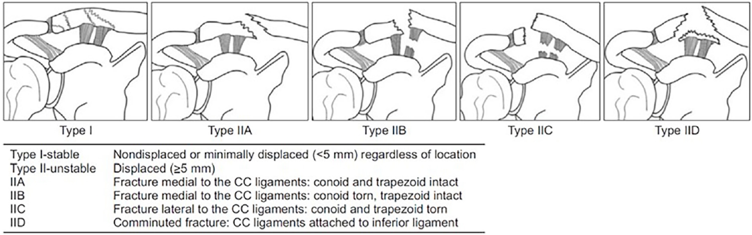
Cho classification system for distal clavicle fractures. Type I fractures are stable, nondisplaced or minimally displaced fractures (<5 mm), regardless of location. Type II fractures are unstable, displaced fractures (≥5 mm): type IIA, fracture medial to the coracoclavicular (CC) ligaments with intact conoid and trapezoid ligaments (TLs); type IIB, fracture medial to the CC ligaments with a torn conoid ligament and intact TL; type IIC, fracture lateral to the CC ligaments with torn conoid and TLs; and type IID, comminuted fracture with the CC ligaments attached to the inferior fragment. Reprinted from Cho et al. [15] according to the Elsevier user license.
Fig. 2.
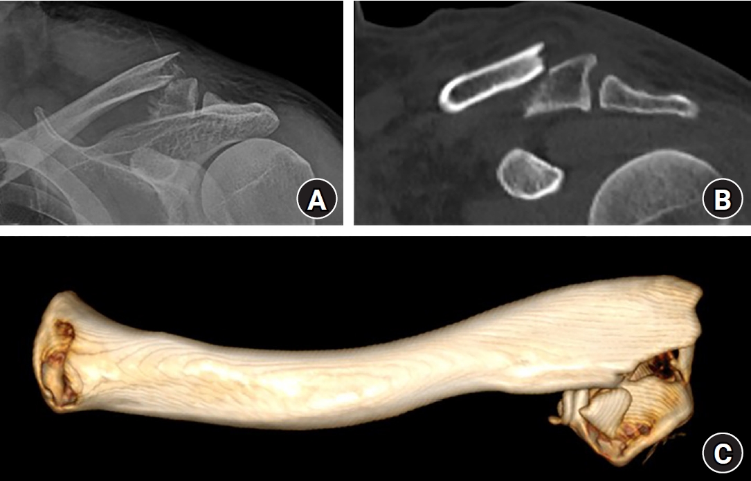
Computed tomography (CT) demonstrating horizontal displacement of a distal clavicle fracture that is not appreciable on standard plain radiographs. (A) Plain radiograph, (B) two-dimensional CT, and (C) three-dimensional CT.
Fig. 3.
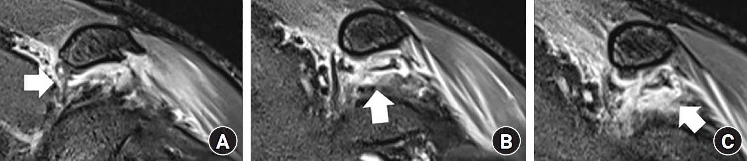
(A–C) Magnetic resonance imaging demonstrating direct visualization of coracoclavicular ligament disruption (arrow), which cannot be reliably assessed on plain radiographs alone.
Fig. 4.
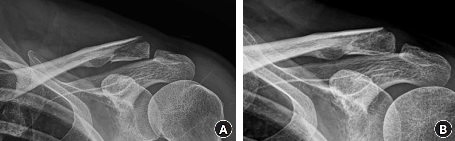
(A) A 53-year-old female patient with a minimally displaced distal clavicle fracture (displacement <5 mm; Neer type I, Cho type I) managed conservatively. (B) Radiographic union confirmed at 5 months of follow-up.
Fig. 5.

(A) A 27-year-old female patient with an unstable distal clavicle fracture (Cho type IIC). (B, C) Surgical fixation was performed using coracoclavicular stabilization with a mini-fragment plate, achieving anatomic reduction and stable fixation.
Table 1.
- 1. Twomey-Kozak J, Whitlock KG, O'Donnell JA, Klifto CS, Anakwenze O. Epidemiology of sports-related clavicle fractures in the United States: injuries from 2015 to 2019. Orthop J Sports Med 2022;10:23259671221126553.ArticlePubMedPMCPDF
- 2. Sayed M, Sutar S, Kamel F, Faleel A, Dwyer AJ. Temporal trends in the management of midshaft clavicle fractures: a systematic review. Cureus 2025;17:e96532. ArticlePubMedPMC
- 3. Tryggedsson I, Viberg B, Gundtoft PH, et al. Increasing incidences and changes in treatment trends of clavicle fractures in adults during 2 decades in Denmark: a nationwide study on data from the Danish National Patient Registry. Acta Orthop 2025;96:135-41.ArticlePMCPDF
- 4. Ziroglu N, Ciftci MU, Duramaz A, Kural C, Koluman AC. Epidemiological changes in clavicle fractures during the COVID-19 pandemic: a six-year analysis from a large single-center cohort. J Orthop Surg Res 2026;21:209.ArticlePubMedPMCPDF
- 5. Hop JC, Richman EH, Marchetti DC, Royster BW, Gorman MA, Alfonso NA. Surgical fixation of clavicle fractures in the elderly: utilization and compensation over two decades. Eur J Orthop Surg Traumatol 2026;36:85.ArticlePubMedPDF
- 6. Bishop JY, Jones GL, Lewis B, MOON Shoulder Group. Intra- and interobserver agreement in the classification and treatment of distal third clavicle fractures. Am J Sports Med 2015;43:979-84.ArticlePubMedPDF
- 7. Stenson J, Baker W. Classifications in brief: the modified Neer classification for distal-third clavicle fractures. Clin Orthop Relat Res 2021;479:205-9.ArticlePubMedPMC
- 8. Uittenbogaard SJ, van Es LJ, den Haan C, van Deurzen DF, van den Bekerom MP. Outcomes, union rate, and complications after operative and nonoperative treatments of Neer type II distal clavicle fractures: a systematic review and meta-analysis of 2284 patients. Am J Sports Med 2023;51:534-44.ArticlePubMedPDF
- 9. Thurston D, Jordan RW, Thangarajah T, et al. Are displaced distal clavicle fractures associated with inferior clinical outcomes following nonoperative management? A systematic review. J Shoulder Elbow Surg 2024;33:1847-57.ArticlePubMed
- 10. Yue L, Huang C, Zhang J, Wang Z, Wang S, Sun H. Treatment strategies for distal clavicle fractures: a narrative review. Orthop Res Rev 2025;17:221-7.ArticlePubMedPMCPDF
- 11. Peebles LA, Aman ZS, Kraeutler MJ, Mulcahey MK. Qualitative and quantitative anatomic descriptions of the coracoclavicular and acromioclavicular ligaments: a systematic review. Arthrosc Sports Med Rehabil 2022;4:e1545-e55.ArticlePubMedPMCPDF
- 12. Chung ST, Yoo JH, Joh JW, Kim SY, Bak DJ. Structural analysis of the coracoclavicular ligaments in Koreans: a cadaveric study. J Korean Orthop Assoc 2010;45:222-7.Article
- 13. Lin J, Sun WJ, Chen JH, et al. Comparison of interobserver agreement of four classification systems for lateral clavicle fractures between two groups of surgeons: a multicenter study. Orthop Surg 2023;15:2138-43.ArticlePubMedPMCPDF
- 14. Fleischhacker E, Siebenburger G, Gleich J, Bocker W, Gilbert F, Helfen T. The accuracy of distal clavicle fracture classifications-do we need an amendment to imaging modalities or fracture typing. J Clin Med 2022;11:5638.ArticlePubMedPMC
- 15. Cho CH, Kim BS, Kim DH, Choi CH, Dan J, Lee H. Distal clavicle fractures: a new classification system. Orthop Traumatol Surg Res 2018;104:1231-5.ArticlePubMed
- 16. Sandstrom CK, Gross JA, Kennedy SA. Distal clavicle fracture radiography and treatment: a pictorial essay. Emerg Radiol 2018;25:311-9.ArticlePubMedPDF
- 17. Liu J, Mi J, Jin Y, et al. Three-dimensional mapping of distal clavicle fractures: displacement patterns and clinical implications for surgical management. Orthop Surg 2025;17:1656-68.ArticlePubMedPMC
- 18. Hall JA, Schemitsch CE, Vicente MR, et al. Operative versus nonoperative treatment of acute displaced distal clavicle fractures: a multicenter randomized controlled trial. J Orthop Trauma 2021;35:660-6.ArticlePubMed
- 19. Andersen K, Jensen PO, Lauritzen J. Treatment of clavicular fractures: figure-of-eight bandage versus a simple sling. Acta Orthop Scand 1987;58:71-4.PubMed
- 20. Malik SS, Tahir M, Remtulla M, Malik S, Jordan RW. A systematic review and meta-analysis comparing the use of hook plates and superior plates in the treatment of displaced distal clavicle fractures. Arch Orthop Trauma Surg 2023;143:329-52.ArticlePubMedPDF
- 21. Stegeman SA, Nacak H, Huvenaars KH, Stijnen T, Krijnen P, Schipper IB. Surgical treatment of Neer type-II fractures of the distal clavicle: a meta-analysis. Acta Orthop 2013;84:184-90.PubMedPMC
- 22. Elrih M, Quinlan J. Hook plate versus distal locking plate for the fixation of unstable distal clavicle injuries, outcomes and complications: a meta-analysis. Cureus 2022;14:e30806. ArticlePubMedPMC
- 23. Asadollahi S, Bucknill A. Hook plate fixation for acute unstable distal clavicle fracture: a systematic review and meta-analysis. J Orthop Trauma 2019;33:417-22.ArticlePubMed
- 24. Li L, Li TY, Jiang P, et al. Clavicle hook plate versus distal clavicle locking plate for Neer type II distal clavicle fractures. J Orthop Surg Res 2019;14:472.ArticlePubMedPMCPDF
- 25. Beisemann N, Spiller YM, Schnetzke M, Grutzner PA, Nolte PC. Hook plate fixation of Neer type II distal clavicle fractures results in satisfactory patient-reported outcomes but complications and revisions are high. BMC Musculoskelet Disord 2023;24:850.ArticlePubMedPMCPDF
- 26. Fox HM, Ramsey DC, Thompson AR, Hoekstra CJ, Mirarchi AJ, Nazir OF. Neer type-ii distal clavicle fractures: a cost-effectiveness analysis of fixation techniques. J Bone Joint Surg Am 2020;102:254-61.PubMed
- 27. Erdle B, Izadpanah K, Jaeger M, et al. Comparative analysis of locking plate versus hook plate osteosynthesis of Neer type IIB lateral clavicle fractures. Arch Orthop Trauma Surg 2017;137:651-62.ArticlePubMedPDF
- 28. Liu ZQ, Zhang MS, Zhou ZF, Zhang L, Zheng LP. Comparative study of three different fixation techniques for the treatment of Neer type IIb distal clavicle fractures: a retrospective cohort study. Front Surg 2023;10:1100720.ArticlePubMedPMC
- 29. Xu H, Chen WJ, Zhi XC, Chen SC. Comparison of the efficacy of a distal clavicular locking plate with and without a suture anchor in the treatment of Neer IIb distal clavicle fractures. BMC Musculoskelet Disord 2019;20:503.ArticlePubMedPMCPDF
- 30. Memon K, Awan M, Abro AA, Ahmad S, Shehata Abdelmesih SR. Outcomes of fixation of lateral-end clavicle fractures using locking clavicular plate with acromioclavicular TightRope augmentation: a meta-analysis. Cureus 2025;17:e100119. ArticlePubMedPMC
- 31. Xu Y, Guo X, Peng H, Dai H, Huang Z, Zhao J. Different internal fixation methods for unstable distal clavicle fractures in adults: a systematic review and network meta-analysis. J Orthop Surg Res 2022;17:43.ArticlePubMedPMCPDF
- 32. Boonard M, Sumanont S, Arirachakaran A, et al. Fixation method for treatment of unstable distal clavicle fracture: systematic review and network meta-analysis. Eur J Orthop Surg Traumatol 2018;28:1065-78.ArticlePubMedPDF
- 33. Malik SS, Malik S, Tahir M, Jordan RW, Laprus H, D'Alessandro P. Open coraco-clavicular ligament reconstruction (CCLR) in treatment of displaced distal clavicle fractures has low complication rate and excellent union rate: a systematic review. Arch Orthop Trauma Surg 2023;143:1459-77.ArticlePubMedPDF
- 34. Panagopoulos A, Solou K, Nicolaides M, Triantafyllopoulos IK, Kouzelis A, Kokkalis ZT. Coracoclavicular fixation techniques for Neer IIb and "extralateral" fractures of the distal clavicle: a systematic review. JSES Rev Rep Tech 2024;4:676-83.ArticlePubMed
- 35. Yagnik GP, Seiler JR, Vargas LA, Saxena A, Narvel RI, Hassan R. Outcomes of arthroscopic fixation of unstable distal clavicle fractures: a systematic review. Orthop J Sports Med 2021;9:23259671211001773.ArticlePubMedPMCPDF
- 36. Malik SS, Tahir M, Malik S, Kwapisz A, Jordan RW. Arthroscopically assisted coraco-clavicular ligament reconstruction in treatment of acute displaced distal clavicle fractures provides good to excellent shoulder function despite low union rates and high complication rates: a systematic review. Arthroscopy 2022;38:567-82.ArticlePubMedPDF
- 37. Flinkkila T, Heikkila A, Sirnio K, et al. TightRope versus clavicular hook plate fixation for unstable distal clavicular fractures. Eur J Orthop Surg Traumatol 2015;25:465-9.ArticlePubMedPDF
- 38. Singh A, Schultzel M, Fleming JF, Navarro RA. Complications after surgical treatment of distal clavicle fractures. Orthop Traumatol Surg Res 2019;105:853-9.ArticlePubMed
References
Figure & Data
REFERENCES
Citations
Citations to this article as recorded by 
 ePub Link
 Cite
Cite XML Download
Current concepts in distal clavicle fractures: an evidence-based update
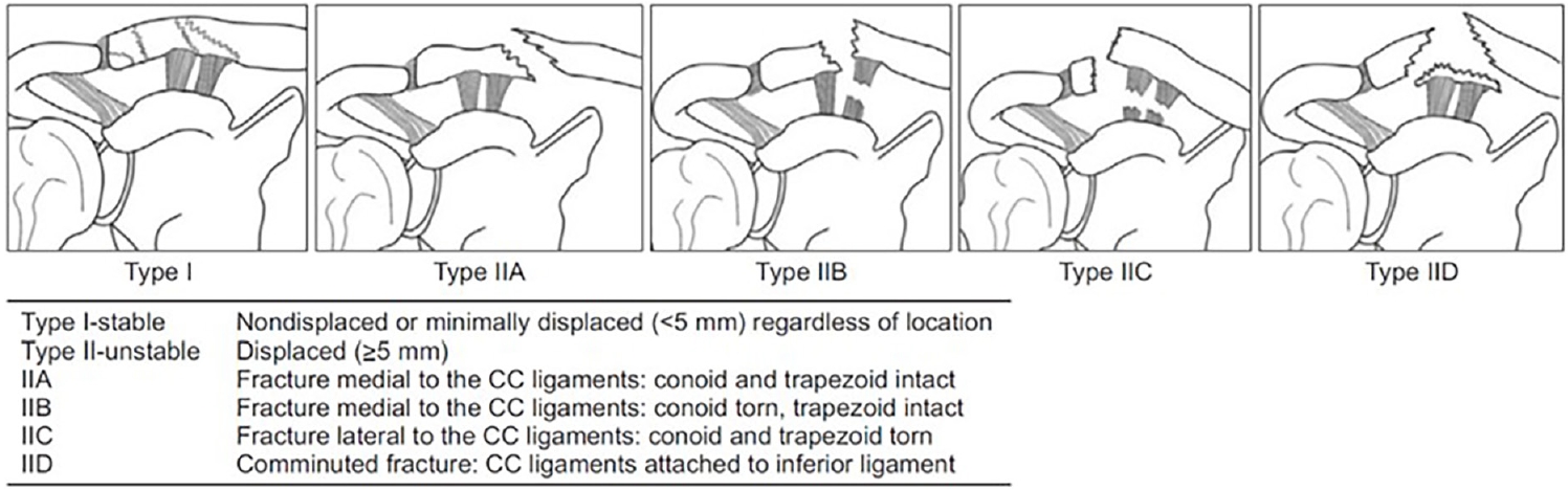
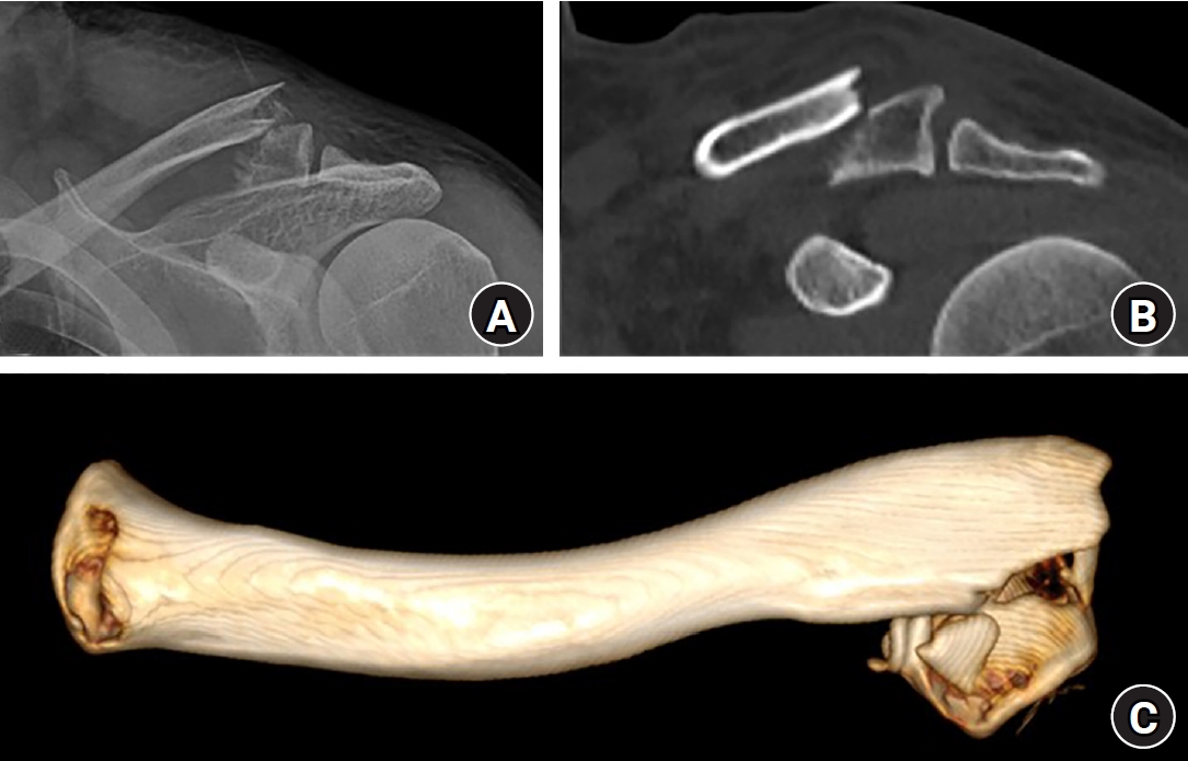
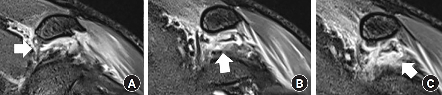
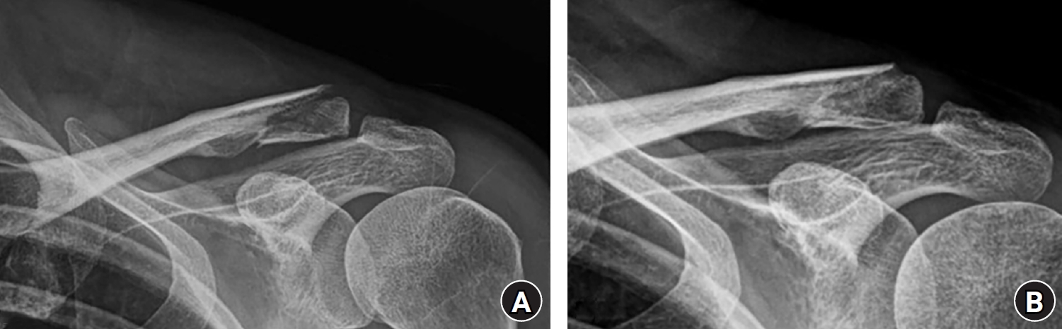

Fig. 1. Cho classification system for distal clavicle fractures. Type I fractures are stable, nondisplaced or minimally displaced fractures (<5 mm), regardless of location. Type II fractures are unstable, displaced fractures (≥5 mm): type IIA, fracture medial to the coracoclavicular (CC) ligaments with intact conoid and trapezoid ligaments (TLs); type IIB, fracture medial to the CC ligaments with a torn conoid ligament and intact TL; type IIC, fracture lateral to the CC ligaments with torn conoid and TLs; and type IID, comminuted fracture with the CC ligaments attached to the inferior fragment. Reprinted from Cho et al. [15] according to the Elsevier user license.
Fig. 2. Computed tomography (CT) demonstrating horizontal displacement of a distal clavicle fracture that is not appreciable on standard plain radiographs. (A) Plain radiograph, (B) two-dimensional CT, and (C) three-dimensional CT.
Fig. 3. (A–C) Magnetic resonance imaging demonstrating direct visualization of coracoclavicular ligament disruption (arrow), which cannot be reliably assessed on plain radiographs alone.
Fig. 4. (A) A 53-year-old female patient with a minimally displaced distal clavicle fracture (displacement <5 mm; Neer type I, Cho type I) managed conservatively. (B) Radiographic union confirmed at 5 months of follow-up.
Fig. 5. (A) A 27-year-old female patient with an unstable distal clavicle fracture (Cho type IIC). (B, C) Surgical fixation was performed using coracoclavicular stabilization with a mini-fragment plate, achieving anatomic reduction and stable fixation.
Fig. 1.
Fig. 2.
Fig. 3.
Fig. 4.
Fig. 5.
Current concepts in distal clavicle fractures: an evidence-based update
| Variable | Hook plate | Anatomic locking plate (±CC augmentation) | CC stabilization (open or arthroscopic) |
|---|---|---|---|
| Indication | Neer V / Cho IID fractures; severe comminution; very small distal fragment | Neer IIA / Cho IIA fractures; adequate distal fragment (≥3 screws) | Neer IIB / Cho IIB/IIC fractures; small or comminuted distal fragment |
| Advantage | Broadly applicable; high union rate; no need for distal fragment purchase | Direct fixation; no subacromial contact; planned hardware removal not required | No distal fragment required; low major complication rate; concomitant shoulder pathology addressable (arthroscopic) |
| Limitation | Planned removal obligatory; subacromial impingement; restricted ROM during healing | Requires adequate distal fragment; limited applicability in severe comminution | Indirect fracture reduction; technically demanding; coracoid exposure risk |
| Union rate | 95%–98% | 95%–98% | 70%–98% |
| Complication rate | Overall, 32.7%; acromial osteolysis, 11.3% | Overall, 12.7% | 0–28.6% |
| Reoperation | ~92.5% (planned implant removal) | Low (~1.3% for suture-button augmentation) | 6%–8% (hardware-related) |
Table 1. Comparison of major fixation techniques for unstable distal clavicle fractures [8,20-36]
CC, coracoclavicular; ROM, range of motion.


