
 E-submission
 KOTA
 TOTA
 TOTS

Articles
- Page Path
- HOME > > Ahead-of print articles > Article
-
Original Article
- Percutaneous Kirschner-wire fixation followed by an opencast to permit protected daily and sports activities in patients with tendinous mallet finger: a case series at a tertiary hospital in Korea
- Hyeon Jun Kim, Ji Woong Kim, Min Kyun Cho, Sung Yoon Jung
-
DOI: https://doi.org/10.12671/jmt.2026.00269
Published online: October 20, 2026
Department of Orthopedic Surgery, Dong-A University Hospital, Dong-A University School of Medicine, Busan, Korea
- Correspondence to: Sung Yoon Jung Department of Orthopedic Surgery, Dong-A University Hospital, 26 Daesingongwon-ro, Seo-gu, Busan 49201, Korea Tel: +82-51-240-5160 Email: jungsy@dau.ac.kr
• Received: August 5, 2026 • Revised: August 30, 2026 • Accepted: September 10, 2026
© 2026 The Korean Orthopaedic Trauma Association
This is an Open Access article distributed under the terms of the Creative Commons Attribution Non-Commercial License (http://creativecommons.org/licenses/by-nc/4.0/) which permits unrestricted noncommercial use, distribution, and reproduction in any medium, provided the original work is properly cited.
- 46 Views
- 1 Download


Abstract
-
Background Dong-A University Hospital’s protocol for tendinous mallet finger requires 12 weeks of continuous full-time distal interphalangeal (DIP) joint splinting, which may restrict activities and complicate hygiene. This study evaluated the feasibility, short-term safety, and 6-month clinical outcomes of buried Kirschner-wire fixation followed by opencast application.
-
Methods This retrospective case series screened 36 patients with mallet finger between January 2024 and October 2025. Twelve met the exclusion criteria, leaving 24 eligible patients; eight elected nonoperative treatment, and 16 chose surgery for acute closed tendinous mallet finger and constituted the study cohort. The DIP joint was fixed in approximately 15° of hyperextension using one or two buried 1.1-mm Kirschner wires. An opencast was applied at 1 week. The wires were routinely removed and active motion was initiated at 8 weeks, followed by a removable brace during outdoor or sports activities for 4 weeks. Outcomes were evaluated at 6 months.
-
Results The mean preoperative extension lag was 30.0°. At 6 months, the mean residual extension lag was 1.9°, no flexion loss was observed, and Crawford outcomes were excellent in 62.5% and good in 37.5%. Two superficial pin-site infections resolved with oral antibiotics and local wound care. A separate case of wire loosening with painful subcutaneous prominence was managed with an aluminum splint. All patients resumed daily activities and showering at 1 week, when protected sports were also permitted; exact return dates and activity intensity were not recorded.
-
Conclusions Buried Kirschner-wire fixation followed by opencast application may be an option for selected patients who wish to avoid prolonged full-time splinting. The protocol permitted protected activities from week 1 and active DIP motion from week 8; these findings support its feasibility but do not establish earlier return to unrestricted activity.
-
Level of evidence IV.
Introduction
Mallet finger is an injury characterized by disruption of the terminal extensor tendon at the distal interphalangeal (DIP) joint, resulting in loss of active extension. Mallet finger is classified as bony or tendinous depending on whether an avulsion fracture fragment is present [1,2]. Tendinous mallet finger falls within Doyle type I and typically results from a sudden flexion force applied to an extended fingertip. It is commonly encountered among individuals participating in sports, particularly ball sports [1,3,4].
The standard treatment for tendinous mallet finger is nonoperative management with continuous immobilization of the DIP joint in extension or slight hyperextension [5,6]. In our institutional protocol, continuous full-time DIP joint splinting is maintained for 12 weeks to allow the terminal extensor tendon to heal through scar formation and acquire sufficient tensile strength. Several studies and systematic reviews have reported no significant difference in final clinical outcomes between nonoperative and operative treatment of mallet finger injuries [7,8]. Considerable variation in treatment strategies among countries and surgeons has also been noted [9]. Accordingly, the rationale for surgery in tendinous mallet finger lies not in achieving superior final clinical outcomes or less residual extension lag, but in reducing the burden of treatment and allowing earlier functional participation. A treatment that maintains healing while permitting hygiene, routine hand use, and protected sports participation may improve convenience, satisfaction, and quality of life during the treatment period.
The problems associated with prolonged splinting can be summarized in three points. First, treatment success depends entirely on patient compliance [6]. If a patient removes the splint for cleaning or because of discomfort and allows the DIP joint to flex, the healing terminal tendon is stretched again, effectively requiring treatment to restart from the beginning. Second, continuous splint wear may cause skin maceration, malodor, contact dermatitis, and even skin ulceration. Stern and Kastrup [10] reported a 45% complication rate among 84 splinted digits; most complications were skin-related and transient. Restrictions on hand washing, routine hand use, and sports participation can delay return to usual activities and reduce patient satisfaction and quality of life during treatment. Third, as swelling subsides, the splint may loosen and the DIP joint immobilization angle may change subtly; consequently, residual extension lag can be difficult to control completely even in compliant patients [11].
Importantly, surgeons’ concerns do not always align with patients’ priorities. Surgeons tend to focus on a few degrees of residual extension lag, whereas patients may place greater importance on the burden of prolonged full-time splinting, including hygiene problems, inability to use the hand normally, and restrictions on sports. When final clinical outcomes are expected to be broadly comparable, these differences in the treatment experience should be considered during shared decision-making.
We therefore adapted the technique of subcutaneous Kirschner wire (K-wire) burial and opencast immobilization, which we previously reported for bony mallet finger, to the treatment of tendinous mallet finger [12].
The purpose of this study was to evaluate the feasibility and short-term safety of this treatment protocol and to report the 6-month clinical outcomes and functional progression permitted during treatment in a consecutive surgical series. We hypothesized that, without necessarily improving the final clinical result compared with nonoperative treatment, the protocol could reduce the restrictions associated with our 12-week institutional nonoperative splinting protocol and facilitate earlier return to daily and sports activities.
Methods
This study was approved by the Institutional Review Board of Dong-A University Hospital (No. DAUHIRB-26-205). Informed consent was obtained from all study participants. Written informed consent for publication of identifiable clinical photographs was obtained from the patients whose images are included in this article.
This study was a retrospective, consecutive case series.
Between January 2024 and October 2025, 36 patients with mallet finger were screened for eligibility at Dong-A University Hospital.
All procedures were performed under digital block anesthesia with mobile fluoroscopic guidance. Prophylactic intravenous antibiotics were administered before K-wire insertion. After sterile preparation, a rubber tourniquet fashioned from the finger of a surgical glove was applied. The DIP joint was manually reduced, and the operating surgeon used a handheld goniometer to position it at a target of approximately 15° of hyperextension, using the longitudinal axes of the middle and distal phalanges as reference lines. The angle was rounded to the nearest 5° and was subsequently confirmed on lateral fluoroscopic imaging. The achieved angle was not recorded separately for each patient, and no serial angular measurement was performed at K-wire removal. The target position was selected to compensate, as far as possible, for the residual extension lag that commonly develops during healing. Care was taken to avoid excessive hyperextension because perfusion of the dorsal skin over the DIP joint decreases as hyperextension increases, potentially causing dorsal skin necrosis.
A 1.1-mm K-wire was then inserted centrally along the longitudinal axis from the tip of the distal phalanx, passed across the DIP joint, and advanced to the proximal one-third of the middle phalanx. One K-wire was used in four patients, whereas two K-wires were used in 12 patients. A second wire was added when rotational instability was a concern or fixation with a single wire was considered insufficient, using a symmetric configuration on the anteroposterior view [13]. Joint congruity and maintenance of approximately 15° of hyperextension were reconfirmed on anteroposterior and lateral fluoroscopic images.
Using a dedicated cutter (Fig. 1), each K-wire was cut sufficiently short to allow burial beneath the skin [12]. The insertion trajectory was kept away from the nail bed and nail matrix. The skin over the wire insertion site was closed with 5-0 nylon sutures. After release of the tourniquet and before completion of the procedure, the operating surgeon, an orthopedic hand specialist, inspected the nail bed and eponychium and performed a capillary refill test of the fingertip. Intact skin circulation was confirmed in all cases. To accommodate postoperative swelling and protect the wound, temporary immobilization with an aluminum splint extending to the proximal interphalangeal joint was applied. An additional aluminum splint was fabricated in the operating room and provided to each patient in case wire loosening or loss of protection occurred.
At the first outpatient visit 1 week postoperatively, the sutures were removed and tissue adhesive was applied. The temporary splint was then replaced with a custom-fitted opencast immobilizing only the DIP joint while leaving the proximal interphalangeal joint free [12]. The opencast was fabricated and applied by an experienced cast technician under physician supervision. Its exact material composition could not be specified because the manufacturer did not disclose the proprietary formulation. Showering and protected daily activities were permitted from that time. Running and protected sports requiring hand use, including golf, basketball, and tennis, were also permitted with the opencast in place. Because swelling reduction or external impact could result in wire or construct loosening, patients were instructed to reapply the spare aluminum splint and present immediately to the outpatient clinic or emergency department if loosening was suspected. Patients were followed at 1-week intervals.
K-wire removal was routinely scheduled at 8 weeks postoperatively. Removal was performed without hospitalization at the ambulatory surgery center under local anesthesia, required approximately 5 minutes, and cost approximately 120,000 Korean won. A short course of oral antibiotics was prescribed after K-wire removal; no additional intravenous antibiotics were administered for the removal procedure. Tissue adhesive was applied to the wire-removal sites, and active DIP joint motion was permitted immediately thereafter. During the subsequent 4 weeks, until 12 weeks postoperatively, continuous immobilization was no longer required for routine daily activities; patients wore a removable mallet brace only during outdoor or sports activities. From 12 weeks onward, unrestricted activities were permitted. When wire loosening or painful subcutaneous prominence was suspected, the spare aluminum splint was applied and the patient was reassessed promptly.
Doyle type I injury was defined as a closed or blunt injury with loss of terminal extensor tendon continuity, with or without a small avulsion fracture [1]. In this study, tendinous mallet finger was defined as a Doyle type I injury with loss of active DIP joint extension and without an avulsion fragment on plain radiographs. Exclusion criteria were an open injury, bony mallet finger with an avulsion fragment, presentation more than 2 weeks after injury, poor dorsal skin condition over the DIP joint, a high risk of delayed wound healing such as diabetes mellitus, and follow-up of less than 6 months. Nonoperative treatment consisting of continuous full-time DIP extension splinting for 12 weeks was recommended to all 24 eligible patients. Surgery was indicated only for acute injuries treated within 2 weeks of injury.
Extension lag was defined as the degree to which the DIP joint failed to reach the neutral position during active extension, and flexion loss was measured in comparison with the contralateral unaffected digit [11]. Outcomes were graded according to the Crawford criteria [14]. Resumption of daily activities was defined as return to routine self-care and occupational hand use, including washing, dressing, eating, and work duties. Showering was assessed separately, with patients advised to avoid soaking or direct friction around the healing wound. Pin-site infection, pin protrusion, wire loosening, nail deformity, and dorsal skin complications were also assessed.
Preoperative extension lag and DIP joint range of motion at follow-up were measured directly with a handheld goniometer by the operating surgeon, an orthopedic hand specialist. Measurements were rounded to the nearest 5° because finer precision was not considered clinically reliable. Residual pain was measured at 6 months using a visual analog scale (VAS), with scores ranging from 0 (no pain) to 10 (worst pain imaginable). Permission to resume sports and subsequent sports participation were assessed by direct patient questioning during follow-up visits; however, the exact date and intensity of return to each sport were not systematically recorded. All angular measurements reported in this study were performed directly by the operating surgeon.
To minimize selection bias, all consecutive patients who underwent surgery during the study period were included without selective omission, although angular measurements by the operating surgeon without an independent assessor may have introduced observer bias.
No a priori sample size calculation was performed because this was a descriptive case series; the study size was determined by all consecutive eligible patients who chose surgical treatment between January 2024 and October 2025.
Because this was a small, single-arm case series, only descriptive analyses were performed. Continuous variables were summarized as means and ranges, and categorical variables as numbers and percentages. No inferential statistical testing was performed.
Results
Twelve patients were excluded: four because of a bony avulsion fragment, four because they presented more than 2 weeks after injury, two because follow-up was less than 6 months (both treated nonoperatively), one because of an open injury, and one because of diabetes mellitus with a high risk of delayed wound healing. This left 24 eligible patients. Eight patients declined surgery and elected nonoperative treatment, whereas 16 chose surgical treatment and constituted a consecutive surgical series; no surgically treated patient was selectively omitted. None of the 16 surgical patients underwent a trial of splint treatment before surgery because surgery was performed within 1–2 days after presentation. The reasons for choosing surgery were an unwillingness to undergo 12 weeks of full-time splinting in four patients, a desire for early return to sports in eight, and the need for continued hand use at work in four. The mean injury-to-surgery interval was approximately 7 days (maximum, 13 days). The mean patient age was 49 years (range, 36–74 years); 12 patients were male and four were female. The index finger was affected in four patients, the middle finger in six, the ring finger in two, and the little finger in four. All injuries were sustained during sports: soccer in eight patients, basketball in four, and dodgeball in four. The dominant hand was involved in 14 patients and the nondominant hand in two.
The preoperative mean extension lag was 30.0° (range, 15°–45°). Before surgery, the deformity in all 16 patients was passively correctable to the neutral position. One K-wire was used in four patients and two K-wires in 12. All patients completed the scheduled follow-up through 12 weeks and underwent final clinical assessment 6 months postoperatively; none was lost before the 6-month assessment.
All patients resumed routine daily activities and showering with wound precautions 1 week postoperatively. With the opencast in place, running and sports requiring hand use, including golf, basketball, and tennis, were permitted from the same time point. Subsequent participation in sports was confirmed by direct patient questioning at follow-up, but the exact return date and intensity were not systematically recorded. Active DIP joint motion was initiated after routine K-wire removal at 8 weeks. During the subsequent period through postoperative week 12, patients wore a removable mallet brace only during outdoor or sports activities. At the 6-month evaluation, the VAS pain score was 0 in all 16 patients. The mean residual extension lag was 1.9° (range, 0°–5°), and no patient had flexion loss compared with the contralateral digit. According to the Crawford criteria [14], outcomes were excellent in 10 patients (62.5%) and good in six (37.5%); there were no fair or poor outcomes (Table 1).
Two patients developed superficial pin-site infections; both resolved with oral antibiotics and local wound care without additional surgery. One separate patient developed wire loosening with painful subcutaneous prominence, which was managed with the spare aluminum splint and close follow-up until routine wire removal at 8 weeks; this event did not overlap with either pin-site infection. After tourniquet release at the end of surgery, the nail bed and eponychium were inspected and capillary refill was confirmed to be intact in all patients. No dorsal skin necrosis, permanent or transient nail deformity, unplanned reoperation, or other major complication was observed.
A 53-year-old man presented 5 days after trauma to the left little finger with inability to extend the DIP joint. Plain radiographs showed no avulsion fragment, and physical examination confirmed acute tendinous mallet finger with loss of active extension of the DIP joint (Fig. 2). Under digital block anesthesia, the DIP joint was reduced in approximately 15° of hyperextension, with the angle directly measured by the operating surgeon, and a K-wire was inserted from the fingertip across the DIP joint. The wire was cut short using the dedicated cutter, buried beneath the skin, and the insertion site was closed with 5-0 nylon sutures (Fig. 3). After tourniquet release, the nail bed and eponychium were inspected and normal capillary refill was confirmed before completion of the procedure. At 1 week postoperatively, the sutures were removed and an opencast was applied; showering, daily activities, and protected sports activities were permitted (Fig. 4). At 8 weeks, the K-wire was removed at the ambulatory surgery center under local anesthesia (Fig. 5), and active DIP joint motion was initiated. Until 12 weeks postoperatively, the patient wore a removable mallet brace only during outdoor or sports activities (Fig. 6).
Discussion
In this study, we evaluated the feasibility and short-term safety of treating acute tendinous mallet finger within 2 weeks of injury using percutaneous K-wire fixation of the DIP joint in approximately 15° of hyperextension, together with the 6-month clinical outcomes. Of 36 patients screened, 12 were excluded according to the prespecified exclusion criteria. All 24 eligible patients were counseled regarding 12 weeks of nonoperative splinting; eight declined surgery and elected nonoperative treatment, whereas 16 underwent the present procedure. The wires were buried beneath the skin, and the temporary splint was replaced with an opencast at 1 week, permitting showering and protected daily and sports activities, including activities requiring hand use. The wires were routinely removed at 8 weeks, after which active DIP motion was initiated; a removable brace was then worn only during outdoor or sports activities for 4 weeks. This approach addresses a different clinical priority from the final extension result. Although operative and nonoperative treatment may achieve comparable final outcomes, the present protocol was designed to reduce restrictions during treatment, facilitate earlier return to daily and sports activities, and potentially improve convenience, satisfaction, and quality of life.
Practical problems arise during splint treatment, the most fundamental of which is compliance. Because the terminal tendon has a limited blood supply and heals through scar formation, interruption of immobilization allows it to stretch again [15]. Even brief DIP joint flexion while washing the hand without the splint may disrupt healing and necessitate restarting the immobilization period. Accordingly, under our institutional nonoperative treatment protocol, all patients were initially counseled to undergo continuous full-time DIP joint splinting for 12 weeks. With the present technique, internal K-wire fixation maintained the DIP position and allowed showering and protected daily and sports activities from 1 week. Routine K-wire removal at 8 weeks enabled active DIP motion 4 weeks earlier than completion of the 12-week full-time nonoperative splinting protocol, although a removable brace was still used during outdoor or sports activities until week 12. This represents earlier functional progression rather than proof of faster tendon healing. Nevertheless, earlier progression from rigid immobilization to active motion and continued participation in daily and protected sports activities are practical advantages that may lessen the effect of treatment on work, recreation, and quality of life.
A second concern is skin and hygiene-related complications. Stern and Kastrup reported a 45% complication rate among 84 splinted digits; most complications were skin-related and transient [10]. A continuously enclosed splint creates a warm, moist environment through perspiration, leading to maceration and malodor; this is also a common reason for patients to remove the splint without medical advice. Thus, hygiene and compliance problems are causally linked rather than independent. With the present technique, the use of an opencast and tissue adhesive from 1 week postoperatively allows direct wound cleansing and showering while reducing occlusion-related perspiration and maceration [12]. The absence of exposed wires and protection with an opencast may make selected sports activities more practical; however, activities involving substantial direct impact to the affected finger should be avoided. By reducing restrictions on washing, routine hand use, and protected activity, this protocol may also improve treatment convenience and indirectly support compliance.
A third concern is control of extension lag. As swelling subsides during splint treatment, the splint may loosen and the actual immobilization position of the DIP joint may gradually shift toward flexion. Over time, these subtle positional changes may contribute to residual extension lag [11]. Because some residual extension loss commonly occurs after treatment, we fixed the DIP joint in approximately 15° of hyperextension rather than neutral in an attempt to compensate for this loss. The mean residual extension lag of 1.9° at 6 months is consistent with this rationale, but the absence of a control group prevents a causal conclusion.
The degree of hyperextension must nevertheless be selected carefully. Rayan and Mullins reported that perfusion of the dorsal skin over the DIP joint decreases in proportion to the degree of hyperextension and that excessive hyperextension may cause skin necrosis [16]. We selected 15° as a compromise that we considered sufficient to compensate for extension loss after K-wire removal without substantially compromising dorsal skin perfusion. In addition, after tourniquet release, the operating orthopedic hand specialist inspected the nail bed and eponychium and confirmed intact capillary refill before completion of each procedure. No dorsal skin necrosis occurred in this study.
K-wire removal was routinely scheduled at 8 weeks based on the time required for the healing terminal tendon to acquire sufficient tensile strength [6]. It was completed at the ambulatory surgery center without hospitalization under local anesthesia in approximately 5 minutes, at a cost of approximately 120,000 Korean won. Active DIP motion was initiated after removal. Compared with our institutional protocol of 12 weeks of continuous full-time nonoperative splinting, this protocol allowed active DIP motion from 8 weeks in routine cases; a removable mallet brace was used only during outdoor or sports activities for the subsequent 4 weeks. Thus, the protocol shortened the period of rigid immobilization and facilitated earlier functional progression. These treatment-period advantages may be meaningful to patients even if final clinical outcomes are similar, although comparative studies are required to quantify their effect on satisfaction and quality of life.
Multiple studies have consistently reported no significant difference in final clinical outcomes between nonoperative and operative treatment of mallet finger injuries [8,17]. A Cochrane systematic review also concluded that the evidence was insufficient to support the superiority of any particular treatment [7].
Conventional techniques that leave K-wires exposed outside the skin have been associated with pin-site infection, soft-tissue injury, and wire migration [18,19]. We cut the wires with a dedicated cutter and buried them beneath the skin [12]. Nevertheless, superficial pin-site infection occurred in two patients and resolved with oral antibiotics and local wound care. One separate patient developed wire loosening with painful subcutaneous prominence, which was managed with the spare aluminum splint and close follow-up. To address this risk proactively, a spare aluminum splint was fabricated in the operating room for each patient, and patients were instructed to apply it and seek immediate assessment if loosening was suspected. These findings indicate that wire-related complications remain possible even with subcutaneous burial, although all events in this series were managed without additional surgery.
For chronic or neglected tendinous mallet finger, reported surgical options include tenodermodesis [20], direct terminal tendon repair, spiral oblique retinacular ligament reconstruction, and, in severe cases, DIP joint arthrodesis [2,5]. These procedures require soft-tissue dissection and reconstruction and are therefore relatively invasive and associated with longer recovery. The present technique is a simple percutaneous procedure that does not require joint exposure or extensive soft-tissue dissection. Moreover, failure does not preclude subsequent use of the aforementioned procedures, making it a potentially useful initial surgical option.
The disadvantages of this technique are clear. First, subcutaneous wire burial requires a second procedure for removal, although in our practice this was completed without hospitalization at an ambulatory surgery center under local anesthesia in approximately 5 minutes and cost approximately 120,000 Korean won. Second, wire-related complications remain possible. Two patients developed superficial pin-site infections, and one separate patient developed wire loosening with painful subcutaneous prominence; all events were managed with oral antibiotics, local wound care, close follow-up, or aluminum-splint application, without additional surgery. Third, wires inserted from the fingertip could theoretically injure the nail apparatus. However, the insertion trajectory did not pass through the nail bed or nail matrix, and no transient or permanent nail abnormality was observed. Fourth, there is a theoretical risk of cartilage injury from wires crossing the articular surface.
This study has several limitations. First, it was a retrospective study with a small, self-selected surgical cohort. Of 36 patients screened, 12 were excluded according to the prespecified criteria. Although nonoperative treatment was recommended to all 24 eligible patients, eight declined surgery and elected nonoperative treatment, introducing selection bias. Outside the study period, we also encountered a number of patients with tendinous mallet finger who declined surgical treatment after counseling, further emphasizing that the decision to undergo surgery is strongly influenced by individual preferences regarding treatment burden and activity restrictions. Second, there was no direct comparison with the patients treated nonoperatively; therefore, the magnitude of any advantage in treatment-period convenience, satisfaction, quality of life, or time to return cannot be quantified. Third, all angular measurements were performed by the operating surgeon without an independent assessor, creating a risk of observer bias. Fourth, the 6-month follow-up was relatively short and did not permit assessment of long-term changes in extension lag or DIP degeneration. Fifth, subsequent sports participation was confirmed only by direct patient questioning; the exact timing and intensity of return to each sport were not systematically recorded or independently verified. Finally, no validated instrument was used to quantify satisfaction regarding hygiene and activity restrictions.
Splinting remains the primary treatment for acute closed mallet finger injuries [5,21], and the present technique should not be recommended universally. Rather, it should be considered selectively for patients who are unable or unwilling to accept the burdens inherent in prolonged splint treatment. In such patients, treatment-period convenience and the ability to continue usual activities may be clinically meaningful even when the expected final range of motion is not superior.
Future studies should directly compare final extension lag at different hyperextension angles.
Prospective comparative studies using validated patient-reported outcome measures are required to confirm the treatment-period benefits of this protocol.
Although final clinical outcomes after operative and nonoperative treatment of acute tendinous mallet finger may not differ substantially, the burden experienced during treatment differs between the two approaches. Under our institutional nonoperative treatment protocol, continuous full-time DIP joint splinting is maintained for 12 weeks, which can cause hygiene problems, restrict routine hand use and sports participation, and delay functional return. In contrast, percutaneous K-wire fixation in approximately 15° of hyperextension with subcutaneous wire burial and opencast application permitted showering and protected daily and sports activities from 1 week. Routine wire removal at 8 weeks allowed active DIP joint motion, with a removable brace required only during outdoor or sports activities for the subsequent 4 weeks. Therefore, this technique may reduce the inconvenience associated with prolonged full-time splinting; however, whether it leads to earlier return to activity or improves satisfaction and quality of life remains to be established. The two superficial pin-site infections and the single separate case of wire loosening were managed without additional surgery. Although this technique should not replace standard nonoperative treatment in all patients, it may be a useful alternative for selected patients who prioritize hygiene, continued hand use, sports participation, and fewer treatment-related restrictions.
-
Author contributions
Conceptualization: SYJ. Data curation: HJK, JWK. Methodology: SYJ. Formal analysis: MKC. Validation: SYJ. Investigation: HJK, JWK. Supervision: SYJ. Writing–original draft: HJK, JWK. Writing–review & editing: HJK, JWK, MKC, SYJ. All authors read and approved the final manuscript.
-
Conflicts of interest
No potential conflict of interest relevant to this article was reported.
-
Funding
None.
-
Data availability
All data supporting the findings of this study are available in the manuscript and Table 1.
-
Acknowledgments
None.
-
Supplementary materials
None.
Article Information
Fig. 1.
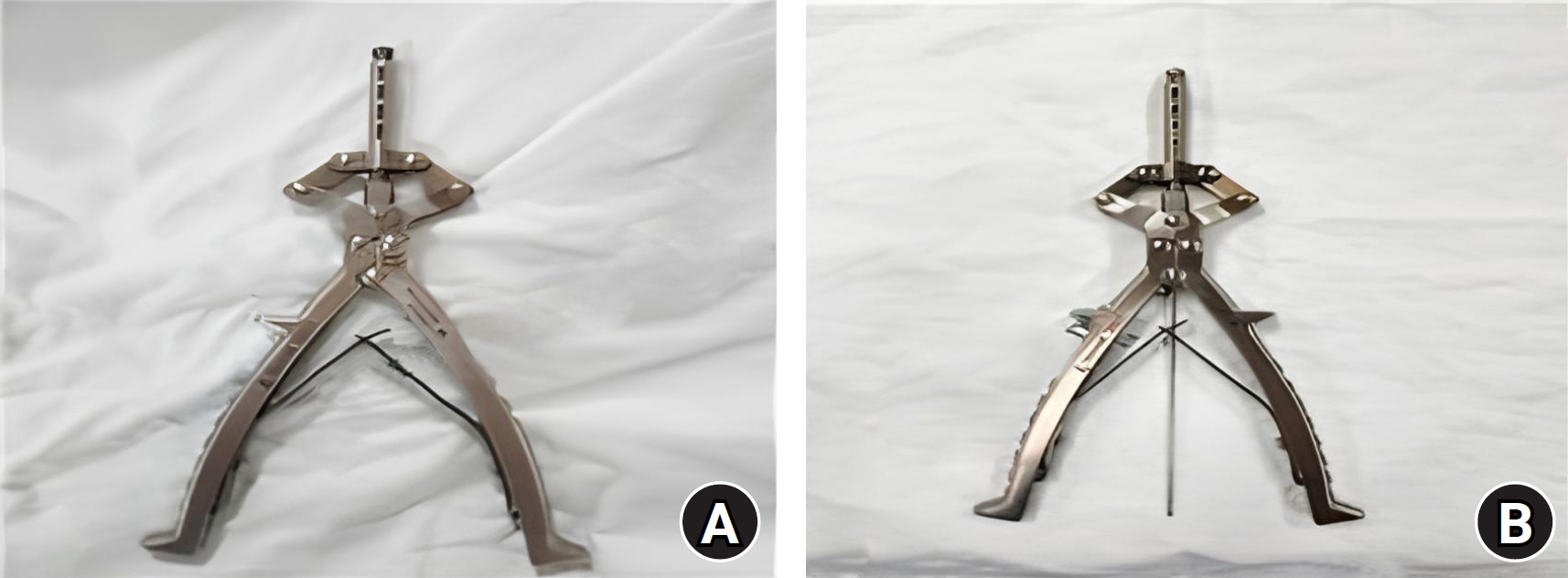
Dedicated Kirschner-wire cutter without (A) and with (B) a Kirschner wire loaded. The cutter allows the wire to be cut close to the bone surface for burial beneath the skin.
Fig. 2.
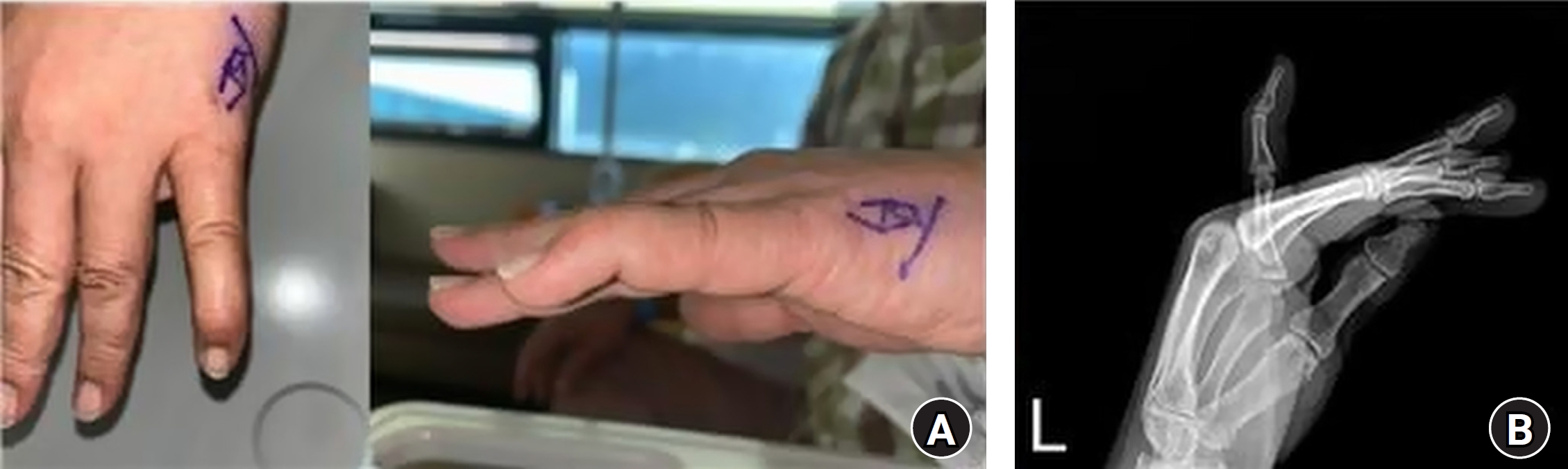
Representative preoperative clinical photographs (A) and radiograph (B) showing loss of active extension of the distal interphalangeal joint of the left little finger.
Fig. 3.
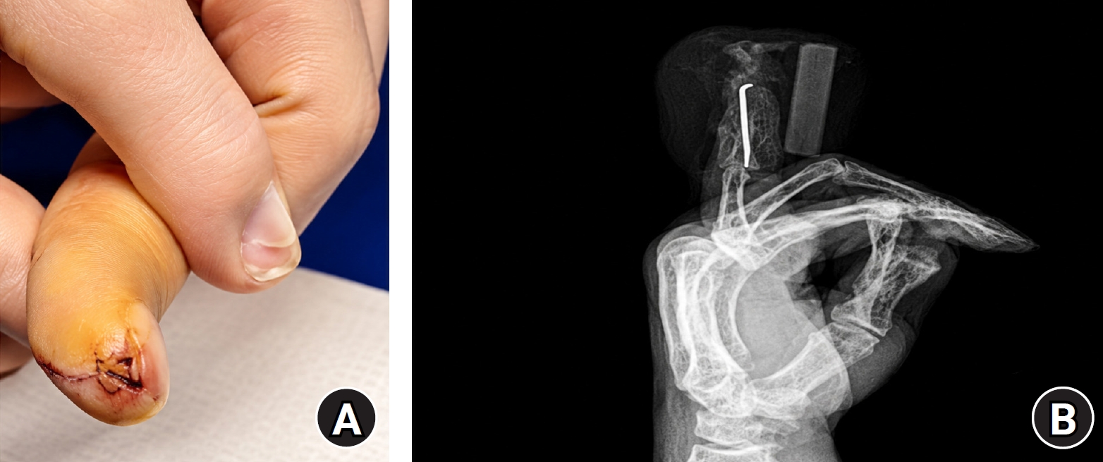
(A) The Kirschner wire was buried beneath the skin, and the overlying insertion site was closed with 5-0 nylon sutures. (B) Immediate postoperative radiograph. The distal interphalangeal joint was fixed in approximately 15° of hyperextension using a Kirschner wire passed from the fingertip across the joint and into the middle phalanx.
Fig. 4.
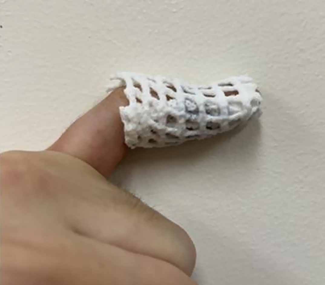
Opencast applied at the first outpatient visit, 1 week postoperatively. Only the distal interphalangeal joint was immobilized, permitting showering and protected daily and sports activities.
Fig. 5.
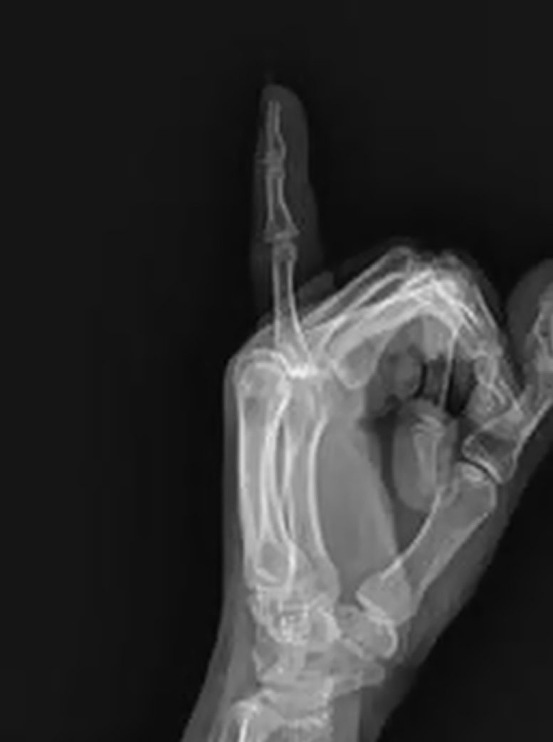
Radiograph obtained 8 weeks postoperatively, immediately after removal of the Kirschner wire.
Fig. 6.
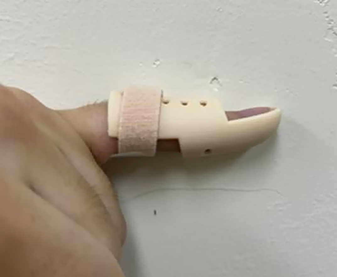
Removable mallet finger brace applied after Kirschner-wire removal and worn until 12 weeks postoperatively.
Table 1.
Summary of the 16 cases
| Case no. | Sex | Age (yr) | Preoperative extension lag (°) | Residual extension lag (°)a) | Flexion loss (°)a) | Outcomeb) |
|---|---|---|---|---|---|---|
| 1 | Male | 36 | 15 | 5 | 0 | Good |
| 2 | Male | 38 | 20 | 0 | 0 | Excellent |
| 3 | Male | 40 | 20 | 5 | 0 | Good |
| 4 | Female | 42 | 25 | 0 | 0 | Excellent |
| 5 | Male | 44 | 25 | 0 | 0 | Excellent |
| 6 | Male | 45 | 25 | 5 | 0 | Good |
| 7 | Female | 46 | 30 | 0 | 0 | Excellent |
| 8 | Male | 47 | 30 | 0 | 0 | Excellent |
| 9 | Male | 48 | 30 | 5 | 0 | Good |
| 10 | Male | 49 | 30 | 0 | 0 | Excellent |
| 11 | Male | 50 | 35 | 0 | 0 | Excellent |
| 12 | Male | 51 | 35 | 5 | 0 | Good |
| 13 | Male | 52 | 35 | 0 | 0 | Excellent |
| 14 | Male | 53 | 40 | 0 | 0 | Excellent |
| 15 | Female | 69 | 40 | 5 | 0 | Good |
| 16 | Female | 74 | 45 | 0 | 0 | Excellent |
a)Measured at the 6-month follow-up. b)Graded according to the Crawford evaluation criteria [14].
- 1. Doyle JR. Extensor tendons: acute injuries. In: Green DP, Hotchkiss RN, Pederson WC, editors. Green’s operative hand surgery. 4th ed. Churchill Livingstone; 1999. p. 1950-87.
- 2. Alla SR, Deal ND, Dempsey IJ. Current concepts: mallet finger. Hand (N Y) 2014;9:138-44.ArticlePubMedPMCPDF
- 3. Bachoura A, Ferikes AJ, Lubahn JD. A review of mallet finger and jersey finger injuries in the athlete. Curr Rev Musculoskelet Med 2017;10:1-9.ArticlePubMedPMCPDF
- 4. Bourland B, Astacio E, Bachoura A, Lubahn JD. Fingertip injuries in athletes, musicians, and other special cases. Hand Clin 2021;37:117-23.ArticlePubMed
- 5. Salazar Botero S, Hidalgo Diaz JJ, Benaïda A, Collon S, Facca S, Liverneaux PA. Review of acute traumatic closed mallet finger injuries in adults. Arch Plast Surg 2016;43:134-44.ArticlePubMedPMCPDF
- 6. Valdes K, Naughton N, Algar L. Conservative treatment of mallet finger: a systematic review. J Hand Ther 2015;28:237-45.ArticlePubMed
- 7. Handoll HH, Vaghela MV. Interventions for treating mallet finger injuries. Cochrane Database Syst Rev 2004;(3):CD004574.Article
- 8. Yoon JO, Baek H, Kim JK. The outcomes of extension block pinning and nonsurgical management for mallet fracture. J Hand Surg Am 2017;42:387.e1-7.ArticlePubMed
- 9. Tang JB, Giddins G, Omokawa S, Boeckstyns ME, Tay SC, Giesen T. Common hand problems with different treatments in countries in Asia and Europe. Hand Clin 2017;33:561-9.ArticlePubMed
- 10. Stern PJ, Kastrup JJ. Complications and prognosis of treatment of mallet finger. J Hand Surg Am 1988;13:329-34.Article
- 11. Kim JY, Lee SH. Factors related to distal interphalangeal joint extension loss after extension block pinning of mallet finger fractures. J Hand Surg Am 2016;41:414-9.ArticlePubMed
- 12. Lee KH, Jung SY, Kim JW. Alternative technique using opencast for early return to play after operation in patients with bony mallet finger by sports injury: a retrospective study in a tertiary hospital in South Korea. Korean J Sports Med 2024;42:113-8.Article
- 13. Lee SH, Lee JE, Lee KH, Pyo SH, Kim MB, Lee YH. Supplemental method for reduction of irreducible mallet finger fractures by the 2-extension block technique: the dorsal counterforce technique. J Hand Surg Am 2019;44:695.e1-8.ArticlePubMed
- 14. Crawford GP. The molded polythene splint for mallet finger deformities. J Hand Surg Am 1984;9:231-7.ArticlePubMed
- 15. Warren RA, Kay NR, Norris SH. The microvascular anatomy of the distal digital extensor tendon. J Hand Surg Br 1988;13:161-3.ArticlePDF
- 16. Rayan GM, Mullins PT. Skin necrosis complicating mallet finger splinting and vascularity of the distal interphalangeal joint overlying skin. J Hand Surg Am 1987;12:548-52.ArticlePubMed
- 17. Lin JS, Samora JB. Surgical and nonsurgical management of mallet finger: a systematic review. J Hand Surg Am 2018;43:146-63.e2.ArticlePubMed
- 18. Ishiguro T, Itoh Y, Yabe Y, Hashizume N. Extension block with Kirschner wire for fracture dislocation of the distal interphalangeal joint. Tech Hand Up Extrem Surg 1997;1:95-102.ArticlePubMed
- 19. King HJ, Shin SJ, Kang ES. Complications of operative treatment for mallet fractures of the distal phalanx. J Hand Surg Br 2001;26:28-31.ArticlePubMedPDF
- 20. Kardestuncer T, Bae DS, Waters PM. The results of tenodermodesis for severe chronic mallet finger deformity in children. J Pediatr Orthop 2008;28:81-5.ArticlePubMed
- 21. Kalainov DM, Hoepfner PE, Hartigan BJ, Carroll C 4th, Genuario J. Nonsurgical treatment of closed mallet finger fractures. J Hand Surg Am 2005;30:580-6.ArticlePubMed
References
Figure & Data
REFERENCES
Citations
Citations to this article as recorded by 
 ePub Link
 Cite
 XML Download
Percutaneous Kirschner-wire fixation followed by an opencast to permit protected daily and sports activities in patients with tendinous mallet finger: a case series at a tertiary hospital in Korea
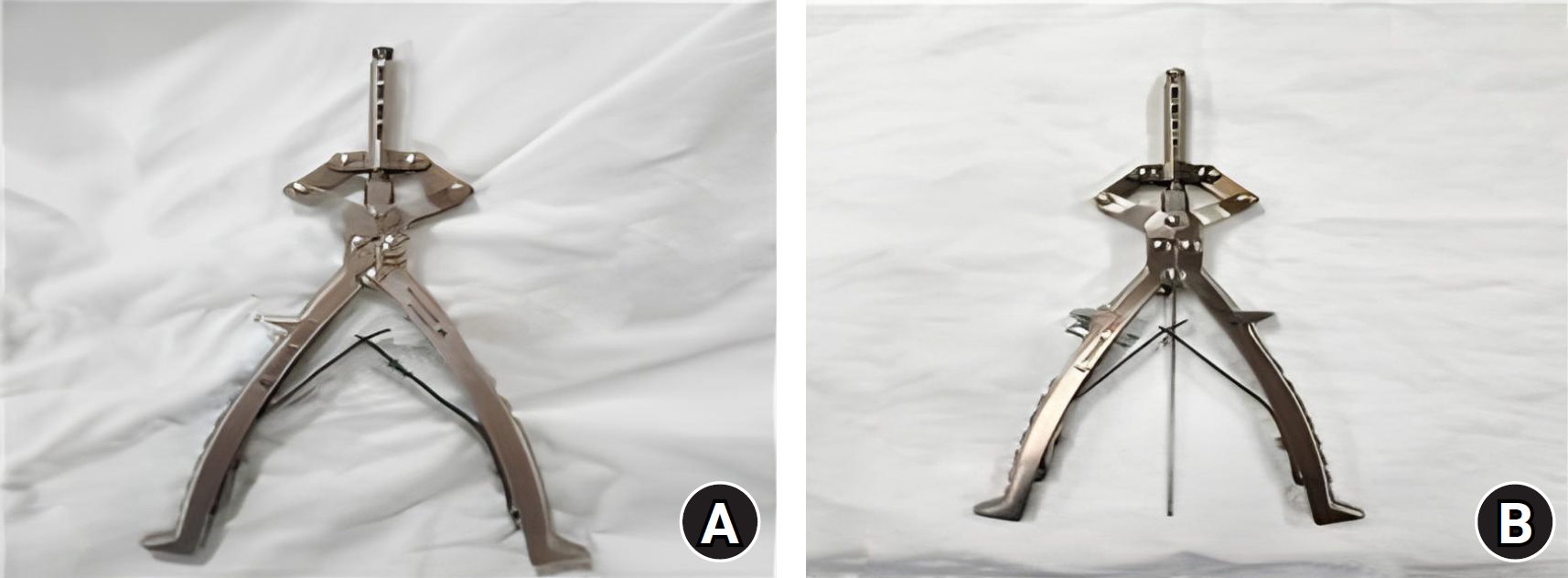
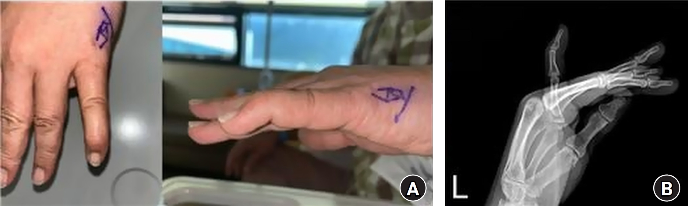
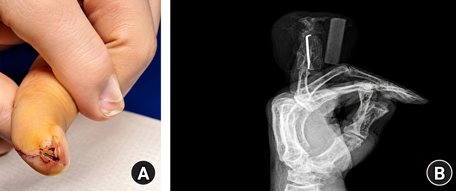
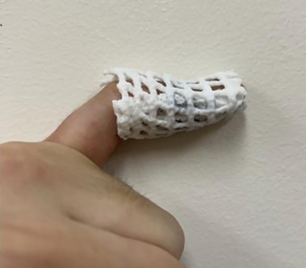
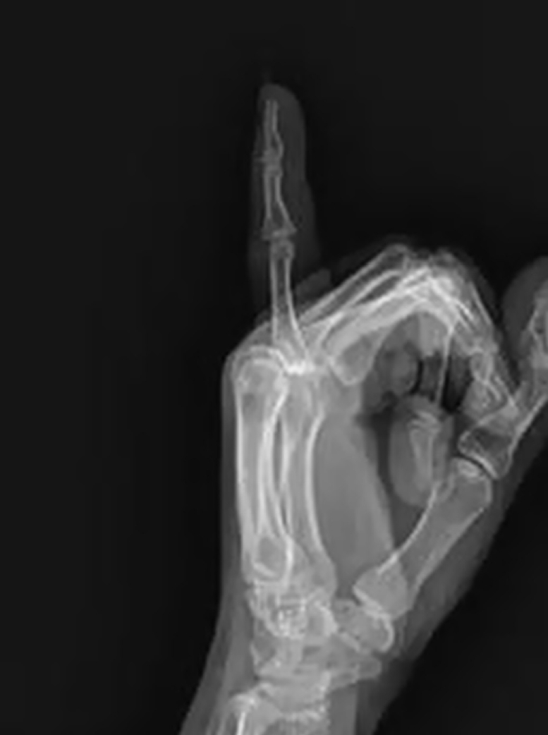
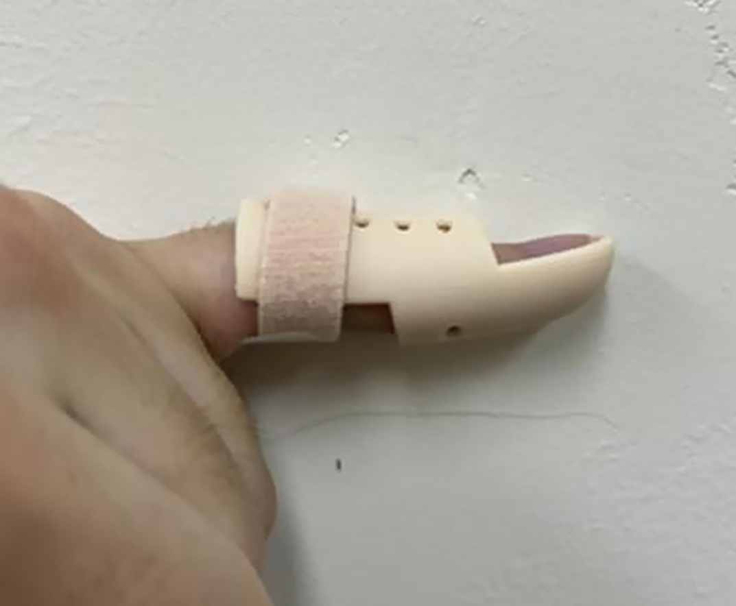
Fig. 1. Dedicated Kirschner-wire cutter without (A) and with (B) a Kirschner wire loaded. The cutter allows the wire to be cut close to the bone surface for burial beneath the skin.
Fig. 2. Representative preoperative clinical photographs (A) and radiograph (B) showing loss of active extension of the distal interphalangeal joint of the left little finger.
Fig. 3. (A) The Kirschner wire was buried beneath the skin, and the overlying insertion site was closed with 5-0 nylon sutures. (B) Immediate postoperative radiograph. The distal interphalangeal joint was fixed in approximately 15° of hyperextension using a Kirschner wire passed from the fingertip across the joint and into the middle phalanx.
Fig. 4. Opencast applied at the first outpatient visit, 1 week postoperatively. Only the distal interphalangeal joint was immobilized, permitting showering and protected daily and sports activities.
Fig. 5. Radiograph obtained 8 weeks postoperatively, immediately after removal of the Kirschner wire.
Fig. 6. Removable mallet finger brace applied after Kirschner-wire removal and worn until 12 weeks postoperatively.
Fig. 1.
Fig. 2.
Fig. 3.
Fig. 4.
Fig. 5.
Fig. 6.
Percutaneous Kirschner-wire fixation followed by an opencast to permit protected daily and sports activities in patients with tendinous mallet finger: a case series at a tertiary hospital in Korea
| Case no. | Sex | Age (yr) | Preoperative extension lag (°) | Residual extension lag (°) |
Flexion loss (°) |
Outcome |
|---|---|---|---|---|---|---|
| 1 | Male | 36 | 15 | 5 | 0 | Good |
| 2 | Male | 38 | 20 | 0 | 0 | Excellent |
| 3 | Male | 40 | 20 | 5 | 0 | Good |
| 4 | Female | 42 | 25 | 0 | 0 | Excellent |
| 5 | Male | 44 | 25 | 0 | 0 | Excellent |
| 6 | Male | 45 | 25 | 5 | 0 | Good |
| 7 | Female | 46 | 30 | 0 | 0 | Excellent |
| 8 | Male | 47 | 30 | 0 | 0 | Excellent |
| 9 | Male | 48 | 30 | 5 | 0 | Good |
| 10 | Male | 49 | 30 | 0 | 0 | Excellent |
| 11 | Male | 50 | 35 | 0 | 0 | Excellent |
| 12 | Male | 51 | 35 | 5 | 0 | Good |
| 13 | Male | 52 | 35 | 0 | 0 | Excellent |
| 14 | Male | 53 | 40 | 0 | 0 | Excellent |
| 15 | Female | 69 | 40 | 5 | 0 | Good |
| 16 | Female | 74 | 45 | 0 | 0 | Excellent |
Table 1. Summary of the 16 cases
a)Measured at the 6-month follow-up. b)Graded according to the Crawford evaluation criteria [


