
 E-submission
 KOTA
 TOTA
 TOTS

Articles
- Page Path
- HOME > > Ahead-of print articles > Article
-
Systematic Review
- Impact of posterior wall involvement and fixation on clinical and radiographic outcomes of associated both-column acetabular fractures: a systematic review and meta-analysis
-
Chul-Ho Kim1
 , Eic Ju Lim2
-
DOI: https://doi.org/10.12671/jmt.2026.00248
Published online: October 19, 2026
1Department of Orthopedic Surgery, Asan Medical Center, University of Ulsan College of Medicine, Seoul, Korea
2Department of Orthopedic Surgery, Chungbuk National University Hospital, Chungbuk National University College of Medicine, Cheongju, Korea
- Correspondence to: Eic Ju Lim Department of Orthopedic Surgery, Chungbuk National University Hospital, Chungbuk National University College of Medicine, 776 1Sunhwan-ro, Seowon-gu, Cheongju 28644, Korea Tel: +82-43-269-6077 Email: limeicju@gmail.com
• Received: July 15, 2026 • Revised: August 13, 2026 • Accepted: August 20, 2026
© 2026 The Korean Orthopaedic Trauma Association
This is an Open Access article distributed under the terms of the Creative Commons Attribution Non-Commercial License (http://creativecommons.org/licenses/by-nc/4.0/) which permits unrestricted noncommercial use, distribution, and reproduction in any medium, provided the original work is properly cited.
- 77 Views
- 1 Download


Abstract
-
Background Posterior wall (PW) fractures are frequently associated with both-column (ABC) acetabular fractures. However, whether the presence of a PW fragment or the need for internal fixation affects prognosis remains a topic of debate. This study compared postoperative outcomes, including reduction quality and clinical results, according to the presence of an associated PW fracture and whether PW fixation was performed.
-
Methods This systematic review and meta-analysis followed the Preferred Reporting Items for Systematic Reviews and Meta-Analyses (PRISMA) 2020 guidelines. MEDLINE, Embase, and the Cochrane Library were searched for articles published up to December 10, 2025. Comparative studies of ABC fractures were included if they evaluated the presence or absence of PW involvement or compared internal fixation with nonfixation of the PW component. Clinical and radiographic outcome data were extracted.
-
Results Seven retrospective studies including 434 analyzable patients were included. No clear differences were found in the odds of poor reduction (odds ratio [OR], 0.91; 95% confidence interval [CI], 0.43–1.94; P=0.80), osteoarthritis (OR, 1.04; 95% CI, 0.37–2.92; P=0.94), or conversion to total hip arthroplasty (OR, 0.63; 95% CI, 0.23–1.71; P=0.36) according to the presence of a PW fragment in ABC fractures. Among ABC fractures with PW involvement, nonfixation was associated with numerically higher odds of poor reduction than fixation, although the difference was not statistically significant (OR, 1.47; 95% CI, 0.46–4.76; P=0.52). In unadjusted comparisons of non-randomized cohorts, PW fixation was associated with significantly longer operative time (mean difference [MD], 70.38 min; 95% CI, 24.46–116.29; P=0.003) and greater intraoperative blood loss (MD, 351.36 mL; 95% CI, 244.97–457.74; P<0.00001). The final modified Merle d'Aubigné-Postel score did not differ significantly between the fixation and nonfixation groups (MD, 0.58 points; 95% CI, −0.57 to 1.74; P=0.32).
-
Conclusions Current evidence does not clearly support routine fixation of all PW fragments. In unadjusted comparisons of non-randomized cohorts, patients who underwent PW fixation had longer operative time and greater blood loss. These differences cannot be attributed to fixation itself because of confounding by indication and the frequent use of an additional posterior approach. Selective fixation based on residual displacement, joint congruency, and hip stability appears reasonable, although the certainty of evidence remains limited.
-
Level of evidence Not applicable.
Introduction
Acetabular fractures, although relatively uncommon, are typically the result of high-energy trauma and frequently lead to poor functional outcomes if not appropriately managed. Since the 1960s, these injuries have been categorized using the ten fracture patterns described by Judet et al. [1]. However, establishing an accurate diagnosis and surgical plan remains challenging because of intricate anatomical structures and inherent variability in fracture patterns. Among these, associated both-column (ABC) fractures are complex lesions involving detachment of the entire articular surface from the sciatic buttress. Although a posterior wall (PW) fragment is observed in approximately 35% to 39% of ABC cases, this specific combination is notably absent from the original Letournel classification [2].
The presence of a PW component in ABC fractures raises significant clinical concerns regarding joint stability, given its primary role as a weight-bearing surface [3]. Although surgical stabilization of the PW is conventionally recommended to restore hip integrity, emerging evidence suggests that PW fragments with ABC patterns differ etiologically from isolated PW fractures. Although isolated PW fractures often result from direct posterior shear forces during dislocation, PW fragments in ABC fractures are frequently caused by an anterior pull-type or avulsion mechanism through an intact capsulolabral complex during central dislocation [4]. Due to this distinct injury mechanism, the optimal management of the associated PW component remains a subject of significant debate. It has been proposed that these fragments may remain stable after column reconstruction alone and can be ignored, without compromising functional outcomes [5]. However, PW fixation using plates and screws through a sequential Kocher-Langenbeck approach has also been suggested to ensure anatomical restoration [6]. Furthermore, other studies have demonstrated the feasibility of lag screw fixation alone through the lateral extension of a single anterior approach, thereby avoiding the morbidity of a secondary posterior incision [7].
Although several clinical studies have investigated the relationship between PW involvement and outcomes in ABC fractures, these reports are often limited by small sample sizes, leading to inconsistent conclusions. To our knowledge, no systematic review has synthesized these findings to provide consolidated evidence on PW involvement and ABC fracture outcomes. Therefore, we integrated data from multiple institutions to enhance the statistical power of the analysis and overcome the limitations of single-center studies.
The purpose of this study was to evaluate the impact of PW involvement on the clinical prognosis and to determine whether PW fixation is necessary for optimal outcomes in ABC fracture patterns.
Methods
The overall methodology of this study was based on the Cochrane Handbook for Systematic Reviews of Interventions [8]. Furthermore, this systematic review and meta-analysis was reported in accordance with the Preferred Reporting Items for Systematic Reviews and Meta-Analyses (PRISMA) 2020 guidelines [9].
This is a literature-based study; therefore, neither institutional review board approval nor informed consent was required.
The selected studies directly compared (1) the clinical outcomes of ABC fractures according to the presence or absence of a concomitant PW fragment, and (2) the surgical results between fixation and nonfixation of the PW component when present. The exclusion criteria were nonclinical or secondary research, including biomechanical and cadaveric studies, review articles, meta-analyses, and expert opinions. Technical notes, letters to the editor, conference abstracts, and case reports were also excluded. Additionally, duplicate publications that used identical patient cohorts or multiple publications that should have been reported as a single study were removed.
We searched the MEDLINE, Embase, and Cochrane Library databases to identify comparative studies that evaluated the surgical outcomes of ABC fractures with concurrent PW fragments.
A systematic search was conducted for articles published from inception until December 10, 2025, using an a priori search strategy. The search terms included synonyms and related terms for ABC and PW as follows: ("both columns" OR "two columns") AND ("posterior wall") AND ("acetabular fracture"). Relevant studies were identified through a systematic search of the databases, with the scope limited to articles published in English. In addition, the reference lists of the retrieved studies were manually reviewed to ensure a comprehensive search.
Study titles and abstracts were independently reviewed by two fellowship-trained board-certified orthopedic surgeons specializing in hip and pelvic trauma. In cases where the abstract provided inadequate details, a comprehensive full-text assessment was conducted. To quantify the level of agreement between reviewers, the κ-value was calculated for each phase of the study selection. Agreement levels were categorized as follows: perfect (κ=1), almost perfect (0.8≤κ<1.0), substantial (0.6≤κ<0.8), moderate (0.4≤κ<0.6), fair (0.2≤κ<0.4), and slight (κ<0.2). Disagreements were resolved by mutual consensus or, if necessary, by a third independent board-certified orthopedic surgeon.
For the qualitative synthesis, we performed a systematic extraction and comprehensive review of essential parameters from the recruited studies. Data extraction was performed independently by two reviewers and discrepancies were resolved through discussion.
We extracted the following data: study design, number of patients in each group, mean patient age, sex distribution, follow-up duration, indications for PW fixation, whether stability was assessed after column fixation, surgical approach, operation time, blood loss, overall acetabular/articular reduction quality assessed according to the Matta criteria, as reported in each original study [10], functional outcomes, presence of osteoarthritis, conversion to total hip arthroplasty (THA), and surgical site infection (SSI). Because the grading systems and thresholds varied across studies, osteoarthritis and reduction quality were dichotomized according to the definitions reported in each original study. Accordingly, the pooled estimates were interpreted cautiously because the underlying definitions were not fully equivalent. Given the heterogeneity in the surgical indications for PW fixation across the included studies, we specifically analyzed the clinical criteria for fixation and the methodology of intraoperative stability assessment following column reconstruction.
The methodological quality and risk of bias of the included studies were assessed using the Risk of Bias in Non-randomized Studies of Interventions (ROBINS-I) tool [11]. This tool evaluates seven domains: bias due to confounding factors, bias in the selection of participants, bias in the classification of interventions, bias due to deviations from intended interventions, bias due to missing data, bias in the measurement of outcomes, and bias in the selection of reported results. Each domain was rated as follows: low, moderate, serious, critical, or no information.
Odds ratios (ORs) with 95% confidence intervals (CIs) were calculated for dichotomous outcomes, whereas mean differences (MDs) with 95% CIs were used for continuous outcomes. The I2 statistic was used to assess study heterogeneity, where values of 25%, 50%, and 75% represent low, moderate, and high levels of inconsistency, respectively.
Comparative analyses were stratified into two primary subgroups: (1) presence or absence of PW fracture and (2) fixation or nonfixation of the PW fragment. This meta-analysis focused on variables reported in at least two studies. We included data on operation time, blood loss, reduction quality, osteoarthritis, THA conversion, and functional outcome. For the pooled reduction-quality analyses, only overall acetabular/articular reduction was included; PW-specific displacement measurements were not pooled. Individual study findings, pooled effect sizes, and overall estimates were visualized using forest plots. Statistical significance was defined as P<0.05. The data were synthesized using random-effects models because clinical and methodological heterogeneity across studies was anticipated, including differences in indications for PW fixation, surgical approaches, outcome assessment, and perioperative management [12]. Between-study variance (τ²) was estimated using the DerSimonian-Laird method. Fixed-effect analyses using the same eligible studies were performed as sensitivity analyses. All computational analyses were conducted using Review Manager (RevMan v.5.3; The Cochrane Collaboration).
Following Cochrane recommendations, publication bias was not formally assessed because the total number of included studies was less than ten [13].
Results
The study selection and identification procedures are shown in Fig. 1. An initial systematic search of electronic databases yielded 149 records (MEDLINE, 70; Embase, 75; and Cochrane, 4). After removing 69 duplicates, 80 unique citations were retained for further assessment. Although a manual search was conducted to identify additional relevant literature, no further studies were found (n=0). During the initial screening of titles and abstracts, 66 records were excluded. The remaining 14 articles underwent comprehensive full-text eligibility assessment. Of these, seven were excluded for the following reasons: lack of relevance to the specific field of interest (n=3) and insufficient data to allow for a comparative analysis (n=4). Seven studies met the inclusion criteria and were incorporated into both the qualitative synthesis and quantitative meta-analysis. Reviewer consistency was evaluated using κ statistics. The levels of agreement were substantial during the title and abstract screening phases (κ=0.720 and κ=0.777, respectively). Furthermore, the reviewers achieved perfect consensus during the full-text review stage (κ=1.0).
All seven studies were retrospective cohort studies [3,5-7,14-16]. Among the included studies, four compared outcomes based on the presence or absence of a PW fragment [3,5,7,15]. Two studies focused specifically on patients with PW fragments and compared those who received fixation to those who did not [14,16]. Additionally, one study conducted a three-arm comparative analysis of ABC fractures without PW, ABC fractures with fixed PW, and ABC fractures with non-fixed PW [6]. Seven studies contributed comparative data on 434 patients: 211 without PW involvement, 126 with PW involvement treated with fixation, and 97 with PW involvement managed without fixation. No patient-cohort overlap was identified among the included studies based on the reported institutions, enrollment periods, author groups, and cohort characteristics. The mean patient age ranged from 41.7–62.7 years. The proportion of female patients ranged from 18.4%–40.3%. The follow-up periods ranged from 12–54 months (Table 1) [3,5-7,14-16].
Regarding the surgical approaches for ABC fractures, five studies used either the ilioinguinal or Stoppa approaches [5,7,14-16]. One study utilized either the ilioinguinal or extended iliofemoral approach [3], whereas another study used the supra-ilioinguinal approach [6]. Of the approaches used for open reduction and PW fixation, the Kocher-Langenbeck approach was the predominant choice [3,5,6,14,16]. The operative time ranged from 163–241 minutes in the absence of PW fixation and from 197–271 minutes in cases where PW fixation was performed. The estimated intraoperative blood loss ranged from 703–889 mL in the absence of PW fixation, and varied from 957–1,177 mL when PW fixation was performed. The rate of suboptimal reduction varied across the individual study cohorts (Table 2) [3,5-7,14-16].
Heterogeneity in functional outcomes was observed across the included studies. Five studies [6,7,14-16] utilized the Merle d'Aubigné-Postel (MAP) score as the primary assessment tool [17], whereas one study employed the Harris Hip Score [18]. The mean MAP scores reported in three studies ranged from 15.4–16.2. In two studies reporting categorical functional outcomes, the proportion of patients achieving excellent or good results ranged from 78.3%–88.0%, according to the criteria used in the original studies. Four studies reported the presence of osteoarthritis during follow-up. The incidence of post-traumatic osteoarthritis reported in the included studies ranged from 2.9%–20.7%. The conversion rate to THA ranged from 0%–22.2% in patients who underwent PW fixation. The incidence of SSI ranged from 0% in patients who did not undergo PW fixation to 8.7% in those who underwent PW fixation (Table 3) [3,5-7,14-16].
The risk of bias assessment using ROBINS-I showed an overall moderate risk of bias in five studies and a serious risk in two studies [6,14]. The principal concern in the two studies with serious risk of bias was confounding by indication, because PW fixation was determined by residual displacement or surgeon discretion without adequate adjustment for these treatment-selection factors. None of the studies was considered to have a critical risk of bias (Table 4).
Individual study characteristics, including patient demographics, surgical approaches, operative time, and blood loss, are presented in Tables 1 and 2. Functional outcomes, osteoarthritis incidence, and conversion to THA for each study are detailed in Table 3. Forest plots (Figs. 2 and 3) visualize the individual study effect estimates for each analyzed outcome.
Meta-analysis results stratified by the presence of PW fragment
Reduction quality
Four studies compared reduction quality between patients with ABC fractures without PW involvement (n=146) and those with PW involvement (n=116) [5-7,15]. No clear difference was demonstrated in the odds of poor reduction between the two groups (OR, 0.91; 95% CI, 0.43–1.94; P=0.80; I²=0%) (Fig. 2A).
Osteoarthritis
Three studies compared osteoarthritis between patients with ABC fractures without PW involvement (n=118) and those with PW involvement (n=72) [5,7,15]. The mean follow-up durations were 37 months in Shin et al. [5], 45 months in Wang et al. [7], and 54 months in Min et al. [15]. No clear difference was demonstrated in the odds of osteoarthritis between the two groups (OR, 1.04; 95% CI, 0.37–2.92; P=0.94; I²=0%) (Fig. 2B).
THA conversion
Four studies compared conversion to THA between patients with ABC fractures without PW involvement (n=144) and those with PW involvement (n=90) [3,5,7,15]. The mean follow-up durations were 43 months in Reahl et al. [3], 37 months in Shin et al. [5], 45 months in Wang et al. [7], and 54 months in Min et al. [15]. No clear difference was demonstrated in the odds of THA conversion between the two groups (OR, 0.63; 95% CI, 0.23–1.71; P=0.36; I²=0%) (Fig. 2C).
Meta-analysis results according to fixation of PW fragment
Reduction quality
Three studies compared the reduction quality among patients with ABC fractures and PW involvement according to whether the PW fragment was fixed [6,14,16]. The pooled analysis did not demonstrate a clear difference in the odds of poor reduction between the nonfixation and fixation groups (OR, 1.47; 95% CI, 0.46–4.76; P=0.52; I²=0%) (Fig. 3A).
Operation time
Two studies directly compared operative time between PW fixation and nonfixation among patients with PW involvement [6,16]. The PW fixation and nonfixation groups included 44 and 49 patients, respectively. The pooled analysis showed PW fixation was associated with a longer operative time than nonfixation (MD, 70.38 min; 95% CI, 24.46–116.29; P=0.003). Substantial between-study heterogeneity was observed (I²=82%) (Fig. 3B).
Blood loss
Two studies directly compared intraoperative blood loss between PW fixation and nonfixation among patients with PW involvement [6,16]. The PW fixation and nonfixation groups included 44 and 49 patients, respectively. The pooled analysis showed greater intraoperative blood loss in the PW fixation group than in the nonfixation group (MD, 351.36 mL; 95% CI, 244.97–457.74; P<0.00001; I²=0%) (Fig. 3C).
Functional outcome
Two studies involving 70 patients directly compared final functional outcomes using the modified MAP score between PW fixation and nonfixation groups [14,16]. The PW fixation and nonfixation groups included 30 and 40 patients, respectively. The pooled analysis did not demonstrate a clear difference in the final MAP score between the two groups (MD, 0.58 points; 95% CI, −0.57 to 1.74; P=0.32; I²=0%) (Fig. 3D).
Sensitivity analysis
Fixed-effect sensitivity analyses using the same eligible studies yielded results that were materially consistent with the primary random-effects analyses, without changes in the direction or statistical significance of the pooled estimates (Supplementary Figs. 1, 2).
Indications for fixation and stability assessment
The criteria for internal fixation of the PW fragment and methods used to assess hip stability varied among the included studies (Table 5). Some studies applied fragment size or residual displacement as key criteria for fixation, whereas others relied on the surgeon's discretion, intraoperative stability assessment, treatment period, or preoperative virtual simulation. Min et al. [15] used a 20% PW involvement threshold combined with an assessment of joint stability, whereas Shin et al. [5] emphasized residual incongruity, gross instability on stress examination, and attachment of the fragment to the labrocapsular complex. In addition, intraoperative stability assessment following reconstruction of the main columns was reported by Shin et al. [5], Wang et al. [7], and Tian et al. [16], although the role of assessment differed among studies, ranging from confirmation of stability after fixation to decision-making for selective nonfixation. Wan et al. [6] used computer-assisted virtual reduction and selected a posterior approach when >3 mm of displacement remained after simulated reduction of the main columns. These differences highlight the absence of standardized indications for PW fixation in patients with ABC fractures.
Publication bias assessment was not formally conducted due to fewer than ten included studies, limiting formal evaluation of potential reporting bias. However, the retrospective design of all studies and heterogeneity in outcome reporting across studies potentially introduces selective reporting bias, though the direction and magnitude of such bias remain uncertain.
Discussion
The principal finding of this study was that no clear association was demonstrated between PW involvement and inferior overall clinical or radiographic outcomes in ABC fractures. In the included studies, PW fragments were managed using different strategies; some authors adopted selective nonfixation when the fragment was considered stable after column reconstruction, whereas others performed PW fixation when residual displacement, incongruity, or instability was suspected. In analyses limited to ABC fractures with PW involvement, PW fixation was associated with a greater operative burden, including longer operative time and greater intraoperative blood loss. However, the available analyses did not demonstrate clear differences in the reported clinical and radiographic outcomes between the fixation and nonfixation groups. Among the meta-analytic results, nonfixation was associated with numerically higher odds of poor reduction than fixation, although the difference was not statistically significant (OR, 1.47; 95% CI, 0.46–4.76; P=0.52).
The findings should not be interpreted as supporting either routine nonfixation or mandatory fixation of PW fragments. Rather, they suggest that the presence of a PW component itself does not necessarily worsen prognosis when it is appropriately assessed and managed. Selective fixation in the indicated cases may mitigate the potential adverse effects of unstable or incongruent PW fragments, although routine fixation may not be mandatory. Given that several of the included studies incorporated intraoperative stability testing or assessment of residual incongruity after column reconstruction, individualized management based on hip stability, fragment displacement, and joint congruency appears to be a reasonable strategy.
Heterogeneity in the indications for PW fixation is a key consideration in interpreting the present findings, as it represents both a limitation of the quantitative synthesis and an important finding of the qualitative analysis. Because PW fixation was generally selected for larger, more displaced, incongruent, or unstable fragments, the fixation groups may have included more complex injuries, potentially biasing operative and radiographic outcomes against fixation. Conversely, the younger age observed in some fixation cohorts may reflect a greater tendency to pursue PW fixation in patients for whom long-term joint preservation was considered particularly important. These baseline imbalances and treatment-selection factors limit causal interpretation of comparisons according to PW fixation status. Previous morphological studies suggested that PW involvement in ABC fractures tends to represent a larger and more extensive posterior element injury, with transverse or oblique fracture lines present on the retroacetabular surface, rather than a typical posterior rim fracture-dislocation pattern [19,20]. In this context, reconstruction of the main ABC components may indirectly reduce or stabilize the associated PW fragment through capsulolabral tension or traction of the surrounding soft tissues [5,15,16]. This suggests that after the reduction and fixation of the main ABC components, the assessment of residual displacement, joint congruency, and intraoperative hip stability may provide a clinically meaningful indication for additional PW fixation.
In the analysis stratified by the presence of a PW fracture, clear associations with reduction quality, osteoarthritis, or conversion to THA were not demonstrated. Because the quality of reduction is a major determinant of long-term clinical and radiographic outcomes after acetabular fracture surgery, the absence of a significant difference in reduction quality may partly explain the comparable rates of osteoarthritis and THA conversion between patients with and without PW involvement [21]. However, long-term outcomes in ABC fractures may be more strongly influenced by the quality of reduction of the main acetabular columns and other established predictors of conversion to THA, such as age, increased body mass index, and dome impaction, than by the presence of a PW fragment [21,22].
In the analysis according to PW fixation status, the pooled reduction outcome reflected overall acetabular/articular reduction based on the Matta criteria, which may not be sufficiently sensitive to residual displacement confined to an unfixed PW fragment. Therefore, the present analysis may have underestimated PW-specific differences in reduction between the fixation and nonfixation groups. Nevertheless, whether improved PW reduction translates into clinically meaningful long-term benefits remains unclear because subsequent outcomes, such as osteoarthritis and THA conversion, were not reported according to PW fixation status.
This study found that operative time and intraoperative blood loss differed significantly according to the PW fixation status. This finding may be partly explained by the surgical approach used for the PW fixation. In several of the included studies, PW fixation required an additional posterior approach, most commonly the Kocher-Langenbeck approach, which may have increased operative time and blood loss due to patient repositioning, additional exposure, and greater soft-tissue dissection [5,6,14,16]. Given that the fixation of ABC fractures alone is associated with substantial intraoperative blood loss, often in the range of ~700–900 mL in the included studies, the addition of a posterior approach may further increase the invasiveness of the surgical procedure [6,7].
Previous systematic reviews identified PW involvement as a risk factor for conversion to THA after acetabular fracture surgery (reported ORs, approximately 1.81 [23] and 1.82 [24]). In contrast, our analysis restricted to ABC fractures did not demonstrate a clear association between PW involvement and THA conversion. This apparent difference may reflect the narrower fracture population, selective fixation, small sample size, and variable follow-up; it should not be interpreted as evidence that PW involvement has no impact on long-term outcomes.
This study had several limitations that should be acknowledged. First, the number of included studies was small, limiting the statistical power of the meta-analysis and precluding further subgroup analyses. Because only two to four studies contributed to each pooled analysis, estimates of between-study heterogeneity and random-effects summary effects may also be imprecise and should be interpreted cautiously. In addition, high heterogeneity was observed in the analysis of operative time, limiting the precision of the pooled estimate. Accordingly, low I² values should not be considered evidence of true clinical or methodological homogeneity. Second, the indications for PW fixation varied substantially among the included studies, including differences in surgical approach, fixation strategy, and use of intraoperative stability assessment. Therefore, although this study summarized the available evidence, it could not determine a unified indication for PW fixation or provide definitive treatment guidelines. Third, all included studies were retrospective in design and were therefore subject to inherent methodological limitations. Long-term outcomes such as osteoarthritis and conversion to THA were not consistently reported according to PW fixation status, and follow-up duration varied across the included studies, with some patients having relatively short follow-up. Because these outcomes may develop over a prolonged period, their incidence may have been underestimated, limiting the ability to determine whether differences in reduction quality translate into clinically meaningful long-term outcomes. A sensitivity analysis restricted to patients with at least 24 months of follow-up was not feasible because patient-level follow-up and time-to-event data were unavailable. Fourth, the definitions of osteoarthritis and poor reduction varied across studies in terms of grading systems, imaging modalities, and dichotomization thresholds. Therefore, the pooled estimates may have combined outcomes that were not fully clinically equivalent and should be interpreted cautiously.
Future studies should evaluate the clinical impact of PW involvement and fixation, while adjusting for fracture severity, reduction quality, and other relevant covariates.
The presence of a PW fragment in ABC acetabular fractures showed no clear association with inferior reduction quality, osteoarthritis, or conversion to THA, although the osteoarthritis and THA findings should be interpreted cautiously because of variable follow-up. Among patients with ABC fractures and PW involvement, a clear difference in reduction quality was not demonstrated between PW fixation and nonfixation, although nonfixation showed numerically higher odds of poor reduction. In unadjusted comparisons of non-randomized cohorts, patients who underwent PW fixation had longer operative time and greater blood loss. These differences cannot be attributed to fixation itself because of confounding by indication and the frequent use of an additional posterior approach. Current evidence does not clearly support routine fixation of all PW fragments. Selective fixation based on residual displacement, joint congruency, and hip stability appears reasonable, although the certainty of the available evidence remains limited. Additional fixation should be considered selectively based on residual displacement, joint congruency, and hip stability after reconstruction of the main ABC components.
-
Author contributions
Conceptualization: CHK. Data curation: CHK, EJL. Formal analysis: CHK, EJL. Methodology: CHK, EJL. Writing–original draft: EJL. Writing–review & editing: CHK. All authors read and approved the final manuscript.
-
Conflicts of interest
No potential conflict of interest relevant to this article was reported.
-
Funding
This work was supported by a 2026 research grant from the Korean Orthopaedic Trauma Association (KOTA).
-
Data availability
The data analyzed in this systematic review and meta-analysis were derived from previously published studies. All relevant data are available in the original articles cited in this manuscript.
-
Acknowledgments
None.
Article Information
Supplementary materials
Supplementary materials related to this article can be found online at https://doi.org/10.12671/jmt.2026.00248.
Supplementary Fig. 1.
Comparison according to presence of PW fragment.
Supplementary Fig. 2.
Comparison according to fixation of PW fragment among ABC+PW.
Fig. 1.
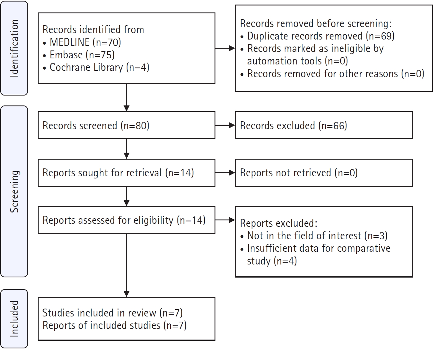
Flow diagram of the study selection process according to the Preferred Reporting Items for Systematic Reviews and Meta-Analyses (PRISMA) 2020 guidelines. No additional records were identified through manual searches of the reference lists of the retrieved studies.
Fig. 2.
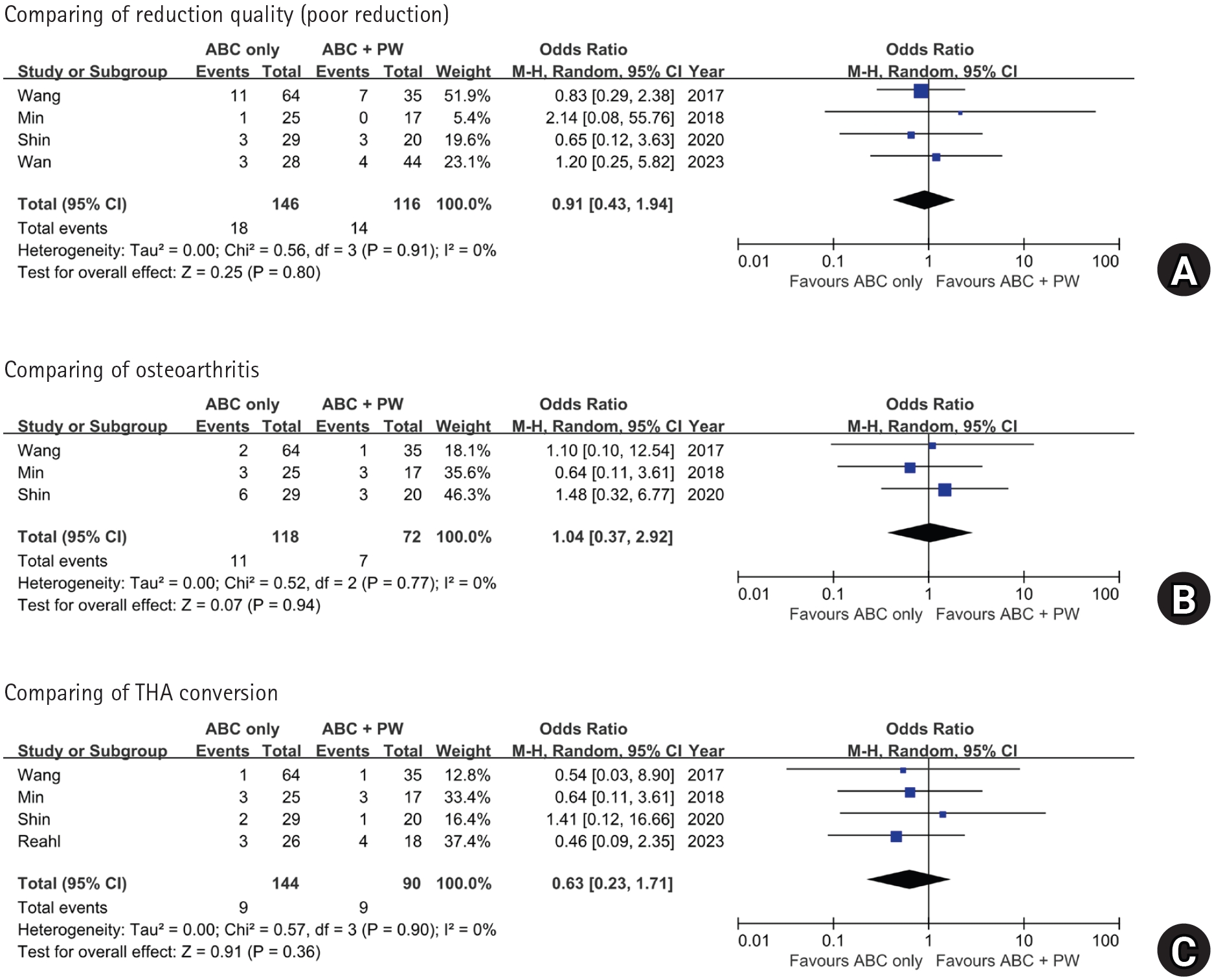
Forest plots comparing (A) poor reduction, (B) osteoarthritis, and (C) conversion to THA according to the presence of a PW fragment. ABC, associated both-column; PW, posterior wall; M-H, Mantel-Haenszel; CI, confidence interval; THA, total hip arthroplasty.
Fig. 3.
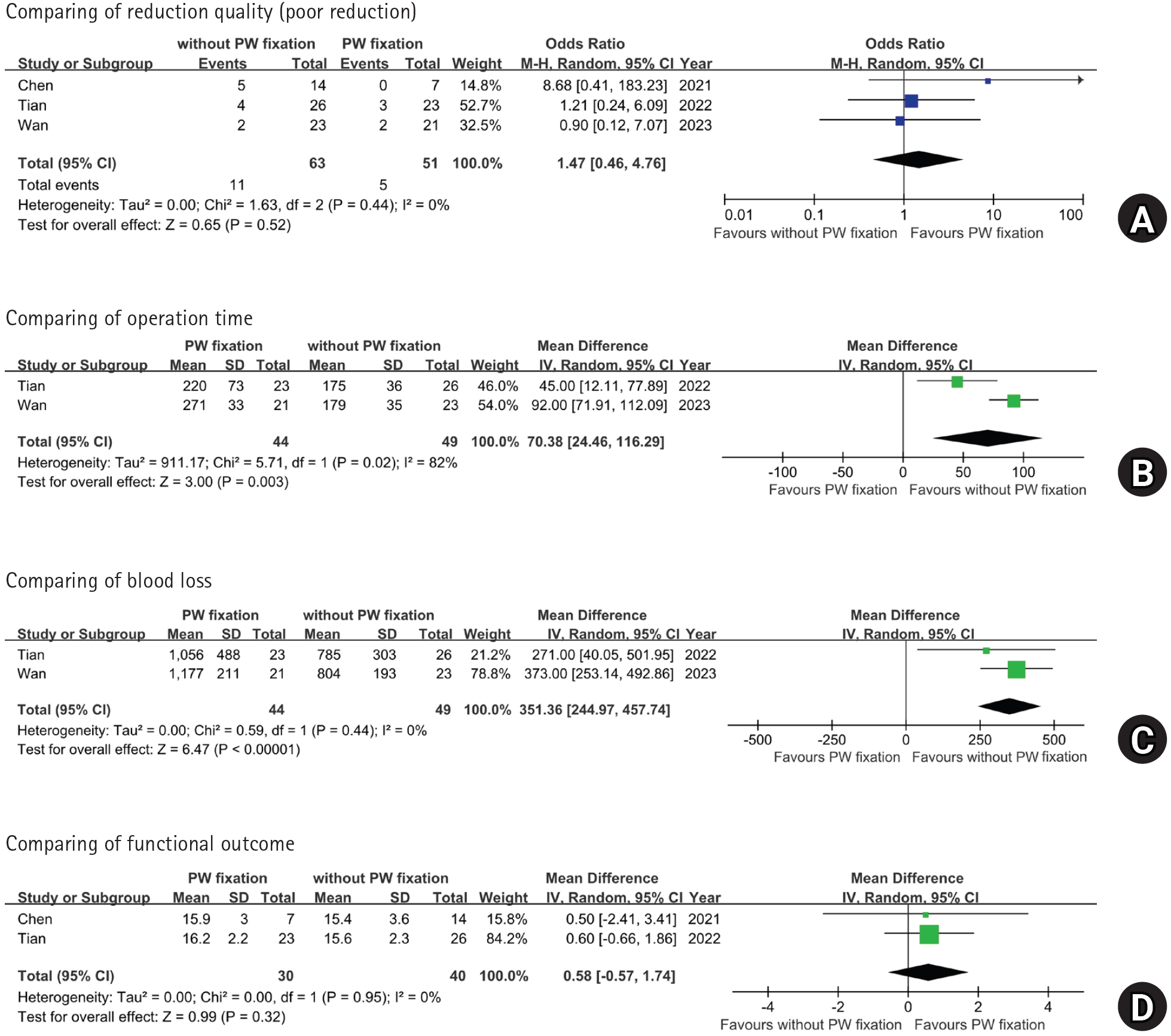
Forest plots comparing (A) poor reduction, (B) operative time, (C) intraoperative blood loss, and (D) functional outcome according to PW fixation status. M-H, Mantel-Haenszel; CI, confidence interval; PW, posterior wall; SD, standard deviation; IV, inverse variance.
Table 1.
Summary of patient demographic data from the included studies
| Study | Study design | Number of patients | Mean age (yr) | Female sex | Follow-up (mo)b) | ||||||
|---|---|---|---|---|---|---|---|---|---|---|---|
| PW− | PW+Fix+ | PW+Fix− | PW− | PW+Fix+ | PW+Fix− | PW− | PW+Fix+ | PW+Fix− | |||
| Wang et al. [7] (2017) | RCS | 64 | 35 | NA | 48.3±11.8 | 44.4±12.5 | NA | 17 | 10 | NA | 44.7±18.5 |
| Min et al. [15] (2018) | RCS | 25 | 15 | 2 | 46.2 | 45.7c) | 8 | 4c) | 54.0 | ||
| Shin et al. [5] (2020) | RCS | 29 | 3 | 17 | 51.1±23.3 | 48.1±23.6c) | 5 | 4c) | 36.6±18.5 | ||
| Chen et al.a) [14] (2021) | RCS | 39 | 11 | 29 | 50.9±18.8 | 51.2±16.8 | 62.7±16.7 | 12 | 1 | 7 | 49.9±39.2 |
| Tian et al. [16] (2022) | RCS | NA | 23 | 26 | NA | 45.5±11.2 | 46.1±11.8 | NA | 5 | 5 | 27.1±6.9 |
| Reahl et al. [3] (2023) | RCS | 26 | 18 | NA | 48.5±16.7 | 51.4±12.0 | NA | 6 | 4 | NA | 43.0 |
| Wan et al. [6] (2023) | RCS | 28 | 21 | 23 | 44.8±14.1 | 41.7±12.3 | 42.5±13.1 | 12 | 8 | 9 | Min 12 mo |
PW, posterior wall; Fix, fixation; PW−, associated both-column fractures without posterior wall involvement; PW+Fix+, associated both-column fractures with posterior wall involvement treated with fixation; PW+Fix−, associated both-column fractures with posterior wall involvement treated without fixation; RCS, retrospective cohort study; NA, not applicable; Min, minimum.
a)Chen et al. [14] initially identified 88 patients; nine patients with insufficient follow-up were excluded, leaving 79 analyzable patients.
b)Follow-up periods are presented as mean±standard deviation when available. Studies without reported standard deviations are shown as mean values only. Wan et al. [6] reported a minimum follow-up of 12 months without a mean value.
c)For Min et al. [15] and Shin et al. [5], baseline age and sex data were reported for the combined PW-positive cohort and were not separately available according to PW fixation status.
Table 2.
Perioperative profile
| Study | Approach | Operation time (min) | Blood loss (mL) | Poor reduction | Assessment modality | ||||||||
|---|---|---|---|---|---|---|---|---|---|---|---|---|---|
| PW− | PW+Fix+ | PW+Fix− | PW− | PW+Fix+ | PW+Fix− | PW− | PW+Fix+ | PW+Fix− | PW− | PW+Fix+ | PW+Fix− | ||
| Wang et al. [7] (2017) | Ilioinguinal | 241±49 | 258±60 | NA | 889±154 | 957±259 | NA | 11 | 7 | NA | X-ray | ||
| Min et al. [15] (2018) | Ilioinguinal | NR | NR | NR | NR | NR | NR | 1 | - | 0 | X-ray & CT | ||
| Shin et al. [5] (2020) | Ilioinguinal | Ilioinguinal, KL | Ilioinguinal | NR | NR | NR | NR | NR | NR | 3 | - | 3 | X-raya) |
| Chen et al. [14] (2021) | Ilioinguinal, Stoppa | Ilioinguinal, Stoppa, KL | Ilioinguinal, Stoppa | NR | NR | NR | NR | NR | NR | NR | 0 | 5 | X-rayb) |
| Tian et al. [16] (2022) | NA | Stoppa, KL | Stoppa | NA | 220±73 | 175±36 | NA | 1,056±488 | 785±303 | NA | 3 | 4 | NR |
| Reahl et al. [3] (2023) | Ilioinguinal, KL, extended iliofemoral | 215±94 | 197±63 | NA | NR | NR | NA | NR | NR | NA | NA | ||
| Wan et al. [6] (2023) | Supra-ilioinguinal | Supra-ilioinguinal, KL | Supra-ilioinguinal | 163±21 | 271±33 | 179±35 | 703±143 | 1,177±211 | 804±193 | 3 | 2 | 2 | X-ray |
PW, posterior wall; Fix, fixation; PW−, associated both-column fractures without posterior wall involvement; PW+Fix+, associated both-column fractures with posterior wall involvement treated with fixation; PW+Fix−, associated both-column fractures with posterior wall involvement treated without fixation; NA, not applicable; NR, not reported; CT, computed tomography; KL, Kocher-Langenbeck.
a)Shin et al. [5] assessed reduction quality using both postoperative radiographs and CT; radiograph-based Matta grading was used for the present analysis.
b)For Chen et al. [14], overall articular reduction according to the Matta criteria was assessed in the subset with ≥1-year follow-up (PW+Fix+, 7; PW+Fix−, 14).
Table 3.
Clinical outcomes
| Study | Functional outcomea) | Osteoarthritisb) | Conversion to THA | SSI | ||||||||
|---|---|---|---|---|---|---|---|---|---|---|---|---|
| PW− | PW+Fix+ | PW+Fix− | PW− | PW+Fix+ | PW+Fix− | PW− | PW+Fix+ | PW+Fix− | PW− | PW+Fix+ | PW+Fix− | |
| Wang et al. [7] (2017) | 15.7±1.9 | 15.9±1.8 | NA | 2 | 1 | NA | 1 | 1 | NA | 1 | 2 | NA |
| Min et al. [15] (2018) | 22 | 14 | 3 | 3 | 3 | 3 | 0 | 1 | ||||
| Shin et al. [5] (2020) | 89.0±6.6 | 90.5±6.8 | 6 | 3 | 2 | 1 | 0 | 1 | ||||
| Chen et al. [14] (2021) | NR | 15.9±3.0 | 15.4±3.6 | NR | 1.1±0.7 | 1.3±0.9 | NR | 0 | 3 | NR | NR | NR |
| Tian et al. [16] (2022) | NA | 16.2±2.2 | 15.6±2.3 | NA | NR | NR | NA | NR | NR | NA | 2 | 0 |
| Reahl et al. [3] (2023) | NR | NR | NA | NR | NR | NA | 3 | 4 | NA | 2 | 1 | NA |
| Wan et al. [6] (2023) | 24 | 18 | 18 | NR | NR | NR | NR | NR | NR | NR | NR | NR |
THA, total hip arthroplasty; SSI, surgical site infection; PW, posterior wall; Fix, fixation; PW−, associated both-column fractures without posterior wall involvement; PW+Fix+, associated both-column fractures with posterior wall involvement treated with fixation; PW+Fix−, associated both-column fractures with posterior wall involvement treated without fixation; NA, not applicable; NR, not reported; MAP, modified Merle d'Aubigné-Postel score; HHS, Harris Hip Score.
a)Functional outcomes are presented as mean MAP scores for Wang et al. [7], Chen et al. [14], and Tian et al. [16]; as mean HHS values for Shin et al. [5]; and as the numbers of patients with excellent or good outcomes according to the MAP criteria for Min et al. [15] and Wan et al. [6].
b)Wang et al. [7] used the Kellgren-Lawrence grading system. Min et al. [15] and Shin et al. [5] used Matta’s radiographic grading system; Min et al. [15] reported poor outcomes, whereas Shin et al. [5] reported fair or poor outcomes. Chen et al. [14] reported mean Tonnis grades.
Table 4.
Risk of bias assessment using the ROBINS-I tool
| Study | Confounding | Selection | Intervention classification | Deviation from intervention | Missing data | Measurement of outcome | Selection of reported result | Overall |
|---|---|---|---|---|---|---|---|---|
| Wang et al. [7] (2017) | Moderate | Low | Low | Low | Low | Moderate | Moderate | Moderate |
| Min et al. [15] (2018) | Moderate | Low | Low | Low | Low | Low | Moderate | Moderate |
| Shin et al. [5] (2020) | Moderate | Low | Low | Low | Low | Low | Moderate | Moderate |
| Chen et al. [14] (2021) | Serious | Moderate | Low | Low | Moderate | Moderate | Moderate | Serious |
| Tian et al. [16] (2022) | Moderate | Moderate | Low | Low | Low | Moderate | Moderate | Moderate |
| Reahl et al. [3] (2023) | Moderate | Moderate | Low | Low | Moderate | Moderate | Moderate | Moderate |
| Wan et al. [6] (2023) | Serious | Low | Low | Low | Low | Moderate | Moderate | Serious |
Table 5.
Indications for fixation of PW
| Study | Comparisons | Indications for fixation of PW | Stability assessment after column fixation |
|---|---|---|---|
| Wang et al. [7] (2017) | PW− vs. PW+Fix+ | No specific clinical threshold (such as size or mm) was provided. | Performed (under fluoroscopy) |
| Min et al. [15] (2018) | PW− vs. PW+ | Left unfixed if <20% and the joint was stable. Fixed if the fragment represented >20% of the fracture surface on CT. | Not performed (acknowledged as limitation) |
| Shin et al. [5] (2020) | PW− vs. PW+ | Fixed if gross instability was present on stress exam, or if there was joint incongruency (step-off >2 mm or gap >3 mm), or if the fragment lacked attachment to the labrocapsular complex. | Performed (90° flexion with axial load) |
| Chen et al. [14] (2021) | PW+Fix+ vs. PW+Fix− | Performed at the surgeon’s discretion. | Not performed (not routinely assessed) |
| Tian et al. [16] (2022) | PW+Fix+ vs. PW+Fix− | Treatment was based on the study period: PWs were routinely fixed in the early group (via the KL approach) but were no longer fixed in the recent group due to evolving understanding. | Performed (under anesthesia) |
| Reahl et al. [3] (2023) | PW− vs. PW+Fix+ | No specific clinical threshold (like size or mm) was provided. | Not reported |
| Wan et al. [6] (2023) | PW− vs. PW+Fix− vs. PW+Fix+ | A posterior approach was required if more than 3 mm of displacement remained in the reduced 3D model after virtual reduction of the main columns. | Not performed (relied on preoperative 3D virtual simulation) |
PW, posterior wall; Fix, fixation; PW−, associated both-column fractures without posterior wall involvement; PW+, associated both-column fractures with posterior wall involvement; PW+Fix+, associated both-column fractures with posterior wall involvement treated with fixation; PW+Fix−, associated both-column fractures with posterior wall involvement treated without fixation; CT, computed tomography; KL, Kocher-Langenbeck; 3D, three-dimensional.
- 1. Judet R, Judet J, Letournel E. Fractures of the acetabulum: classification and surgical approaches for open reduction. Preliminary report. J Bone Joint Surg Am 1964;46:1615-46.PubMed
- 2. Gansslen A, Frink M, Hildebrand F, Krettek C. Both column fractures of the acetabulum: epidemiology, operative management and long-term-results. Acta Chir Orthop Traumatol Cech 2012;79:107-13.ArticlePubMed
- 3. Reahl GB, McTague MF, Suneja N, Weaver MJ, Smith M, von Keudell AG. Outcomes of associated both-column acetabular fractures with and without a posterior wall fracture: a retrospective observational cohort study. J Orthop 2023;36:132-6.ArticlePubMedPMC
- 4. Tian S, Liang S, Wang Z, et al. Morphological characteristics of the posterior wall associated with complex acetabular fractures: a radiological study using 3D software and fracture mapping technique. Biomed Res Int 2022;2022:9212895.ArticlePubMedPMCPDF
- 5. Shin KH, Choi JH, Han SB. Posterior wall fractures associated with both-column acetabular fractures can be skillfully ignored. Orthop Traumatol Surg Res 2020;106:885-92.ArticlePubMed
- 6. Wan Y, Xue P, Chen K, et al. Both-column acetabular fractures with posterior wall involved can be managed through single anterior approach by evaluation of computer-assisted virtual surgery technique. Orthop Surg 2023;15:2400-9.ArticlePubMedPMC
- 7. Wang H, Utku K, Zhuang Y, et al. Post wall fixation by lag screw only in associated both column fractures with posterior wall involvement. Injury 2017;48:1510-7.ArticlePubMed
- 8. Higgins JP, Thomas J, Chandler J, et al. Cochrane handbook for systematic reviews of interventions. 2nd ed. John Wiley & Sons; 2019.
- 9. Page MJ, McKenzie JE, Bossuyt PM, et al. The PRISMA 2020 statement: an updated guideline for reporting systematic reviews. BMJ 2021;372:n71.ArticlePubMedPMC
- 10. Matta JM. Fractures of the acetabulum: accuracy of reduction and clinical results in patients managed operatively within three weeks after the injury. J Bone Joint Surg Am 1996;78:1632-45.PubMed
- 11. Sterne JA, Hernán MA, Reeves BC, et al. ROBINS-I: a tool for assessing risk of bias in non-randomized studies of interventions. BMJ 2016;355:i4919.ArticlePubMedPMC
- 12. Borenstein M, Hedges LV, Higgins JP, Rothstein HR. A basic introduction to fixed-effect and random-effects models for meta-analysis. Res Synth Methods 2010;1:97-111.ArticlePubMedPDF
- 13. Higgins JP, Green S, editors. Cochrane handbook for systematic reviews of interventions version 5.1.0. [updated March 2011; cited 2026 Aug 20]. The Cochrane Collaboration; 2011. Available from: https://handbook.cochrane.org/
- 14. Chen MJ, Hollyer I, Wadhwa H, et al. Management of the posterior wall fracture in associated both column fractures of the acetabulum. Eur J Orthop Surg Traumatol 2021;31:1047-54.ArticlePubMedPDF
- 15. Min BW, Lee KJ, Jung JW, et al. Outcomes are equivalent for two-column acetabular fractures either with or without posterior-wall fractures. Arch Orthop Trauma Surg 2018;138:1223-34.ArticlePubMedPDF
- 16. Tian S, Zhang R, Liang S, et al. Is plating fixation through the Kocher-Langenbeck approach for associated posterior wall fragment indispensable in both-column acetabular fractures? Orthop Surg 2022;14:513-21.ArticlePubMedPMC
- 17. Ugino FK, Righetti CM, Alves DP, Guimarães RP, Honda EK, Ono NK. Evaluation of the reliability of the modified Merle d'Aubigné and Postel method. Acta Ortop Bras 2012;20:213-7.ArticlePubMedPMC
- 18. Wamper KE, Sierevelt IN, Poolman RW, Bhandari M, Haverkamp D. The Harris hip score: do ceiling effects limit its usefulness in orthopedics? Acta Orthop 2010;81:703-7.ArticlePubMedPMC
- 19. Cho JW, Cho WT, Sakong S, et al. Mapping of acetabular posterior wall fractures using a three-dimensional virtual reconstruction software. Injury 2021;52:1403-9.ArticlePubMed
- 20. Ye K, Shen L, Tang J, An Z. Morphological analysis of the posterior elements of associated both-column acetabular fractures. Acta Radiol 2023;64:1556-65.ArticlePubMedPDF
- 21. Lim EJ, Shon HC, Yang JY, Ahn J, Kim JJ, Kim JW. Surgical outcomes of acetabular fracture of elderly patients with superomedial dome impaction. Sci Rep 2023;13:19091.ArticlePubMedPMCPDF
- 22. Christiaans C, Hoogmoet S, Rijnen W, Stirler V, Hermans E. Risk factors for conversion to total hip arthroplasty after acetabular fractures. Eur J Trauma Emerg Surg 2024;50:2955-61.ArticlePubMedPMCPDF
- 23. Schemitsch GW, Lameire DL, Hoit G, et al. Predictors of conversion total hip arthroplasty after surgically managed acetabulum fractures: a prognostic factor systematic review and meta-analysis. JBJS Rev 2025;13:e25.00086.Article
- 24. Esmaeili S, Shaker F, Ghaseminejad-Raeini A, Baghchi M, Sajadi SM, Shafiei SH. Risk factors for acetabular fracture treatment failure: a systematic review and meta-analysis. BMC Musculoskelet Disord 2024;25:976.ArticlePubMedPMCPDF
References
Figure & Data
REFERENCES
Citations
Citations to this article as recorded by 
 ePub Link
 Cite
 XML Download
Impact of posterior wall involvement and fixation on clinical and radiographic outcomes of associated both-column acetabular fractures: a systematic review and meta-analysis
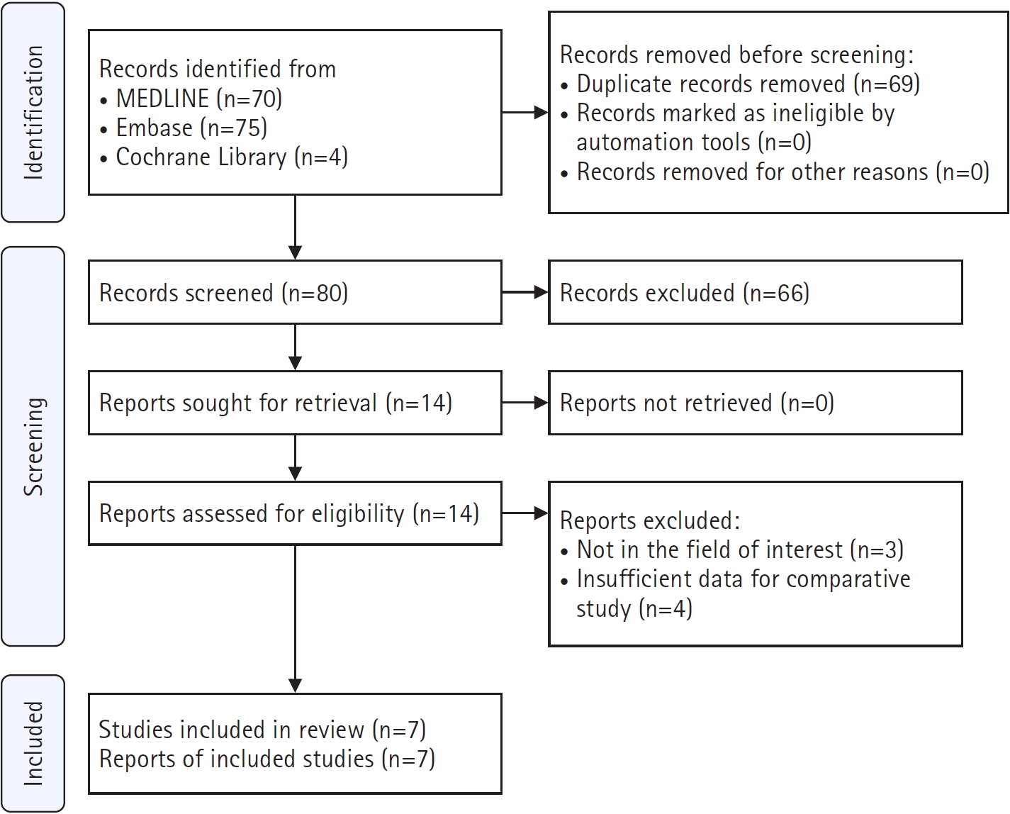
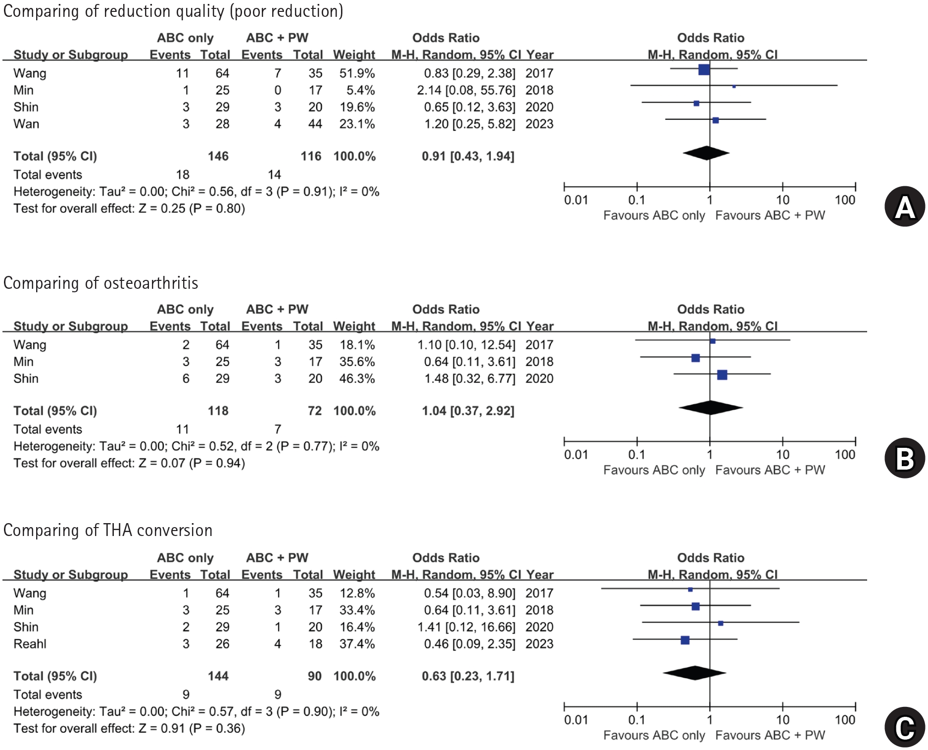
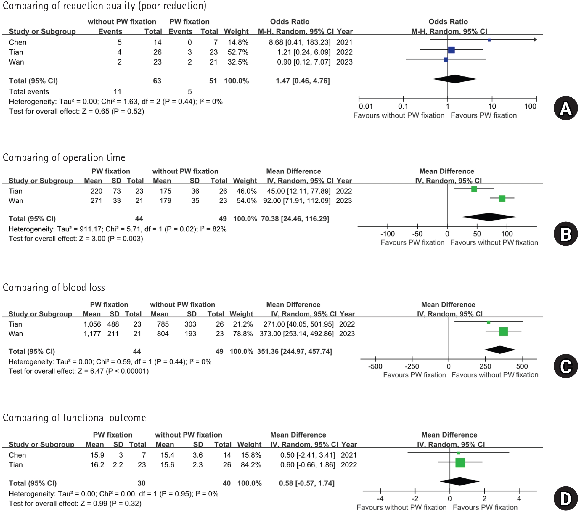
Fig. 1. Flow diagram of the study selection process according to the Preferred Reporting Items for Systematic Reviews and Meta-Analyses (PRISMA) 2020 guidelines. No additional records were identified through manual searches of the reference lists of the retrieved studies.
Fig. 2. Forest plots comparing (A) poor reduction, (B) osteoarthritis, and (C) conversion to THA according to the presence of a PW fragment. ABC, associated both-column; PW, posterior wall; M-H, Mantel-Haenszel; CI, confidence interval; THA, total hip arthroplasty.
Fig. 3. Forest plots comparing (A) poor reduction, (B) operative time, (C) intraoperative blood loss, and (D) functional outcome according to PW fixation status. M-H, Mantel-Haenszel; CI, confidence interval; PW, posterior wall; SD, standard deviation; IV, inverse variance.
Fig. 1.
Fig. 2.
Fig. 3.
Impact of posterior wall involvement and fixation on clinical and radiographic outcomes of associated both-column acetabular fractures: a systematic review and meta-analysis
| Study | Study design | Number of patients | Mean age (yr) | Female sex | Follow-up (mo) |
||||||
|---|---|---|---|---|---|---|---|---|---|---|---|
| PW− | PW+Fix+ | PW+Fix− | PW− | PW+Fix+ | PW+Fix− | PW− | PW+Fix+ | PW+Fix− | |||
| Wang et al. [7] (2017) | RCS | 64 | 35 | NA | 48.3±11.8 | 44.4±12.5 | NA | 17 | 10 | NA | 44.7±18.5 |
| Min et al. [15] (2018) | RCS | 25 | 15 | 2 | 46.2 | 45.7 |
8 | 4 |
54.0 | ||
| Shin et al. [5] (2020) | RCS | 29 | 3 | 17 | 51.1±23.3 | 48.1±23.6 |
5 | 4 |
36.6±18.5 | ||
| Chen et al. |
RCS | 39 | 11 | 29 | 50.9±18.8 | 51.2±16.8 | 62.7±16.7 | 12 | 1 | 7 | 49.9±39.2 |
| Tian et al. [16] (2022) | RCS | NA | 23 | 26 | NA | 45.5±11.2 | 46.1±11.8 | NA | 5 | 5 | 27.1±6.9 |
| Reahl et al. [3] (2023) | RCS | 26 | 18 | NA | 48.5±16.7 | 51.4±12.0 | NA | 6 | 4 | NA | 43.0 |
| Wan et al. [6] (2023) | RCS | 28 | 21 | 23 | 44.8±14.1 | 41.7±12.3 | 42.5±13.1 | 12 | 8 | 9 | Min 12 mo |
| Study | Approach | Operation time (min) | Blood loss (mL) | Poor reduction | Assessment modality | ||||||||
|---|---|---|---|---|---|---|---|---|---|---|---|---|---|
| PW− | PW+Fix+ | PW+Fix− | PW− | PW+Fix+ | PW+Fix− | PW− | PW+Fix+ | PW+Fix− | PW− | PW+Fix+ | PW+Fix− | ||
| Wang et al. [7] (2017) | Ilioinguinal | 241±49 | 258±60 | NA | 889±154 | 957±259 | NA | 11 | 7 | NA | X-ray | ||
| Min et al. [15] (2018) | Ilioinguinal | NR | NR | NR | NR | NR | NR | 1 | - | 0 | X-ray & CT | ||
| Shin et al. [5] (2020) | Ilioinguinal | Ilioinguinal, KL | Ilioinguinal | NR | NR | NR | NR | NR | NR | 3 | - | 3 | X-ray |
| Chen et al. [14] (2021) | Ilioinguinal, Stoppa | Ilioinguinal, Stoppa, KL | Ilioinguinal, Stoppa | NR | NR | NR | NR | NR | NR | NR | 0 | 5 | X-ray |
| Tian et al. [16] (2022) | NA | Stoppa, KL | Stoppa | NA | 220±73 | 175±36 | NA | 1,056±488 | 785±303 | NA | 3 | 4 | NR |
| Reahl et al. [3] (2023) | Ilioinguinal, KL, extended iliofemoral | 215±94 | 197±63 | NA | NR | NR | NA | NR | NR | NA | NA | ||
| Wan et al. [6] (2023) | Supra-ilioinguinal | Supra-ilioinguinal, KL | Supra-ilioinguinal | 163±21 | 271±33 | 179±35 | 703±143 | 1,177±211 | 804±193 | 3 | 2 | 2 | X-ray |
| Study | Functional outcome |
Osteoarthritis |
Conversion to THA | SSI | ||||||||
|---|---|---|---|---|---|---|---|---|---|---|---|---|
| PW− | PW+Fix+ | PW+Fix− | PW− | PW+Fix+ | PW+Fix− | PW− | PW+Fix+ | PW+Fix− | PW− | PW+Fix+ | PW+Fix− | |
| Wang et al. [7] (2017) | 15.7±1.9 | 15.9±1.8 | NA | 2 | 1 | NA | 1 | 1 | NA | 1 | 2 | NA |
| Min et al. [15] (2018) | 22 | 14 | 3 | 3 | 3 | 3 | 0 | 1 | ||||
| Shin et al. [5] (2020) | 89.0±6.6 | 90.5±6.8 | 6 | 3 | 2 | 1 | 0 | 1 | ||||
| Chen et al. [14] (2021) | NR | 15.9±3.0 | 15.4±3.6 | NR | 1.1±0.7 | 1.3±0.9 | NR | 0 | 3 | NR | NR | NR |
| Tian et al. [16] (2022) | NA | 16.2±2.2 | 15.6±2.3 | NA | NR | NR | NA | NR | NR | NA | 2 | 0 |
| Reahl et al. [3] (2023) | NR | NR | NA | NR | NR | NA | 3 | 4 | NA | 2 | 1 | NA |
| Wan et al. [6] (2023) | 24 | 18 | 18 | NR | NR | NR | NR | NR | NR | NR | NR | NR |
| Study | Confounding | Selection | Intervention classification | Deviation from intervention | Missing data | Measurement of outcome | Selection of reported result | Overall |
|---|---|---|---|---|---|---|---|---|
| Wang et al. [7] (2017) | Moderate | Low | Low | Low | Low | Moderate | Moderate | Moderate |
| Min et al. [15] (2018) | Moderate | Low | Low | Low | Low | Low | Moderate | Moderate |
| Shin et al. [5] (2020) | Moderate | Low | Low | Low | Low | Low | Moderate | Moderate |
| Chen et al. [14] (2021) | Serious | Moderate | Low | Low | Moderate | Moderate | Moderate | Serious |
| Tian et al. [16] (2022) | Moderate | Moderate | Low | Low | Low | Moderate | Moderate | Moderate |
| Reahl et al. [3] (2023) | Moderate | Moderate | Low | Low | Moderate | Moderate | Moderate | Moderate |
| Wan et al. [6] (2023) | Serious | Low | Low | Low | Low | Moderate | Moderate | Serious |
| Study | Comparisons | Indications for fixation of PW | Stability assessment after column fixation |
|---|---|---|---|
| Wang et al. [7] (2017) | PW− vs. PW+Fix+ | No specific clinical threshold (such as size or mm) was provided. | Performed (under fluoroscopy) |
| Min et al. [15] (2018) | PW− vs. PW+ | Left unfixed if <20% and the joint was stable. Fixed if the fragment represented >20% of the fracture surface on CT. | Not performed (acknowledged as limitation) |
| Shin et al. [5] (2020) | PW− vs. PW+ | Fixed if gross instability was present on stress exam, or if there was joint incongruency (step-off >2 mm or gap >3 mm), or if the fragment lacked attachment to the labrocapsular complex. | Performed (90° flexion with axial load) |
| Chen et al. [14] (2021) | PW+Fix+ vs. PW+Fix− | Performed at the surgeon’s discretion. | Not performed (not routinely assessed) |
| Tian et al. [16] (2022) | PW+Fix+ vs. PW+Fix− | Treatment was based on the study period: PWs were routinely fixed in the early group (via the KL approach) but were no longer fixed in the recent group due to evolving understanding. | Performed (under anesthesia) |
| Reahl et al. [3] (2023) | PW− vs. PW+Fix+ | No specific clinical threshold (like size or mm) was provided. | Not reported |
| Wan et al. [6] (2023) | PW− vs. PW+Fix− vs. PW+Fix+ | A posterior approach was required if more than 3 mm of displacement remained in the reduced 3D model after virtual reduction of the main columns. | Not performed (relied on preoperative 3D virtual simulation) |
Table 1. Summary of patient demographic data from the included studies
PW, posterior wall; Fix, fixation; PW−, associated both-column fractures without posterior wall involvement; PW+Fix+, associated both-column fractures with posterior wall involvement treated with fixation; PW+Fix−, associated both-column fractures with posterior wall involvement treated without fixation; RCS, retrospective cohort study; NA, not applicable; Min, minimum. Chen et al. [ Follow-up periods are presented as mean±standard deviation when available. Studies without reported standard deviations are shown as mean values only. Wan et al. [ For Min et al. [
Table 2. Perioperative profile
PW, posterior wall; Fix, fixation; PW−, associated both-column fractures without posterior wall involvement; PW+Fix+, associated both-column fractures with posterior wall involvement treated with fixation; PW+Fix−, associated both-column fractures with posterior wall involvement treated without fixation; NA, not applicable; NR, not reported; CT, computed tomography; KL, Kocher-Langenbeck. Shin et al. [ For Chen et al. [
Table 3. Clinical outcomes
THA, total hip arthroplasty; SSI, surgical site infection; PW, posterior wall; Fix, fixation; PW−, associated both-column fractures without posterior wall involvement; PW+Fix+, associated both-column fractures with posterior wall involvement treated with fixation; PW+Fix−, associated both-column fractures with posterior wall involvement treated without fixation; NA, not applicable; NR, not reported; MAP, modified Merle d'Aubigné-Postel score; HHS, Harris Hip Score. Functional outcomes are presented as mean MAP scores for Wang et al. [ Wang et al. [
Table 4. Risk of bias assessment using the ROBINS-I tool
ROBINS-I, Risk of Bias in Non-randomized Studies of Interventions.
Table 5. Indications for fixation of PW
PW, posterior wall; Fix, fixation; PW−, associated both-column fractures without posterior wall involvement; PW+, associated both-column fractures with posterior wall involvement; PW+Fix+, associated both-column fractures with posterior wall involvement treated with fixation; PW+Fix−, associated both-column fractures with posterior wall involvement treated without fixation; CT, computed tomography; KL, Kocher-Langenbeck; 3D, three-dimensional.


