
 E-submission
 KOTA
KOTA TOTA
TOTA TOTS
TOTS
Articles
- Page Path
- HOME > > Ahead-of print articles > Article
-
Case Report
- Surgical management of a displaced proximal fibular fracture with peroneal nerve injury in Korea: a case report
- Sang-Gon Kim, Sang-Heon Oh, Ki-Bong Park
-
DOI: https://doi.org/10.12671/jmt.2026.00213
Published online: September 21, 2026
Department of Orthopedic Surgery, Ulsan University Hospital, University of Ulsan College of Medicine, Ulsan, Korea
- Correspondence to: Ki-Bong Park Department of Orthopedic Surgery, Ulsan University Hospital, University of Ulsan College of Medicine, 25 Daehakbyeongwon-ro, Dong-gu, Ulsan 44033, Korea Tel: +82-52-250-7129 Email: kbpark@uuh.ulsan.kr
• Received: May 15, 2026 • Revised: June 30, 2026 • Accepted: July 1, 2026
© 2026 The Korean Orthopaedic Trauma Association
This is an Open Access article distributed under the terms of the Creative Commons Attribution Non-Commercial License (http://creativecommons.org/licenses/by-nc/4.0/) which permits unrestricted noncommercial use, distribution, and reproduction in any medium, provided the original work is properly cited.
- 72 Views
- 4 Download


Abstract
-
This case report describes a rare inferomedially displaced proximal fibular fracture associated with acute common peroneal nerve injury in a 73-year-old man after an industrial crush injury. The patient presented with severe knee pain and a subjective sensory deficit on the dorsum of the right foot. Given the marked inferomedial displacement of the fracture fragment and the associated neurological symptoms, surgical intervention was selected to achieve anatomical reduction and permit direct nerve assessment. After four days of soft-tissue management, open reduction and internal fixation were performed via a posterior approach with the patient in the prone position. The fragment was securely fixed with 4.5-mm cannulated screws, and nerve exploration confirmed mild nerve sheath injury without complete disruption; therefore, neurorrhaphy was not required. This case illustrates that operative intervention may be considered in selected patients with markedly inferomedially displaced proximal fibular fractures, particularly when acute neurological deficits are present. In this specific displacement pattern, a posterior approach in the prone position may provide a useful surgical option for achieving anatomical reduction while allowing direct neurovascular protection.Level of evidence: V.
Introduction
Proximal fibular fractures are commonly associated with tibial plateau fractures or multi-ligamentous knee injuries; however, severe displacement of the fracture fragments poses substantial clinical and surgical challenges [1]. The fibular head serves as the critical attachment site for the biceps femoris tendon and the lateral collateral ligament, contributing significantly to posterolateral knee stability [2]. Although isolated or minimally displaced proximal fibular fractures are frequently treated conservatively with satisfactory outcomes, high-energy trauma around the proximal fibula carries a considerable risk of injury to the common peroneal nerve due to its close anatomical relationship [3]. As the nerve courses superficially around the fibular neck, it is highly susceptible to entrapment or laceration in crush injuries. Therefore, careful neurological examination and detailed radiological evaluation are essential [4].
The displacement pattern of proximal fibular fractures is generally dictated by traction forces of attached musculotendinous structures, typically resulting in superior or lateral migration [5]. In contrast, high-energy industrial crush mechanisms may overcome these usual biomechanical forces and produce unusual displacement configurations. Inferomedial displacement of the proximal fibular fragment toward the tibial shaft and posterior compartment is especially uncommon. In this patient, the inferomedially displaced fragment extended into the posterior compartment, raising concern that a conventional lateral approach might provide limited direct access to the fragment; therefore, an alternative approach was selected based on the displacement pattern [6].
This report describes a 73-year-old man with an inferomedially displaced proximal fibular fracture and associated common peroneal nerve injury caused by a rolling steel pipe crush injury. This case demonstrates the successful application of a tailored surgical approach to achieve stable fixation while ensuring neurovascular protection.
Case report
This study received approval from the Institutional Review Board (IRB) of our institution (IRB No. 2026-04-023). Informed consent was waived due to the retrospective design and unsuccessful attempts to contact the patient. All procedures were conducted in accordance with the principles of the Declaration of Helsinki.
A 73-year-old man presented to the emergency department with severe pain in the right knee after his leg became caught between two rolling steel pipes on the day of admission.
On physical examination, multiple abrasions and marked swelling were observed over the anterolateral aspect of the right lower leg. Pronounced tenderness was elicited over the proximal fibular region. Neurological examination revealed decreased sensation over the dorsum of the right foot. Light-touch sensation over the dorsum of the right foot was subjectively rated as 7/10 compared with 10/10 on the contralateral side, raising suspicion of an acute common peroneal nerve injury. No motor weakness or foot drop was noted at initial evaluation.
Plain radiographs and computed tomography scans identified a non-displaced fracture of the lateral proximal tibia along with a proximal fibular fracture fragment displaced inferomedially toward the tibial shaft (Fig. 1). The non-displaced proximal tibial fracture was managed conservatively without internal fixation and did not require modification of the planned fibular fixation. Magnetic resonance imaging (MRI) demonstrated that the posterolateral corner structures, including the lateral collateral ligament and the biceps femoris tendon, maintained intact continuity without high-grade tearing or avulsion from their insertions. These findings showed no MRI evidence of high-grade injury to posterolateral corner structures, supporting the interpretation that the displaced osseous fragment and associated nerve symptoms were the principal acute concerns. Notably, preoperative electrodiagnostic studies were not performed due to the acute nature of the injury.
Surgical intervention was performed four days after injury to allow improvement of the soft tissue condition. Under general anesthesia, the patient was positioned prone, and a pneumatic tourniquet was applied. A longitudinal incision approximately 10 cm in length was created over the proximal fibula. The fracture site was exposed by splitting of the lateral head of the gastrocnemius and the soleus muscle. The inferomedially displaced proximal fibular fragment was mobilized with a Kirschner wire (K-wire) used as a joystick and guided back to its anatomic position under direct visualization of the common peroneal nerve. Temporary stabilization was achieved with two 1.6-mm K-wires inserted in the posteroanterior direction, ensuring they bypassed the nerve, which was followed by definitive fixation using two 4.5-mm half-threaded cannulated screws. The screws were inserted in a posterior-to-anterior trajectory, providing maximum compression and rigid stability. After fixation, exploration of the common peroneal nerve and its branching region into the superficial and deep peroneal nerves demonstrated mild injury to the nerve sheath without complete disruption; therefore, neurorrhaphy was unnecessary. A suction drain was placed, and layered closure was performed.
Postoperative radiographs demonstrated satisfactory anatomical restoration with stable internal fixation (Fig. 2). Sensory function over the dorsum of the foot recovered to 8/10 by postoperative day 2 and 9/10 at 4 weeks. Radiographic bone union was achieved at 2 months. At the 1-year follow-up, the patient exhibited full knee extension and flexion (0°–130°), with complete recovery of sensation and normal ankle dorsiflexion strength. The patient reported a complete pain-free status and successfully returned to occupational activities. A timeline of the clinical course is presented in Table 1.
Discussion
Proximal fibular fractures are frequently encountered following trauma; however, the marked inferomedial displacement observed in this case represents an uncommon injury pattern. In most literature, proximal fibular fragments exhibit superior or lateral displacement due to the traction forces exerted by the biceps femoris tendon and the lateral collateral ligament [2,7]. Peroneal nerve injury in those typical patterns is frequently secondary to severe traction or avulsion forces [3]. In contrast, our case involved an intense industrial crush mechanism that forced the fragment medially into the posterior compartment. Although direct compression from the fragment was highly suspected, the neurological dysfunction may have involved a combination of direct contusion, traction, localized edema, or transient compression during the injury.
A review of the literature reveals that nerve injuries at this level are most frequently documented in low-energy sports avulsions [2,7] or high-energy tibial plateau fractures where the fibula suffers secondary lateral shearing [1,5]. In both situations, a standard lateral approach is sufficient for fixation [3,6]. In contrast, the clinical implication of our case is entirely distinct; the fragment was driven inferomedially, shifting the entire pathology into the deep posterior aspect. Nonoperative treatment has been reported for a proximal fibular fatigue fracture with secondary common peroneal nerve injury [3]; however, that injury pattern differs substantially from the high-energy displaced fracture in the present case. In the present case, the deep inferomedial position of the fragment raised concern regarding direct access and nerve protection during reduction through a conventional lateral route [6,8]. This expanded comparison underscores that the surgical approach should be tailored to the specific vector of fracture displacement rather than adhering strictly to traditional lateral routes.
The decision for surgical treatment was based on the unusual degree and direction of displacement, which created a substantial risk of persistent nerve compression, malunion, and subsequent impairment of lower-extremity stability. Surgical intervention was delayed for four days to permit improvement of the soft tissue condition, decreasing the likelihood of wound complications associated with crush injuries.
In this case, the posterior approach in the prone position facilitated direct access to the inferomedially displaced fragment and allowed the common peroneal nerve to be visualized during reduction. Although a lateral approach is typically used for fibular surgery, the medial displacement of the fragment made a posterior-to-anterior surgical trajectory more suitable for stable fixation [8]. Prone positioning provided a suitable operative field for identifying and protecting the common peroneal nerve around the fibular neck during reduction and fixation [9].
Marked inferomedial displacement of a proximal fibular fragment accompanied by acute common peroneal nerve symptoms may be considered for operative reduction after appropriate soft-tissue stabilization. In this patient, a prone posterior approach provided direct access to the displaced fragment while allowing direct visualization and protection of the common peroneal nerve. This approach may represent a useful option in carefully selected cases; however, its generalizability and comparative advantages over conventional approaches cannot be established from a single case.
-
Author contributions
Conceptualization: SGK, KBP. Methodology: SHO, KBP. Supervision: KBP. Visualization: SGK, SHO. Writing-original draft: SGK, SHO, KBP. Writing-review & editing: SGK, SHO, KBP. All authors read and approved the final manuscript.
-
Conflicts of interest
No potential conflict of interest relevant to this article was reported.
-
Funding
None.
-
Data availability
Not applicable.
-
Acknowledgments
None.
-
Supplementary materials
None.
Article Information
Fig. 1.
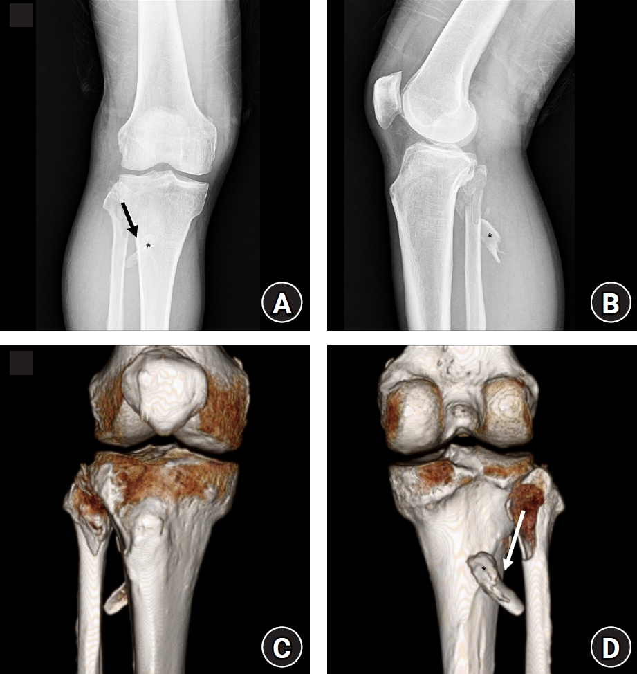
Preoperative imaging. (A) Anteroposterior and (B) lateral plain radiographs and (C) anterior and (D) posterior views of three-dimensional computed tomography reconstruction showing an inferomedially displaced proximal fibular fragment extending into the posterior compartment. The asterisk indicates the displaced fragment, and the arrow indicates the direction of displacement.
Fig. 2.
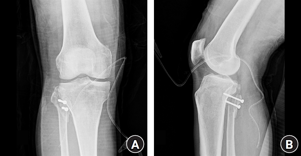
Postoperative radiographs. (A) Anteroposterior and (B) lateral views of the right knee showing definitive internal fixation of the proximal fibular fragment with two 4.5-mm cannulated screws. A surgical suction drain is visible in the immediate postoperative state.
Table 1.
Timeline of clinical events and patient outcomes
- 1. Zheng ZL, Yu YY, Chang HR, Liu H, Zhou HL, Zhang YZ. Establishment of classification of tibial plateau fracture associated with proximal fibular fracture. Orthop Surg 2019;11:97-101.ArticlePubMedPMCPDF
- 2. Cinats DJ, Stone T. Comminuted fibular head fractures with posterolateral knee instability: a novel repair technique using a tension-slide device. J Orthop Trauma 2021;35:e66-70.ArticlePubMed
- 3. Al-Kashmiri A, Delaney JS. Case report: fatigue fracture of the proximal fibula with secondary common peroneal nerve injury. Clin Orthop Relat Res 2007;463:225-8.ArticlePubMed
- 4. Rausch V, Hackl M, Oppermann J, et al. Peroneal nerve location at the fibular head: an anatomic study using 3D imaging. Arch Orthop Trauma Surg 2019;139:921-6.ArticlePubMedPDF
- 5. Liu Y, Yang J, Liu R, et al. Morphological study of proximal fibular fractures with concomitant tibial plateau fractures: an investigation of 223 cases. Sci Prog 2025;108:368504251397508.ArticlePubMedPMCPDF
- 6. AO Foundation. Straight lateral approach to the proximal tibia [Internet]. AO Foundation; [cited 2026 May 13]. Available from: https://surgeryreference.aofoundation.org/orthopedic-trauma/adult-trauma/proximal-tibia/approach/straight-lateral-approach.
- 7. Tornetta P, Ricci WM, Court-Brown CM, editors. Rockwood and Green's fractures in adults. 9th ed. Wolters Kluwer; 2019.
- 8. Kim JM, Park BM, Lee SH, Jeon SJ, Shin JB, Song KS. Surgical management of comminuted avulsion fracture of the proximal fibula with lateral collateral ligament injury: technical note. J Korean Fract Soc 2013;26:77-80.Article
- 9. Souter J, Swong K, McCoyd M, Balasubramanian N, Nielsen M, Prabhu VC. Surgical results of common peroneal nerve neuroplasty at lateral fibular neck. World Neurosurg 2018;112:e465-72.ArticlePubMed
References
Figure & Data
REFERENCES
Citations
Citations to this article as recorded by 
 ePub Link
 Cite
Cite XML Download
Surgical management of a displaced proximal fibular fracture with peroneal nerve injury in Korea: a case report
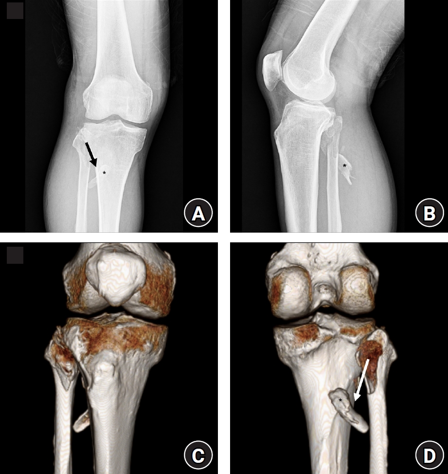
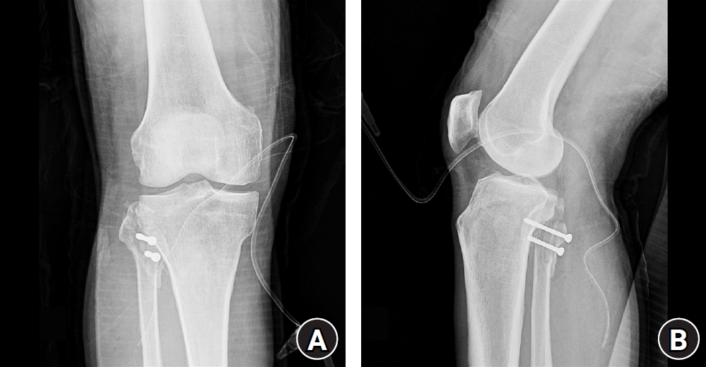
Fig. 1. Preoperative imaging. (A) Anteroposterior and (B) lateral plain radiographs and (C) anterior and (D) posterior views of three-dimensional computed tomography reconstruction showing an inferomedially displaced proximal fibular fragment extending into the posterior compartment. The asterisk indicates the displaced fragment, and the arrow indicates the direction of displacement.
Fig. 2. Postoperative radiographs. (A) Anteroposterior and (B) lateral views of the right knee showing definitive internal fixation of the proximal fibular fragment with two 4.5-mm cannulated screws. A surgical suction drain is visible in the immediate postoperative state.
Fig. 1.
Fig. 2.
Surgical management of a displaced proximal fibular fracture with peroneal nerve injury in Korea: a case report
| Time point / postoperative period | Clinical event / intervention | Key findings and outcomes |
|---|---|---|
| Day 0 (day of admission) | Right knee crush injury between rolling steel pipes | Severe right knee pain, marked anterolateral swelling, exquisite proximal fibular tenderness, and a subjective sensory deficit on the dorsum of the right foot |
| Day 0 | Radiological evaluation using radiographs, computed tomography, and magnetic resonance imaging | Non-displaced lateral proximal tibial fracture and an inferomedially displaced proximal fibular fragment extending toward the tibial shaft; no high-grade ligamentous tear or avulsion on magnetic resonance imaging |
| Days 1–3 | Soft tissue management | Strict bed rest, limb elevation, and ice packing to improve the surrounding soft tissue condition and reduce swelling |
| Day 4 | Surgical intervention with open reduction and internal fixation and nerve exploration | Open reduction and internal fixation via a posterior approach in the prone position using two 4.5-mm cannulated screws; common peroneal nerve exploration confirmed mild nerve sheath injury without complete disruption |
| Postoperative day 2 | Early postoperative assessment | Satisfactory anatomical restoration on radiographs, stable surgical wound condition, and progressive improvement in sensory function on the dorsum of the foot to 8/10 |
| Week 4 | Short-term follow-up | Complete surgical wound healing; recovery of the subjective sensory score to 9/10; initiation of passive and active knee range-of-motion exercises |
| Month 2 | Mid-term follow-up | Successful radiographic bone union; initiation of partial to full weight-bearing ambulation |
| Year 1 (final follow-up) | Long-term functional assessment | Full knee extension and flexion, complete recovery of sensation over the dorsum of the foot, normal ankle dorsiflexion strength, complete pain relief, and full return to daily and occupational activities |
Table 1. Timeline of clinical events and patient outcomes


