
 E-submission
 KOTA
KOTA TOTA
TOTA TOTS
TOTS
Articles
- Page Path
- HOME > J Musculoskelet Trauma > Volume 39(3); 2026 > Article
-
Original Article
- Clinical and radiographic outcomes after surgical treatment of supination-adduction type ankle fractures in older (≥65 years) and younger (<65 years) patients: a retrospective single-center comparative study in Korea
- JungHo Lee1, Bawoo Ko1, Hongik Choi1, Jun-young Lee1,2
-
Journal of Musculoskeletal Trauma 2026;39(3):236-244.
DOI: https://doi.org/10.12671/jmt.2025.00374
Published online: July 24, 2026
1Department of Orthopedic Surgery, Chosun University Hospital, Gwangju, Korea
2Department of Orthopedic Surgery, Chosun University College of Medicine, Gwangju, Korea
- Correspondence to: Jun-young Lee Department of Orthopedic Surgery, Chosun University Hospital, 365 Pilmun-daero, Dong-gu, Gwangju 61453, Korea Tel: +82-62-220-3147 Email: leejy88@chosun.ac.kr
• Received: December 1, 2025 • Revised: July 13, 2026 • Accepted: July 16, 2026
© 2026 The Korean Orthopaedic Trauma Association
This is an Open Access article distributed under the terms of the Creative Commons Attribution Non-Commercial License (http://creativecommons.org/licenses/by-nc/4.0/) which permits unrestricted noncommercial use, distribution, and reproduction in any medium, provided the original work is properly cited.
- 719 Views
- 15 Download


Abstract
-
Background Supination-adduction (SAD) ankle fractures involve a vertical medial malleolar fracture and potential tibial plafond impaction, but pattern-specific evidence in older patients remains limited. We compared postoperative complications and clinical/radiographic outcomes between older and younger patients and explored complication-associated factors within the older group.
-
Methods We retrospectively reviewed 51 patients who underwent surgery for SAD-type ankle fractures (2010–2020): 30 younger (<65 years) and 21 older (≥65 years). The primary outcome was a composite of surgical site infection, loss of reduction, or delayed union. Functional recovery was assessed using Olerud-Molander Ankle Score (OMAS) and American Orthopaedic Foot & Ankle Society (AOFAS) scores through 12 months. Union and posttraumatic osteoarthritis were assessed radiographically at ≥18 months. Preoperative dual-energy X-ray absorptiometry was performed in older patients for exploratory analyses.
-
Results Postoperative complications were higher in the older group (38.1% vs. 10.0%, P=0.035). In older patients, diabetes mellitus was associated with complications (odds ratio [OR], 9.17; 95% confidence interval [CI], 1.15–73.24; P=0.037); smoking was not (OR, 5.50; 95% CI, 0.71–42.60; P=0.103). At 12 months, OMAS and AOFAS scores did not differ significantly. One older patient developed nonunion, and posttraumatic osteoarthritis occurred in one patient per group. Lower T-scores (mean, −2.3±0.5) correlated with greater tibial plafond impaction depth (3.1±1.2 mm; ρ=−0.46, P=0.036) and showed a borderline significant correlation with articular step-off (2.0±0.8 mm; ρ=−0.43, P=0.052).
-
Conclusions Older patients had higher postoperative complication rates than younger patients, although 12-month functional and late radiographic outcomes were comparable. Diabetes was associated with complications, though the small sample warrants cautious interpretation. Findings support careful perioperative optimization and individualized fixation based on fracture morphology and bone quality in older patients, especially those with diabetes.
-
Level of evidence III.
Introduction
Ankle fractures are among the most common fractures treated by orthopedic surgeons, and their incidence increases in older adults as life expectancy and activity levels rise [1-4]. Older patients often have medical comorbidities, impaired soft-tissue conditions, reduced bone quality, and diminished physiologic reserve, all of which may complicate operative treatment and recovery [4-6]. Most studies of geriatric ankle fractures, however, have analyzed heterogeneous fracture patterns, which may obscure pattern-specific challenges.
The supination-adduction (SAD) pattern described by Lauge-Hansen is characterized by a transverse or avulsion injury of the lateral malleolus followed by a vertical medial malleolar fracture [7,8]. Varus loading and axial compression may produce marginal impaction of the medial tibial plafond and an articular step-off [9]. Restoring joint congruity and resisting vertical shear—often with buttress fixation—are therefore central to treatment. In older patients, reduced bone quality may increase articular impaction and make stable fixation more difficult even after low-energy trauma.
Fracture-pattern–specific evidence on surgically treated SAD-type ankle fractures in older patients remains limited. The primary aim of this study was to compare postoperative complications and clinical, functional, and radiographic outcomes between older and younger patients; the secondary aim was to explore clinical and radiographic factors associated with postoperative complications within the older group. We hypothesized that older patients would have a higher postoperative complication rate and slower early functional recovery but would achieve comparable late outcomes when anatomical reduction and stable fixation were obtained.
Methods
This study was conducted in compliance with the principles of the Declaration of Helsinki. The protocol was reviewed and approved by the Institutional Review Board (IRB) of Chosun University Hospital (IRB No. CHOSUN 2025-09-029-002). The requirement for individual informed consent was waived by the IRB because of the retrospective design.
This retrospective comparative study reviewed patients who presented to the emergency department or outpatient clinic of our institution between January 2010 and December 2020 with an ankle fracture. SAD-type fractures were identified on preoperative plain radiographs and computed tomography (CT) according to the Lauge-Hansen pattern. Seventy-three SAD-type ankle fractures were identified; four were treated nonoperatively and 69 underwent surgery. After exclusion of patients with incomplete records or insufficient follow-up (n=8), open fractures (n=4), and multiple trauma (n=6), 51 patients were included (Fig. 1).
Patients were divided into a younger group (<65 years, n=30) and an older group (≥65 years, n=21). The 65-year cutoff was selected because it is widely used in clinical studies of geriatric ankle fractures and permits comparison with prior outcome studies [3,4,10].
Age, sex, body mass index, comorbidities, smoking status, trauma-energy level, fracture characteristics, and follow-up duration were collected from electronic medical records. Diabetes mellitus was defined as a documented diagnosis, current use of glucose-lowering medication, or a preoperative glycated hemoglobin level of ≥6.5%, consistent with current diagnostic criteria [11]. Hypertension was defined as a documented diagnosis or current use of antihypertensive medication.
Trauma was classified according to the documented event. High-energy trauma included motor vehicle, motorcycle, or bicycle collisions with direct impact, crush injuries, falls from a height greater than standing height, and sports injuries involving a documented high-velocity impact or direct collision. Low-energy trauma included falls from standing height, simple twisting or misstep injuries, and noncontact recreational sports injuries without high-velocity impact. Sports injuries were therefore classified individually rather than uniformly categorized as high-energy trauma.
Dual-energy X-ray absorptiometry (DXA) was performed in all 21 older patients, generally during the index admission and before surgery, at the lumbar spine, femoral neck, and total hip. The lowest valid T-score among these sites was recorded as the representative value, and osteoporosis was defined as a T-score ≤−2.5 [12]. Because DXA was not systematically obtained in younger patients, between-group comparisons of T-score and osteoporosis prevalence were not performed; analyses involving T-score were restricted to the older group and were considered exploratory.
All operations were performed by a single orthopedic trauma surgeon. Fixation was individualized according to fracture morphology, fragment size, comminution, bone quality, and intraoperative stability. Simple, noncomminuted lateral malleolar fractures with an adequately sized fragment were treated with cannulated screw fixation, whereas plate fixation was used when the lateral malleolar fracture was comminuted, unstable, or unsuitable for screw fixation alone. Vertical medial malleolar fractures were treated with cannulated screws, a buttress plate, or a combination; buttress plating was preferred in the presence of a vertical shear component, metaphyseal comminution, poor bone quality, or inadequate stability with screws alone.
When tibial plafond impaction was present, the impacted articular fragment was elevated under direct and fluoroscopic guidance to restore joint congruity (Fig. 2), and bone graft or a bone substitute was used for a clinically relevant residual metaphyseal defect. Prophylactic antibiotics were administered within 30 minutes before incision and continued for 24 hours. Postoperative immobilization, range-of-motion exercises, and weight-bearing progression were individualized according to fixation stability, soft-tissue status, and bone quality.
The primary outcome was a composite postoperative complication consisting of superficial incisional surgical site infection (SSI), deep incisional SSI, loss of reduction, or delayed union; each component was also evaluated separately. SSI was classified using the anatomical and clinical principles of the Centers for Disease Control and Prevention National Healthcare Safety Network definitions [13]. Superficial incisional SSI involved only skin or subcutaneous tissue with purulent drainage, a positive superficial culture, localized inflammatory findings with deliberate opening or aspiration, or a clinical diagnosis of superficial SSI. Deep incisional SSI involved the fascial or muscular layers with purulent deep drainage, a positive deep-tissue culture, spontaneous or deliberate dehiscence with compatible findings, or an abscess on examination, surgery, or imaging. Treatment method alone was not used to determine infection depth.
Loss of reduction was defined as new radiographic deterioration relative to immediate postoperative images, including coronal displacement >2 mm, talar tilt >3°, sagittal displacement >5 mm, recurrent articular displacement >2 mm, or clinically relevant implant migration or loosening with fracture displacement [14]. Delayed union was defined as persistent fracture-line visibility with incomplete cortical bridging and insufficient radiographic progression at 6 months. Fracture-related deep infection was interpreted in accordance with the international consensus definition [15]. Nonunion was defined as an ununited fracture at ≥9 months after injury without radiographic progression during the preceding 3 months [16]. Union was defined as bridging or disappearance of the fracture line in at least three of four cortices with resolution of clinically relevant pain or motion at the fracture site.
Preoperative CT was reconstructed using multiplanar reconstruction to obtain standardized coronal and sagittal planes. Tibial plafond impaction depth was measured as the maximum perpendicular distance between the expected intact articular contour and the deepest point of the impacted osteochondral fragment; articular step-off was the maximum vertical displacement between adjacent articular surfaces, measured on the plane of greatest displacement. Because marginal impaction is more reliably identified on CT than on plain radiographs, all measurements were performed on CT [17]. The measurement methods are illustrated in Fig. 3. Final radiographs obtained at ≥18 months were evaluated for union and posttraumatic osteoarthritis, with CT reviewed when union or articular congruity could not be reliably determined on plain films.
Functional outcomes were assessed using the Olerud-Molander Ankle Score (OMAS) and the American Orthopaedic Foot & Ankle Society (AOFAS) ankle-hindfoot score [18-20] at 3, 6, and 12 months postoperatively, with the 12-month assessment designated the principal functional endpoint. Because the interval beyond 12 months was used primarily for radiographic surveillance and functional scores were not systematically recollected at that stage in this retrospective cohort, functional recovery is reported through 12 months, whereas union and posttraumatic osteoarthritis were assessed on radiographs obtained at ≥18 months. A visual analog scale for pain was not consistently documented throughout the study period and was therefore not analyzed. Three-month scores are reported as descriptive measures of early recovery and interpreted with caution, as many patients were still undergoing progressive weight-bearing and rehabilitation.
Statistical analyses were performed using IBM SPSS ver. 29.0 (IBM Corp.). Continuous variables were assessed for normality with the Shapiro-Wilk test and compared using the independent-samples t-test or Mann-Whitney U test; categorical variables were compared using the chi-square or Fisher exact test. Correlations between the lowest T-score and radiographic measurements in the older group were assessed using Spearman rank correlation. Because postoperative complication is a binary outcome, associations between individual clinical variables and complications were explored using univariable logistic regression and reported as odds ratio (OR) with 95% confidence interval (CI). With only eight complication events in the older group, a multivariable model was not fitted, to avoid overfitting. A post hoc power analysis for the primary comparison of overall complication rates was performed using G*Power ver. 3.1.9.7 (Heinrich Heine University Dusseldorf). Based on a two-sided Fisher exact test, an α level of 0.05, group sizes of 30 and 21, and the observed complication rates of 10.0% and 38.1%, respectively, the achieved statistical power was approximately 57.8%. Statistical significance was set at a two-sided P<0.05.
Results
The cohort comprised 30 younger and 21 older patients. Mean age was 49.3±8.2 years (younger) and 72.4±5.7 years (older; P<0.001). The proportions of female sex, diabetes, hypertension, and smoking did not differ significantly between groups. High-energy trauma occurred in 11 younger patients (36.7%) and three older patients (14.3%; P=0.113). No open fractures were included. The mean follow-up duration was 24.5 months overall; group-specific values are presented in Table 1.
Complications occurred in three younger patients (10.0%) and eight older patients (38.1%; Fisher exact P=0.035) (Table 2). The younger group had two superficial SSIs and one deep SSI, with no loss of reduction or delayed union; the older group had two superficial SSIs, two deep SSIs, one loss of reduction, and three delayed unions. No patient experienced more than one of the listed complications; therefore, the component counts equaled the number of patients with any postoperative complication in each group.
In exploratory analyses restricted to the older group, diabetes was present in five of eight patients with complications and two of 13 without (OR, 9.17; 95% CI, 1.15–73.24; P=0.037). Smoking was present in four of eight versus two of 13 (OR, 5.50; 95% CI, 0.71–42.60; P=0.103). Hypertension, female sex, and osteoporosis were not significantly associated with complications (Table 3). The wide CIs reflect the small number of events and indicate that these estimates are exploratory.
All 21 older patients underwent preoperative DXA. The mean lowest T-score was −2.3±0.5, and 10 patients (47.6%) met the densitometric criterion for osteoporosis. The mean tibial plafond impaction depth was 3.1±1.2 mm, and the mean articular step-off was 2.0±0.8 mm. Lower T-scores were associated with greater impaction depth (Spearman ρ=−0.46, P=0.036), with a similar but nonsignificant association for articular step-off (ρ=−0.43, P=0.052) (Table 4).
OMAS and AOFAS scores improved over time in both groups (Table 5). At 3 and 6 months, the older group had lower scores; these early time-point differences are presented descriptively and should be interpreted cautiously because many patients were still progressing through weight-bearing and rehabilitation. At 12 months, the principal functional endpoint, the between-group differences were no longer significant for OMAS (85.7±5.6 vs. 83.2±6.2; P=0.181) or AOFAS score (91.4±5.1 vs. 89.6±5.4; P=0.234).
No younger patient developed nonunion, whereas one older patient did (0% vs. 4.8%; P=0.412). Posttraumatic osteoarthritis was identified in one patient in each group (3.3% vs. 4.8%; P=1.000) (Table 6). The same representative older patient shown in Fig. 2 demonstrated solid bony union without posttraumatic osteoarthritis at 18 months postoperatively (Fig. 4).
Discussion
The principal finding was that older patients with surgically treated SAD-type ankle fractures had a higher overall postoperative complication rate than younger patients. Early functional recovery was slower in the older group, but the differences in OMAS and AOFAS scores were no longer significant by 12 months, and late nonunion and posttraumatic osteoarthritis were uncommon in both groups. These findings suggest that age and systemic vulnerability influence the early postoperative course, whereas satisfactory longer-term recovery may still be achieved when anatomical reduction and stable fixation are obtained.
The higher complication rate in older patients is consistent with studies showing that age, diabetes, soft-tissue vulnerability, and comorbidities affect outcomes after ankle fracture treatment [4,5,10,21-23]. SAD-type fractures pose additional mechanical challenges because the vertical medial malleolar component is exposed to shear and may coexist with marginal plafond impaction [9,17]. In our cohort, loss of reduction and delayed union occurred only in the older group, supporting fixation strategies that resist vertical shear and account for fragment size, comminution, and bone quality rather than a uniform screw-only technique.
Diabetes showed an exploratory, unadjusted association with complications, although the CI was wide. Diabetes may impair leukocyte function, microvascular perfusion, soft-tissue healing, and bone repair, and complicated diabetes is particularly associated with infection, nonunion, and revision after ankle fracture fixation [21-23]. Smoking was more common among older patients with complications but did not reach significance in this small cohort and should be regarded as a clinically plausible signal rather than a confirmed independent predictor.
DXA was analyzed only within the older group because younger patients were not systematically tested, which avoids an invalid age-group comparison. The exploratory association between lower T-score and greater plafond impaction or step-off suggests that reduced systemic bone density may influence fracture morphology. However, lumbar spine and proximal femoral DXA do not directly measure local subchondral bone quality at the ankle; degenerative change at the spine, site-specific variation, and use of the lowest T-score may further limit precision. The absence of a clear association between T-score and complications therefore does not exclude a role for local bone quality in fixation stability.
The comparable 12-month functional outcomes and the low frequency of late nonunion or posttraumatic osteoarthritis are clinically relevant. Posttraumatic osteoarthritis after SAD-type ankle fracture is closely related to the quality of reduction and articular congruity, and plafond malreduction or residual impaction accelerates arthrosis [24-27]. The low rate observed here is consistent with the emphasis on anatomical restoration and elevation of impacted articular fragments. The lower scores in the older group at 3 and 6 months are descriptive measures of early recovery and may reflect delayed weight-bearing, reduced muscle strength, comorbidities, or slower rehabilitation. Because the 12-month assessment was the principal functional endpoint, the convergence of scores by that time indicates that early differences should not be interpreted as inevitable long-term disability [28].
These results have practical implications. Older patients with an SAD-type fracture, particularly those with diabetes or compromised bone quality, require careful soft-tissue evaluation, perioperative medical optimization, anatomical elevation of impacted articular fragments, and fixation capable of resisting vertical shear. Buttress plating or combined fixation may be preferable when screw fixation alone is inadequate, although the construct should remain individualized to fracture morphology and intraoperative stability.
This study has several limitations. Its retrospective design introduces selection bias and information bias, and rehabilitation adherence and activity level could not be fully controlled. The sample size and number of events were small, and the achieved post hoc power for the primary comparison was approximately 57.8%, indicating limited statistical power; only eight complication events in the older group also precluded reliable multivariable modeling. DXA was obtained systematically only in the older group, so age-group comparisons of bone density were not possible, and spine or proximal femoral T-scores may not represent local ankle bone quality. Functional assessment was standardized only through 12 months, and a validated patient-reported pain outcome measure (e.g., visual analog scale) was not consistently available in this retrospective cohort; the ≥18-month interval was used for radiographic rather than functional evaluation. Finally, the study was performed at a single institution by one surgeon, which improves technical consistency but may limit generalizability. Larger prospective studies with standardized final functional follow-up and local bone-quality measurements are warranted.
Older patients with surgically treated SAD-type ankle fractures had a higher postoperative complication rate than younger patients, although 12-month functional outcomes and late radiographic outcomes were broadly comparable. In the exploratory within-group analysis, diabetes mellitus was associated with postoperative complications; however, the wide CI reflects the limited sample and warrants cautious interpretation. This finding is consistent with the recognized adverse effects of diabetes on soft-tissue and bone healing. Older patients with diabetes may therefore represent a higher-risk subgroup warranting preoperative glycemic optimization, vigilant soft-tissue management, anatomical restoration of the articular surface, and fixation designed to resist vertical shear while accommodating comminution and bone quality. The role of smoking remains uncertain and warrants further study.
-
Author contributions
Conceptualization: JHL, JL. Data curation: BK, HC. Formal analysis: HC. Methodology: JHL, BK. Writing-original draft: JHL, BK. Writing-review & editing: JHL, BK, HC, JL. All authors read and approved the final manuscript.
-
Conflicts of interest
No potential conflict of interest relevant to this article was reported.
-
Funding
This study was supported by the Chosun University Hospital Research Fund (2024).
-
Data availability
Contact the corresponding author for data availability.
-
Acknowledgments
None.
-
Supplementary materials
None.
Article Information
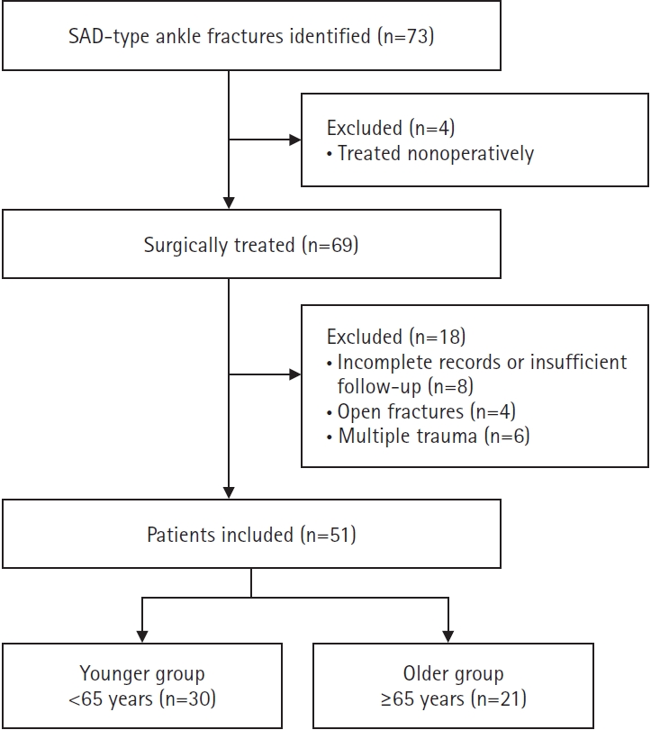
Fig. 2.
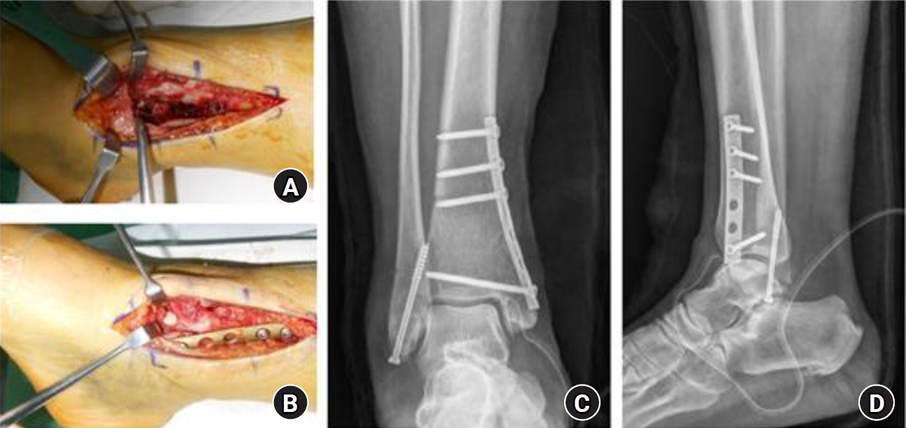
Representative elderly patient with a supination–adduction-type ankle fracture in whom articular congruity was restored by elevation of the impacted articular fragment and fixation with a medial buttress plate and screws. (A) Intraoperative exposure of the impacted articular fragment of the medial tibial plafond. (B) Restoration of articular congruity after elevation and reduction of the impacted fragment, followed by fixation with a medial buttress plate and screws. (C, D) Immediate postoperative anteroposterior and lateral radiographs showing stable fixation of the medial and lateral malleoli.
Fig. 3.
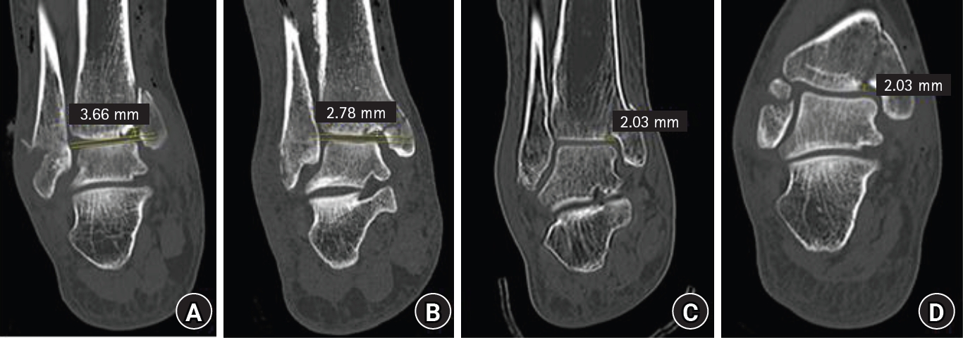
Schematic illustration of radiographic measurements. (A, B) Tibial plafond impaction depth, measured as the maximum perpendicular distance from the expected intact articular contour to the deepest point of the impacted fragment. (C, D) Articular step-off, measured as the maximum vertical displacement between adjacent articular surfaces.
Fig. 4.
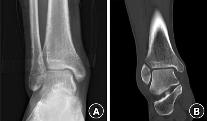
Eighteen-month postoperative images of the patient shown in Fig. 2. (A) Anteroposterior radiograph and (B) coronal computed tomography image demonstrate solid bony union without posttraumatic osteoarthritis, residual marginal impaction of the medial tibial plafond, articular step-off, or other notable abnormalities.
Table 1.
Baseline demographic and clinical characteristics
Table 2.
Postoperative complications according to age group
Table 3.
Exploratory univariable associations with postoperative complications in older patients
Table 4.
Dual-energy X-ray absorptiometry and radiographic parameters in older patients
Table 5.
Functional outcomes during follow-up
Values are presented as mean±standard deviation. The 12-month assessment was designated as the principal functional endpoint. The 3- and 6-month values are descriptive measures of early recovery and should be interpreted cautiously because weight-bearing progression and rehabilitation were ongoing. The visual analog scale was not consistently recorded and is not reported.
OMAS, Olerud-Molander Ankle Score; AOFAS, American Orthopaedic Foot & Ankle Society ankle-hindfoot score.
Table 6.
Final radiographic outcomes at ≥18 months
| Outcome | Younger (n=30) | Older (n=21) | P-value |
|---|---|---|---|
| Nonunion | 0 (0.0) | 1 (4.8) | 0.412 |
| Posttraumatic osteoarthritis | 1 (3.3) | 1 (4.8) | 1.000 |
- 1. Court-Brown CM, Caesar B. Epidemiology of adult fractures: a review. Injury 2006;37:691-7.ArticlePubMed
- 2. Kannus P, Palvanen M, Niemi S, Parkkari J, Jarvinen M. Increasing number and incidence of low-trauma ankle fractures in elderly people: Finnish statistics during 1970-2000 and projections for the future. Bone 2002;31:430-3.ArticlePubMed
- 3. Koval KJ, Lurie J, Zhou W, et al. Ankle fractures in the elderly: what you get depends on where you live and who you see. J Orthop Trauma 2005;19:635-9.ArticlePubMed
- 4. Koval KJ, Zhou W, Sparks MJ, et al. Complications after ankle fracture in elderly patients. Foot Ankle Int 2007;28:1249-55.ArticlePubMedPDF
- 5. SooHoo NF, Krenek L, Eagan MJ, et al. Complication rates following open reduction and internal fixation of ankle fractures. J Bone Joint Surg Am 2009;91:1042-9.ArticlePubMed
- 6. Wukich DK, Kline AJ. The management of ankle fractures in patients with diabetes. J Bone Joint Surg Am 2008;90:1570-8.ArticlePubMed
- 7. Lauge-Hansen N. Fractures of the ankle. II. Combined experimental-surgical and experimental-roentgenologic investigations. Arch Surg (1920) 1950;60:957-85.ArticlePubMed
- 8. Ebraheim NA, Mekhail AO, Haman SP. External rotation-lateral view of the ankle in the assessment of the posterior malleolus. Foot Ankle Int 1999;20:379-83.ArticlePubMedPDF
- 9. McConnell T, Tornetta P. Marginal plafond impaction in association with supination-adduction ankle fractures: a report of eight cases. J Orthop Trauma 2001;15:447-9.ArticlePubMed
- 10. Gauthé R, Desseaux A, Rony L, Tarissi N, Dujardin F. Ankle fractures in the elderly: treatment and results in 477 patients. Orthop Traumatol Surg Res 2016;102(4 Suppl):S241-4.Article
- 11. American Diabetes Association Professional Practice Committee for Diabetes. 2. Diagnosis and classification of diabetes: standards of care in diabetes—2026. Diabetes Care 2026;49(Suppl 1):S27-49.Article
- 12. International Society for Clinical Densitometry (ISCD). Official positions 2023 [Internet]. ISCD; 2023 [cited 2026 Jun 16]. Available from: https://iscd.org/official-positions-2023/
- 13. Centers for Disease Control and Prevention (CDC). National Healthcare Safety Network patient safety component manual: surgical site infection event [Internet]. CDC; 2026 [cited 2026 Jun 16]. Available from: https://www.cdc.gov/nhsn/pdfs/pscmanual/9pscssicurrent.pdf
- 14. Gitajn IL, Natoli RM, Spitler CA, et al. Radiographic assessment of ankle fracture displacement: a validation study. Foot Ankle Int 2022;43:1269-76.ArticlePubMedPDF
- 15. Metsemakers WJ, Morgenstern M, McNally MA, et al. Fracture-related infection: a consensus on definition from an international expert group. Injury 2018;49:505-10.ArticlePubMed
- 16. Hak DJ, Fitzpatrick D, Bishop JA, et al. Delayed union and nonunions: epidemiology, clinical issues, and financial aspects. Injury 2014;45 Suppl 2:S3-7.ArticlePubMed
- 17. Alluri RK, Hill JR, Donohoe S, et al. Radiographic detection of marginal impaction in supination-adduction ankle fractures. Foot Ankle Int 2017;38:1005-10.ArticlePubMedPDF
- 18. Olerud C, Molander H. A scoring scale for symptom evaluation after ankle fracture. Arch Orthop Trauma Surg (1978) 1984;103:190-4.ArticlePDF
- 19. Kitaoka HB, Alexander IJ, Adelaar RS, et al. Clinical rating systems for the ankle-hindfoot, midfoot, hallux, and lesser toes. Foot Ankle Int 1994;15:349-53.ArticlePubMedPDF
- 20. McKeown R, Parsons H, Ellard DR, Kearney RS. An evaluation of the measurement properties of the Olerud Molander Ankle Score in adults with an ankle fracture. Physiotherapy 2021;112:1-8.ArticlePubMed
- 21. Wukich DK, Joseph A, Ryan M, Ramirez C, Irrgang JJ. Outcomes of ankle fractures in patients with uncomplicated versus complicated diabetes. Foot Ankle Int 2011;32:120-30.ArticlePubMedPDF
- 22. Wukich DK, Lowery NJ, McMillen RL, Frykberg RG. Postoperative infection rates in foot and ankle surgery: a comparison of patients with and without diabetes mellitus. J Bone Joint Surg Am 2010;92:287-95.ArticlePubMed
- 23. Jones KB, Maiers-Yelden KA, Marsh JL, et al. Ankle fractures in patients with diabetes mellitus. J Bone Joint Surg Br 2005;87:489-95.ArticlePubMedPDF
- 24. Stufkens SA, van den Bekerom MP, Kerkhoffs GM, Hintermann B, van Dijk CN. Long-term outcome after 1822 operatively treated ankle fractures: a systematic review of the literature. Injury 2011;42:119-27.ArticlePubMed
- 25. Horisberger M, Valderrabano V, Hintermann B. Posttraumatic ankle osteoarthritis after ankle-related fractures. J Orthop Trauma 2009;23:60-7.ArticlePubMed
- 26. Stufkens SA, Knupp M, Horisberger M, Lampert C, Hintermann B. Cartilage lesions and the development of osteoarthritis after internal fixation of ankle fractures: a prospective study. J Bone Joint Surg Am 2010;92:279-86.ArticlePubMed
- 27. Githens MF, DeBaun MR, Jacobsen KA, et al. Plafond malreduction and talar dome impaction accelerates arthrosis after supination-adduction ankle fracture. Foot Ankle Int 2021;42:1245-53.ArticlePubMedPDF
- 28. Egol KA, Tejwani NC, Walsh MG, Capla EL, Koval KJ. Predictors of short-term functional outcome following ankle fracture surgery. J Bone Joint Surg Am 2006;88:974-9.ArticlePubMed
References
Figure & Data
REFERENCES
Citations
Citations to this article as recorded by 
 ePub Link
 Cite
Cite XML Download
Clinical and radiographic outcomes after surgical treatment of supination-adduction type ankle fractures in older (≥65 years) and younger (<65 years) patients: a retrospective single-center comparative study in Korea
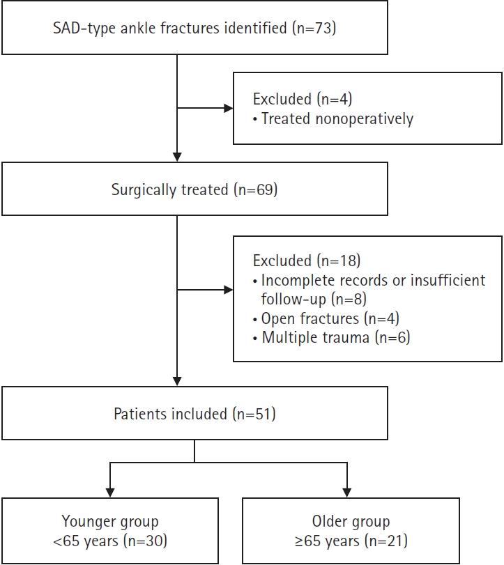
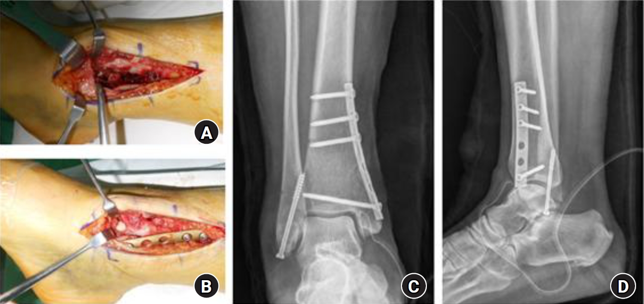
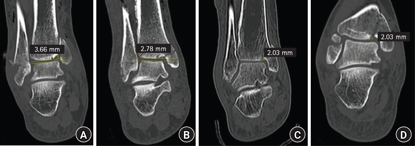
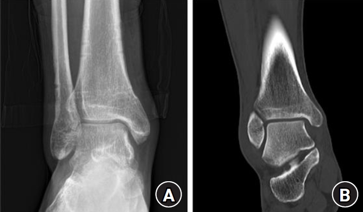
Fig. 1. Patient selection flow diagram. SAD, supination-adduction.
Fig. 2. Representative elderly patient with a supination–adduction-type ankle fracture in whom articular congruity was restored by elevation of the impacted articular fragment and fixation with a medial buttress plate and screws. (A) Intraoperative exposure of the impacted articular fragment of the medial tibial plafond. (B) Restoration of articular congruity after elevation and reduction of the impacted fragment, followed by fixation with a medial buttress plate and screws. (C, D) Immediate postoperative anteroposterior and lateral radiographs showing stable fixation of the medial and lateral malleoli.
Fig. 3. Schematic illustration of radiographic measurements. (A, B) Tibial plafond impaction depth, measured as the maximum perpendicular distance from the expected intact articular contour to the deepest point of the impacted fragment. (C, D) Articular step-off, measured as the maximum vertical displacement between adjacent articular surfaces.
Fig. 4. Eighteen-month postoperative images of the patient shown in Fig. 2. (A) Anteroposterior radiograph and (B) coronal computed tomography image demonstrate solid bony union without posttraumatic osteoarthritis, residual marginal impaction of the medial tibial plafond, articular step-off, or other notable abnormalities.
Fig. 1.
Fig. 2.
Fig. 3.
Fig. 4.
Clinical and radiographic outcomes after surgical treatment of supination-adduction type ankle fractures in older (≥65 years) and younger (<65 years) patients: a retrospective single-center comparative study in Korea
| Variable | Younger (<65 yr) (n=30) | Older (≥65 yr) (n=21) | P-value |
|---|---|---|---|
| Age (yr) | 49.3±8.2 | 72.4±5.7 | <0.001 |
| Female sex | 18 (60.0) | 14 (66.7) | 0.771 |
| Diabetes mellitus | 5 (16.7) | 7 (33.3) | 0.196 |
| Hypertension | 9 (30.0) | 8 (38.1) | 0.563 |
| Current smoking | 8 (26.7) | 6 (28.6) | 1.000 |
| High-energy trauma | 11 (36.7) | 3 (14.3) | 0.113 |
| Open fracture | 0 (0.0) | 0 (0.0) | NA |
| Follow-up (mo) | 25.9±7.5 | 22.5±7.0 | 0.110 |
| Complication | Younger (n=30) | Older (n=21) | P-value |
|---|---|---|---|
| Superficial incisional SSI | 2 (6.7) | 2 (9.5) | 1.000 |
| Deep incisional SSI | 1 (3.3) | 2 (9.5) | 0.561 |
| Loss of reduction | 0 (0.0) | 1 (4.8) | 0.412 |
| Delayed union | 0 (0.0) | 3 (14.3) | 0.064 |
| Any postoperative complication | 3 (10.0) | 8 (38.1) | 0.035 |
| Variable | Complication (+) (n=8) | Complication (−) (n=13) | Odds ratio (95% CI) | P-value |
|---|---|---|---|---|
| Diabetes mellitus | 5 (62.5) | 2 (15.4) | 9.17 (1.15–73.24) | 0.037 |
| Smoking | 4 (50.0) | 2 (15.4) | 5.50 (0.71–42.60) | 0.103 |
| Hypertension | 4 (50.0) | 4 (30.8) | 2.25 (0.37–13.87) | 0.382 |
| Female sex | 6 (75.0) | 8 (61.5) | 1.88 (0.27–13.20) | 0.528 |
| Osteoporosis | 5 (62.5) | 5 (38.5) | 2.67 (0.43–16.39) | 0.290 |
| Variable | Value | Correlation with lowest T-score, ρ | P-value |
|---|---|---|---|
| Lowest T-score | −2.3±0.5 | NA | NA |
| Osteoporosis | 10 (47.6) | NA | NA |
| Tibial plafond impaction depth (mm) | 3.1±1.2 | −0.46 | 0.036 |
| Articular step-off (mm) | 2.0±0.8 | −0.43 | 0.052 |
| Follow-up (mo) | OMAS | AOFAS | ||||
|---|---|---|---|---|---|---|
| <65 yr | ≥65 yr | P-value | <65 yr | ≥65 yr | P-value | |
| 3 | 58.3±8.7 | 50.9±9.1 | 0.012 | 71.6±7.5 | 64.8±8.2 | 0.015 |
| 6 | 73.4±7.2 | 67.5±6.8 | 0.021 | 82.3±6.5 | 76.9±7.4 | 0.027 |
| 12 | 85.7±5.6 | 83.2±6.2 | 0.181 | 91.4±5.1 | 89.6±5.4 | 0.234 |
| Outcome | Younger (n=30) | Older (n=21) | P-value |
|---|---|---|---|
| Nonunion | 0 (0.0) | 1 (4.8) | 0.412 |
| Posttraumatic osteoarthritis | 1 (3.3) | 1 (4.8) | 1.000 |
Table 1. Baseline demographic and clinical characteristics
Values are presented as mean±standard deviation or number (%). Categorical variables were compared using the Fisher exact test when appropriate. NA, not applicable.
Table 2. Postoperative complications according to age group
Values are presented as number (%). P-values by Fisher exact test. No patient experienced more than one of the listed complications; therefore, the component counts equal the patient-level total for any postoperative complication in each group. SSI, surgical site infection.
Table 3. Exploratory univariable associations with postoperative complications in older patients
Values are presented as number (%) unless otherwise indicated. Odds ratios were estimated using univariable logistic regression. CI, confidence interval.
Table 4. Dual-energy X-ray absorptiometry and radiographic parameters in older patients
Values are presented as mean±standard deviation or number (%). Correlations were assessed using Spearman rank correlation.
Table 5. Functional outcomes during follow-up
Values are presented as mean±standard deviation. The 12-month assessment was designated as the principal functional endpoint. The 3- and 6-month values are descriptive measures of early recovery and should be interpreted cautiously because weight-bearing progression and rehabilitation were ongoing. The visual analog scale was not consistently recorded and is not reported. OMAS, Olerud-Molander Ankle Score; AOFAS, American Orthopaedic Foot & Ankle Society ankle-hindfoot score.
Table 6. Final radiographic outcomes at ≥18 months
Values are presented as number (%). P-values by Fisher exact test.


