 E-submission
 TOTA
TOTA TOTS
TOTS
Search
- Page Path
- HOME > Search
Review Article
- Nonoperative management of distal radius fractures: when and how?
- Shin Woo Choi, Jae Kwang Kim
- J Musculoskelet Trauma 2026;39(2):93-102. Published online March 10, 2026
- DOI: https://doi.org/10.12671/jmt.2026.00024
-
 Abstract
 PDF
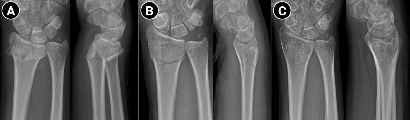
PDF - Distal radius fractures are among the most common injuries of the upper extremity, particularly in the elderly population. Although the use of volar locking plate fixation has increased in recent years, evidence from randomized and prospective studies demonstrates that, while operative treatment may achieve superior radiographic alignment and enable more rapid early recovery, these advantages tend to diminish over time and do not result in superior long-term patient-reported functional outcomes in elderly patients. In addition, radiographic parameters show only a limited correlation with functional recovery. Consequently, nonoperative treatment remains a valid and important treatment option for distal radius fractures. The decision to pursue nonoperative management should be based on a comprehensive assessment of radiographic parameters—including dorsal tilt, radial shortening, and intraarticular displacement—together with patient-specific factors such as age, activity level, comorbidities, and functional expectations. For stable or minimally displaced fractures, an immobilization period of 3‒4 weeks is generally recommended, whereas displaced fractures typically require immobilization for 5‒6 weeks. In cases requiring manual reduction, traditional treatment protocols recommend weekly radiographic follow-up during the first 2‒3 weeks to monitor for secondary displacement. Successful nonoperative management should also emphasize effective swelling control through limb elevation, as well as the initiation of early finger exercises to prevent hand stiffness. After removal of the cast or splint, active wrist mobilization is essential for restoring optimal range of motion and achieving functional recovery.
- 1,814 View
- 25 Download

Original Articles
- Epidemiological changes and surgical trends of distal radius fractures in adults over 50 years during the COVID-19 pandemic in Korea: a nationwide repeated cross-sectional study
- Han-Kook Yoon, So Ra Yoon, Kee-Bum Hong, Youngsu Jung, SeongJu Choi, Jun-Ku Lee
- J Musculoskelet Trauma 2026;39(1):12-19. Published online January 25, 2026
- DOI: https://doi.org/10.12671/jmt.2025.00297

-
Abstract
PDF
 Supplementary Material
 - Background
The COVID-19 pandemic is likely to have affected bone health in older adults in Korea. This study aimed to analyze changes in the epidemiology and management of distal radius fractures (DRFs) in older adults before and during the COVID-19 pandemic.
Methods
Patients with DRF aged over 50 years in 2017, 2018, 2020, and 2021 were included in this study. Patients were classified into a group with DRF occurring between 2017 and 2018 (before COVID-19) and a group with DRF occurring between 2020 and 2021 (during COVID-19). We calculated the incidence rates of DRF and compared them between the two groups. We also analyzed and compared demographic data (age, sex, income, residence) and the operation rate for DRF between the two groups. Patient selection and treatment were based on International Classification of Diseases, 10th revision codes.
Results
A total of 140,634 patients with DRF (before COVID-19, 69,794; during COVID-19, 70,840) were included. The incidence of DRF before COVID-19 (184.4/100,000 person-years) was higher than during COVID-19 (169.8/100,000 person-years). The operation rate was higher during COVID-19 (86.9%) than before COVID-19 (83.3%).
Conclusion
During the COVID-19 pandemic, the incidence of DRF decreased in South Korea. However, the rate of surgical treatment increased and exceeded the global surgical rate. Level of evidence: III.
- 828 View
- 28 Download
- Hook plate versus periarticular-type volar locking plate for distal radius fractures involving the volar lunate facet in Korea: a retrospective cohort study
- Hyun-Jae Park, Joo-Hak Kim
- J Musculoskelet Trauma 2025;38(4):221-228. Published online October 24, 2025
- DOI: https://doi.org/10.12671/jmt.2025.00241
-
Abstract
PDF
- Background
This study investigated the clinical and radiographic outcomes of hook plate (HP) fixation for volar lunate facet fractures, comparing them with periarticular-type volar locking plates (PVLPs).
Methods
A retrospective review was conducted on 24 patients with distal radius fractures involving volar lunate facet fragments who underwent surgery between January 2016 and April 2021. Patients were divided into two groups: HP (n=12) and PVLP (n=12). Radiographic union, wrist range of motion, Disabilities of the Arm, Shoulder and Hand (DASH) scores, and implant-related complications were compared. Statistical analyses included the Mann-Whitney U test and Fisher exact test.
Results
Radiographic union was achieved in all patients (100%), without secondary displacement or hardware failure. No significant differences were observed between the two groups in wrist flexion (P=0.152), extension (P=0.832), pronation (P=0.792), or supination (P=0.328). The mean DASH scores were 12.8±5.5 in the HP group and 14.6±6.0 in the volar plate group (P=0.449). One patient in the HP group experienced mild flexor tendinopathy that resolved with conservative management. No cases of tendon rupture or early reoperation were reported.
Conclusions
Fixation of volar lunate facet fractures using a HP yielded clinical and radiographic outcomes comparable to those of PVLPs, with a low rate of complications and reliable bony union. Due to its mechanical stability, compatibility with standard surgical approaches, and low risk of flexor tendon irritation, the HP may serve as a valuable alternative for managing volar lunate facet fractures. Level of evidence: IV.
- 874 View
- 24 Download
Review Articles
- Surgical Treatment of Distal Radius Fractures and Treatment of Common Accompanying Lesions
- Joo-Hak Kim
- J Korean Fract Soc 2022;35(3):120-127. Published online July 31, 2022
- DOI: https://doi.org/10.12671/jkfs.2022.35.3.120
-
Abstract
PDF
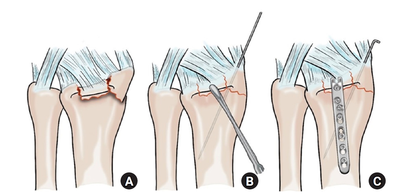
- There have been major advances in the treatment of distal radius fractures over the past 20 years. Specifically, the development of the volar locking plate in 2001 and the subsequent improvements in its design and performance have enabled the treatment of distal radius fractures that were previously considered difficult to treat. The volar plate is used for fractures and shows good results with anatomical reduction and firm fixation. However, when trying to apply it to more complex fractures, there are still difficulties related to the unique anatomical structure of the distal radius, and there are also several factors that can impair joint function and cause pain after surgery. In this review, the factors to be considered to ensure better outcomes during ORIF (open reduction and internal fixation), and external fixation in the treatment of distal radial fractures are described. The review also details the common accompanying injuries and management methods.
- 1,056 View
- 24 Download
- Perilunate Dislocation and Perilunate Fracture-Dislocation
- Jung Il Lee
- J Korean Fract Soc 2022;35(3):114-119. Published online July 31, 2022
- DOI: https://doi.org/10.12671/jkfs.2022.35.3.114
-
Abstract
PDF
- Perilunate dislocations and perilunate fracture-dislocations are one of the most severe forms of wrist injuries and are generally caused by high-energy trauma such as falls from a height or traffic accidents. Prompt recognition and immediate, gentle closed reduction are critical, but diagnosis can often be missed at the initial presentation. The current standard management is open reduction, ligamentous and bony repair, and supplemental fixation for the protection of the repair. The pathomechanics of the injury, diagnosis by plain wrist radiographs, closed reduction techniques, current surgical treatments, and complications are presented in this review.
- 972 View
- 10 Download
- Pediatric Fractures around the Wrist
- Gihun Kim, Kun-Bo Park
- J Korean Fract Soc 2021;34(2):80-86. Published online April 30, 2021
- DOI: https://doi.org/10.12671/jkfs.2021.34.2.80
-
Abstract
PDF
- Fractures around the wrist are the third most common fracture among all pediatric fractures. Furthermore, distal radius fractures, a type of wrist fracture, are the most common fractures in children. Understanding pediatric fractures around the wrist is very important considering their prevalence. There is a specific belief that pediatric fractures can heal easily because of remodeling, but not all fractures can heal without proper treatment. Complications such as growth problems, nonunion can occur if the fracture is not treated properly. This paper reviewed recent articles about distal radius fractures, Galeazzi-equivalent fractures, and carpal bone fractures, including scaphoid fractures in children and adolescents. Successful treatment can be achieved without complications when an accurate diagnosis and proper non-surgical or surgical treatment are performed based on this article.
- 1,933 View
- 43 Download
- Diagnosis and Management of Ligament Injuries of the Wrist
- Ki Tae Na, Joo Yup Lee
- J Korean Fract Soc 2016;29(2):160-170. Published online April 30, 2016
- DOI: https://doi.org/10.12671/jkfs.2016.29.2.160
-
Abstract
PDF
- The wrist joint is formed by the distal end of the radius and ulna proximally, and eight carpal bones distally. It has many ligaments to maintain stability of the complex bony structures. The incidence of ligament injuries of the wrist has increased due to sports activities. However, diagnosis and management of these injuries are sometimes difficult because of the anatomic complexity and variable injury patterns. Among them, scapholunate ligament injury and triangular fibrocartilage tears are the two most common injuries resulting in chronic disabling wrist pain. Thorough understanding of the wrist anatomy and physical and radiologic examination is mandatory for proper diagnosis and management of these conditions. This article will briefly discuss the wrist joint anatomy and biomechanics, and review the diagnosis and management of the scapholunate ligament injury and triangular fibrocartilage injury.
- 1,427 View
- 19 Download
- Biomechanics of the Wrist
- Young Ho Shin, Young Ho Lee
- J Korean Fract Soc 2016;29(1):93-100. Published online January 31, 2016
- DOI: https://doi.org/10.12671/jkfs.2016.29.1.93
-
Abstract
PDF
- The wrist joint is a complicated structure composed of many bones and ligaments. Therefore, understanding the anatomy and the biomechanics of the wrist is important in order to administer proper treatment for patients. To easily understand the complicated structure, there were many trials to unite the complicated structure with a simple group such as the carpal row concept and the carpal column concept. Movement and load transfer along the wrist joint occurs with balanced action between carpal bones. To evaluate this static equilibrium, measuring tools such as carpal height ratio are used. When wrist flexion/extension occurs, each carpal row moves synchronously with action of the scaphoid. In contrast with flexion/extension, when wrist radial deviation/ulnar deviation occurs, the proximal carpal row moves in the sagittal plane, instead of the coronal plane. Recently, the dart throwing motion which occurred from the position of dorsiflexion with radial deviation to volar flexion with ulnar deviation is considered the main movement of the wrist joint.
-
Citations
Citations to this article as recorded by
- Fractal geometry in wrist biomechanics: A preliminary study with implications for arthroplasty and surgery
Lauren Gorelick, Amir Oron, Gil Gannot, Raphael Israeli
Hand Surgery and Rehabilitation.2025; 44(4): 102203. CrossRef - Association between carpal height ratio and ulnar variance in normal wrist radiography
Anas AR Altamimi, Monther A. Gharaibeh, Muntaser Abu Shokor, Moh’d S. Dawod, Mohammad N. Alswerki, Omar M. Al-Odat, Raghda H. Elkhaldi
BMC Musculoskeletal Disorders.2024;[Epub] CrossRef - Reliability and concurrent validity of a new iPhone® goniometric application for measuring active wrist range of motion: a cross‐sectional study in asymptomatic subjects
Mohammad Reza Pourahmadi, Ismail Ebrahimi Takamjani, Javad Sarrafzadeh, Mehrdad Bahramian, Mohammad Ali Mohseni‐Bandpei, Fatemeh Rajabzadeh, Morteza Taghipour
Journal of Anatomy.2017; 230(3): 484. CrossRef
- Fractal geometry in wrist biomechanics: A preliminary study with implications for arthroplasty and surgery
- 4,679 View
- 169 Download
- 3 Crossref
Original Articles
- Results of the Kapandji Procedure in the AO Type C Distal Radius Fracture in Patients over Age 60
- Chul Hong Kim, Sung Soo Kim, Myung Jin Lee, Hyeon Jun Kim, Bo Kun Kim, Young Hoon Lim
- J Korean Fract Soc 2012;25(3):191-196. Published online July 31, 2012
- DOI: https://doi.org/10.12671/jkfs.2012.25.3.191
-
Abstract
PDF
- PURPOSE
To evaluate the clinical and radiologic results of the Kapandji procedure in AO classification type C distal radius fracture patients over 60 years old.
MATERIALS AND METHODS
Twenty-one type C distal radius fracture patients over the age of 60 years who were treated with the Kapandji procedure from June 2004 to June 2009 in our hospital and had a post-operative follow-up period of more than 1 year were enrolled. The volar tilt, radial inclination, and radial length were measured for the radiographic analysis using the modified Lidstrom scoring system about post-operative reduction loss in every follow-up radiogram. The clinical result was assessed with a visual analogue scale (VAS) and Korean Disabilities of the Arm, Shoulder and Hand Questionnaire (DASH) score at the last follow-up.
RESULTS
The mean radiologic loss of volar tilt was 1.1degrees and the mean loss of radial length was 2.6 mm and the mean radial inclination loss was 2.7degrees compared with the immediate post-operative period and last follow-up period. The average VAS and DASH scores were 1.4 and 15.9.
CONCLUSION
The radiologic results of closed reduction and percutaneous pinning using the Kapandji technique for distal radius AO type C fracture patients over 60 years of age was not satisfactory. Nevertheless, the clinical results were satisfactory.
- 599 View
- 4 Download
- TFCC Injury Associated with the Triquetral Dorsal Chip Fracture
- Seoung Joon Lee, Jin Ho Hwang, Min Seok Kang, Jong Woong Park
- J Korean Fract Soc 2009;22(3):179-184. Published online July 31, 2009
- DOI: https://doi.org/10.12671/jkfs.2009.22.3.179
-
Abstract
PDF
- PURPOSE
To evaluate the usefulness of wrist arthroscopic examination in patient with persistent pain after the triquetral dorsal chip fracture and also to determine its relationship with TFCC injury in the triquetral dorsal chip fracture patient manifesting persistent pain.
MATERIALS AND METHODS
This study is based on six cases presenting persistent pain in the ulnar aspect after the triqeutral posterior cord fracture that were treated conservatively. Wrist arthroscopy was carried out for all six cases. All were preoperatively and postoperatively evaluated using VAS pain scale, grip power, ulnar grind test, Kleinman shearing test and lunotriquetral ballottment test.
RESULTS
Preoperatively, ulnar grind test yielded positive results in all six cases, Kleiman shearing test proved positive in three cases and lunotriquetral ballottment test yielded positive result in one case. In the arthroscopic findings, synovitis and TFCC injury were detected in all cases, and based on Palmer classification of TFCC injury, type IA was determined in five cases and type ID in one case. Arthroscopic TFCC partial resection and synovectomy were carried out. VAS pain scale improved from an average 8 points preoperatively to 3 points postoperatively. The difference of grip power between the normal and the other side improved from average of 15 lb preoperatively to 5 lb postoperatively. Based on postoperatively physical examination at 6 weeks, all cases yielded negative results in the ulnar grind test and Kleiman shearing test.
CONCLUSION
We think that TFCC injury is one of the causes of persistent pain after triquetral dorsal chip fracture. We recommend an arthroscopic TFCC partial resection as a valuable treatment option.
- 1,127 View
- 5 Download
- Functional Evaluation of Wrist According to Changes of Length after Operation in Fracture of Both Bones of Forearm
- Seung Suk Seo, Ki Yong Kim, Jang Seok Choi, Young Chang Kim, Jae Keun Park
- J Korean Soc Fract 2003;16(1):74-82. Published online January 31, 2003
- DOI: https://doi.org/10.12671/jksf.2003.16.1.74
-
Abstract
PDF
- PURPOSE
To evaluate the relationship between the length changes of both forearm bones and function of wrist. To know permitted length discrepancy for good wrist function after operation in fracture of both bones of forearm MATERIALS AND METHODS: From Jan. 1995 to Dec. 2000, 21 cases were followed over 1 year, were treated with compression plate and screws due to fracture of both bones of forearm in our hospital. Mean duration of follow-up was 3 years 6 months. The postoperative length difference was compared to preoperative or unaffected side in roentgenography. Four groups were defined to A, B, C and D by postoperative length difference ; < or =1mm, 1 2mm, 2 3mm, and >3mm for comparison. The function of wrist joint was evaluated with the Anderson 's classification and Mayo modified wrist score.
RESULT
Group A were 11 cases(52.3%), B 5 cases(23.8%), C 4 cases(19.0%) and D 1 case(4.8%). By the Anderson 's classification, the number of Excellent were 11 cases(52.3%), Good 7(33.3%), Fair 3(14.3%). In the group of the length difference lesser than 2mm, the number of Excellent were 11, and Good 5. The Mayo modified wrist score was 75.15 in the group of the length difference lesser than 2mm, that was higher than 61.15 in the group of more than 2mm.
CONCLUSION
To obtain a good wrist function after operative treatment of fracture of both bones of forearm the length discrepancy of both bones should be lesser than 2mm.
- 606 View
- 0 Download
- Percutaneous K-wire fixation for Unstable Fracture of distal radius
- Chol Yong Jung, Young Chan Son, Jun Bum Bae, Moon Do Choi
- J Korean Soc Fract 2000;13(4):996-1002. Published online October 31, 2000
- DOI: https://doi.org/10.12671/jksf.2000.13.4.996
-
Abstract
PDF
- PURPOSE
To evaluate the clinical validity of the percutaneous K-wire fixation in applying to unstable extraarticular fracture of distal radius of patients who are older than 50 years. MATERIAL AND METHODS: The validity of K-wire fixation was examined, using subjective study of Cole and Oblelz and objective study of Scheck, on the 20 cases of unstable extraarticular fracture of distal radius of patients older than 50 years, who were treated with percutaneous K-wire fixation and followed up more than 1 year, out of 160 patients with distal radius fracture, treated in the department of orthopedic surgery of our hospital from January 1994 to August 1998.
RESULTS
The result was examined with subjective study of Cole and Oblelz and objective study of Scheck. Combined judgement was made by adding up the scores of both objective and subjective study. 5 excellent cases and 12 good cases were brought forth by subjective study. Objective study achieved the result of average 18 degree of radial angle, 9.8mm of radial length and 3.6 degree of volar angle. Combined judgement achieved a good result of 3 excellent cases, 14 good cases and 3 fair cases.
CONCLUSION
Percutaneous K-wire fixation is expected to be a simple, less invasive, more effective and valuable operation method in the treatment of extraarticular fracture of distal radius with severe comminution
- 486 View
- 0 Download
- Wrist Injury in Juvenile Gymnasts
- Choong Gil Lee, Jin Woo Kwon, Jae Hyum Park, Sang Hoon Lee, Wan Eup Kim, Eun Sik Shin
- J Korean Soc Fract 1996;9(4):1125-1130. Published online October 31, 1996
- DOI: https://doi.org/10.12671/jksf.1996.9.4.1125
-
Abstract
PDF
- In the adolescent gymnasts, recent studios have shown that wrist is particularly vulnerable to chronic stress. In the immature skeleton, growth plate is especially vulnerable to acute or chronic trauma since the joint capsule and ligamentous structures are strong. The purpose of this study is to report the frequency, finding of radiologic abnormalities and the type of sports to cause wrist pain. The authors examined 26 adolescent gymnasts, 20 males and 6 females. The age range was 11 years 10 months to 17 years 5 months for males and 11 years 9 months to 34 years 4 months for females. The results were as follows; 1. The radiologic abnormalities were found in 23 cases(88%), 19 males and 4 females. 2. Wrist pain was most frequently csused by pommel horse exercise in males and by floor exercise in females. 3. Among 23 cases, 18 showed widening of distal radial growth plates and irregularities of the margins of the growth plate(15 cases were bilateral). Widening of distal ulna growth plates were combineti in six cases, ulna styloid process fracture in 3 cases and radial styloid process fracture in 1 case. 4. Among 23 cases, 5 cases showed widening of distal radial metaphysis and increased ulnar tilting.
- 558 View
- 0 Download
- Proximal Row Carpectomy for Disease of the Proximal Carpal Bone
- Seung Koo Rhee, Hyoung Min Kim, Soon Young Kwon, Hwa Sung Lee, Hang Kyu Lee
- J Korean Soc Fract 1996;9(2):311-318. Published online April 30, 1996
- DOI: https://doi.org/10.12671/jksf.1996.9.2.311
-
Abstract
PDF
- The management of pain, stiffness and weakness of the wrist following unsuccessful conservative treatment of fractures of the scaphoid or of Kienbocks disease and so on is a difficult problem. Despite the recommendation by Cotton in 1924 and subsequently by others that the proximal row of carpal bones should be removed in the presence of disease, arthrodesis or various stabilizing procedures continue to be recommended. But, although a radiocarpal fusion, when successful, leads to a painless, stable wrist, the loss of the normal motion of the wrist inevitably results in some loss of function of the hand. The purpose of this study is to evaluate the efficacy of the proximal-row carpectomy. Since 1987, five patients were studied following proximal-row carpectomy. The lesions for which the operation was done included two Kienbocks disease, one crushing injury, one transscaphoid volar lunar dislocation, and one scapholunate dissociation. Their end results after average 74 months of follow-up showed less pain than before operation and a reasonable range of flexion/extension which varied between 65% and 85% of normal, the average being 74%, Postoperative grip strength was from 70 to 90% fo normal, the average being 78%. In conclusion, excision of the proximal row of tile carpus is a useful procedure, with a limited application in patients with Kienbocks disease, dislocation of the lunate bone, scapholunate dissociation and similar injuries which do not respond to conservative management.
- 528 View
- 0 Download
- Treatment of Wrist Frarture-Disluation using the Mini-External Fixator and Internal Fixation
- Chil Soo Kwon, Young Uek Kim, Jin Hyok Kim, Won Ho Choi
- J Korean Soc Fract 1992;5(2):417-425. Published online November 30, 1992
- DOI: https://doi.org/10.12671/jksf.1992.5.2.417
-
Abstract
PDF
- Authors reviewed 8 cases of wrist fracture-dislocation treated with mini-external fixator and internal fixation form Septmeber 1989 to May 1992 with average 6 months follow up. The results were as follows ; 1. Mean ages were 47 years, most patients were young age. 2. We could achieve good results in intra-articular, communited, displaced fracture and open fracture of the wrist by using the mini-externall fixator and internal fixation. 3. Radial length and inclination was maintained mainly by the external fixator. Articular surface restoration and reconstruction was performed by bone graft and the limited internal fixation. We would like to recommend to use the mini-external fixator and limited internal fixation instead of plate and screws for the intraarticular fractue, displaced, communited farcture and open fracture of the wrist.
- 516 View
- 0 Download
- Clinical Application of Wrist Arthrography after Trauma
- Kwon Ick Ha, Sung Ho Han, Min Young Chung, Hee Joong Kim, Tae Won An
- J Korean Soc Fract 1988;1(1):36-42. Published online November 30, 1988
- DOI: https://doi.org/10.12671/jksf.1988.1.1.36
-
Abstract
PDF
- One of the greatest diagnostic challenges that faces both orthopedic surgeons and the radiologists is the patient with a subacute or chronic wrist injury who has no obvious clinical or radiographic abnormality to explain the pain. The wrist arthrography is used to evalute structures that can not be seen on plain radiography. These structures include the synovium, the intraarticular ligaments and the articular cartilage including the triangular fibrocartilage. The most inportant indication is persistent pain or limitation of motion after trauma. We think that the wrist arthrography is to be used widly. We collected and analized the results of wrist arthrographies performed in 33 patients with traumaic painful wrist.
- 572 View
- 1 Download
- A Clinical Experience of Limited Wrist Arthrodesis (Radioscapholunate arthrodesis)
- Yung Khee Chung, Jung Han Yoo, Baek Yong Song
- J Korean Soc Fract 1988;1(1):20-23. Published online November 30, 1988
- DOI: https://doi.org/10.12671/jksf.1988.1.1.20
-
Abstract
PDF
- The principle of treatment in patient with fracture involving articular surface is necessary for anatomical reduction, rigid fixation and early motion. However, on the occasion of the unsatisfactory results such a post-traumatic arthritis of the wrist joint, in 1981, Watson and coworkers reported the good results by limited wrist arthrodesis for relief of pain and allowance of some range of motion. Recently, we have experienced two cases of post-traumatic arthritis of the wrist joint which was treated by limited wrist arthrodesis, especially, radioscapholunate arthrodesis with good results.
- 555 View
- 3 Download
 First
First Prev
Prev

