 E-submission
 TOTA
TOTA TOTS
TOTS
Search
- Page Path
- HOME > Search
Original Articles
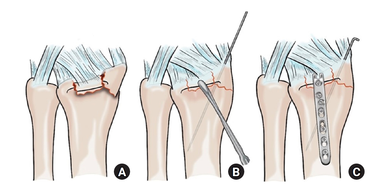
- Hook plate versus periarticular-type volar locking plate for distal radius fractures involving the volar lunate facet in Korea: a retrospective cohort study
- Hyun-Jae Park, Joo-Hak Kim
- J Musculoskelet Trauma 2025;38(4):221-228. Published online October 24, 2025
- DOI: https://doi.org/10.12671/jmt.2025.00241
-
 Abstract
 PDF
PDF - Background
This study investigated the clinical and radiographic outcomes of hook plate (HP) fixation for volar lunate facet fractures, comparing them with periarticular-type volar locking plates (PVLPs).
Methods
A retrospective review was conducted on 24 patients with distal radius fractures involving volar lunate facet fragments who underwent surgery between January 2016 and April 2021. Patients were divided into two groups: HP (n=12) and PVLP (n=12). Radiographic union, wrist range of motion, Disabilities of the Arm, Shoulder and Hand (DASH) scores, and implant-related complications were compared. Statistical analyses included the Mann-Whitney U test and Fisher exact test.
Results
Radiographic union was achieved in all patients (100%), without secondary displacement or hardware failure. No significant differences were observed between the two groups in wrist flexion (P=0.152), extension (P=0.832), pronation (P=0.792), or supination (P=0.328). The mean DASH scores were 12.8±5.5 in the HP group and 14.6±6.0 in the volar plate group (P=0.449). One patient in the HP group experienced mild flexor tendinopathy that resolved with conservative management. No cases of tendon rupture or early reoperation were reported.
Conclusions
Fixation of volar lunate facet fractures using a HP yielded clinical and radiographic outcomes comparable to those of PVLPs, with a low rate of complications and reliable bony union. Due to its mechanical stability, compatibility with standard surgical approaches, and low risk of flexor tendon irritation, the HP may serve as a valuable alternative for managing volar lunate facet fractures. Level of evidence: IV.
- 876 View
- 25 Download

- Restoration of Lateral Tibial Plateau Widening and Articular Depression Is Necessary to Prevent Valgus Deformities after Arthroscopic Reduction and Internal Fixation in AO/OTA 41.B2 or B3 Fractures
- Jun-Ho Kim, Kang-Il Kim, Sang-Hak Lee, Gwankyu Son, Myung-Seo Kim
- J Korean Fract Soc 2024;37(3):125-136. Published online July 31, 2024
- DOI: https://doi.org/10.12671/jkfs.2024.37.3.125
-
Abstract
PDF
- Purpose
This study examined the factors affecting valgus deformities after arthroscopic reduction and internal fixation (ARIF) in lateral joint-depression tibial plateau fractures.
Materials and Methods
Patients with lateral joint-depression tibial plateau fractures treated with ARIF were assessed retrospectively. The radiological evaluations included the articular depression distance (ADD) and the lateral plateau widening distance (LPWD) on preoperative and postoperative computed tomography. A postoperative valgus deformity was defined as valgus malalignment (mechanical axis ≥3°) and valgus deviation (Δmechanical axis of the operated knee from the healthy knee of ≥5°). Subgroup analyses based on a postoperative valgus deformity were performed to compare the clinical outcomes, including the range of motion, patient-reported outcomes measures, and failure and osteoarthritis progression. Furthermore, factors affecting the postoperative mechanical and Δmechanical axes were assessed.
Results
Thirty-nine patients were included with a mean follow-up of 44.6 months (range, 24-106 months). Valgus malalignment and valgus deviation were observed after ARIF in 10 patients (25.6%) and five patients (12.8%), respectively. The clinical outcomes were similar in patients with and without a postoperative valgus deformity. On the other hand, lateral compartment osteoarthritis progression was significantly higher in the valgus deformity group than in the non-valgus deformity group (valgus malalignment group: 50.0% vs 6.9%, p=0.007; valgus deviation group: 60.0% vs 11.8%, p=0.032). One patient with valgus deformity underwent realignment surgery at postoperative five years. The preoperative ADD and postoperative LPWD were significantly associated with the postoperative mechanical (both, p<0.001) and Δmechanical (ADD, p=0.001; LPWD, p=0.025) axes. Moreover, the lateral meniscectomized status during ARIF was significantly associated with the Δmechanical axis (p=0.019).
Conclusion
Osteoarthritis progression was highly prevalent in patients with postoperative valgus deformity. Thus, the restoration of lateral plateau widening and articular depression and preservation of the meniscus are necessary to prevent a valgus deformity after ARIF in lateral joint-depression tibial plateau fractures.
- 3,670 View
- 51 Download
- Computational Simulation of Femoral Neck System and Additional Cannulated Screws Fixation for Unstable Femoral Neck Fractures and the Biomechanical Features for Clinical Applications
- Ju-Yeong Kim
- J Korean Fract Soc 2023;36(1):1-9. Published online January 31, 2023
- DOI: https://doi.org/10.12671/jkfs.2023.36.1.1
-
Abstract
PDF
- Purpose
To identify the biomechanical features for clinical applications through a computational simulation of the fixation of the Femoral Neck System (FNS) with additional cannulated screws for a Pauwels type III femoral neck fractures.
Materials and Methods
Thirty cadaveric femurs underwent computed tomography, and the images were transferred to the Mimics ® program, resulting in three-dimensional proximal femur models. A three-dimensional scan of the FNS and 6.5 mm and 7.0 mm cannulated screws was performed to enable computerized virtual fixation of FNS with additional cannulated screws for unstable femoral neck fractures. Furthermore, the cannulated screw used for additional fixation was modeled and used as a cylinder within the Ansys program. The biomechanical characteristics of these models were investigated by applying a physiological load virtually.
Results
The maximum von Mises stress value at bone was 380.14 MPa in FNS and 297.87 MPa in FNS+7.0 mm full-thread cannulated screw. The maximum von Mises stress value at FNS was 786.83 MPa in FNS and 435.62 MPa in FNS+7.0 mm full-thread cannulated screw. The FNS group showed the highest maximum von Mises stress values at bone and FNS. For total deformation, the maximum deformation value was 10.0420 mm in FNS and 9.2769 mm in FNS+7.0 mm full-thread cannulated screws. The FNS group represented the highest maximum deformation compared to the other groups.
Conclusion
Considering the anatomical spatiality and biomechanical characteristics of the FNS in unstable femoral neck fractures, when one 7.0 mm full thread cannulated screw was also fixed to the anterosuperior portion of the FNS, significant biomechanical stability was demonstrated.
- 1,121 View
- 14 Download
- Clinical Outcomes of Minimally Invasive Surgery in Sanders Type IV Intra-Articular Calcaneal Fractures
- Jun Young Lee, Hyunwoong Jang, Young Wook Kim
- J Korean Fract Soc 2019;32(4):181-187. Published online October 31, 2019
- DOI: https://doi.org/10.12671/jkfs.2019.32.4.181
-
Abstract
PDF
- PURPOSE
This study evaluated the radiologic and clinical results in patients who underwent minimal invasive surgery using sinus tarsi approach in Sanders type IV calcaneal fracture.
MATERIALS AND METHODS
This retrospective study evaluated 13 cases of Sanders type IV calcaneus fractures that were treated by minimal invasive surgery using the sinus tarsi approach from July 2012 to April 2017. Further, these cases could be followed up for more than 12 months. Bone union, radiologic parameters such as Böhler's angle, Gissane's angle, calcaneal height, length, and width, the American Orthopaedic Foot and Ankle Society (AOFAS) ankle-hindfoot score, and the postoperative complications were evaluated.
RESULTS
Bony union was achieved in all the cases at the final follow up, and the mean union time was 5.5 months. One patient underwent reoperation for a surgical site infection, six patients had post traumatic arthritis, and two of them underwent subtalar joint fusion. The mean AOFAS ankle-hindfoot score was 81.2. At the final follow-up, the mean values of Böhler's angle and Gissane's angle were 20° and 119.8°, respectively, and the mean values of the calcaneus height, length, and width were 46.8 mm, 81.8 mm, and 45.6 mm, respectively.
CONCLUSION
Minimal invasive surgery using the sinus tarsi approach for Sanders type IV calcaneal fracture resulted in satisfactory anatomic reduction and stable fixation, and satisfactory clinical and radiologic results were obtained in most of the patients. Minimal invasive surgery is thought to reduce the soft tissue-related complications as compared to surgery using the extensile lateral approach.
- 1,507 View
- 21 Download
- A Comparison of the Results between Internal Fixation and External Fixation in AO C Type Distal Radius Fractures
- Yoon min Lee, Hwa Sung Lee, Seok Whan Song, Jae Hoon Choi, Jong Tae Park
- J Korean Fract Soc 2018;31(3):87-93. Published online July 31, 2018
- DOI: https://doi.org/10.12671/jkfs.2018.31.3.87
-
Abstract
PDF
- PURPOSE
The purpose of this study was to evaluate the radiological and clinical results of plate fixation and external fixation with additional devices for treating distal radius fracture in AO type C subtypes, and propose a treatment method according to the subtypes.
MATERIALS AND METHODS
Two hundred and one AO type C distal radius fracture patients were retrospectively reviewed. Eighty-five patients in group 1 were treated with volar or dorsal plate, and 116 patients in group 2, were treated with external fixation with additional fixation devices. Clinical (range of mtion, Green and O'Brien's score) and radiological outcomes were evaluated.
RESULTS
At the 12-month follow-up, group 1 showed flexion of 64.4°, extension of 68.3°, ulnar deviation of 30.6°, radial deviation of 20.8°, supination of 76.1°, and pronation of 79.4° in average; group 2 showed flexion of 60.5°, extension of 66.9°, ulnar deviation of 25.5°, radial deviation of 18.6°, supination of 73.5°, and pronation of 75.0° in average. The mean Green and O'Brien score was 92.2 in group 1 and 88.6 in group 2. The radial height of group 1 and group 2 was 11.6/11.4 mm; radial inclination was 23.2°/22.5°; volar tilt was 11.6°/8.7°; and the ulnar displacement was 1.27/0.93 mm.
CONCLUSION
Judicious surgical techniques during device application and tips for postoperative management during external fixation can produce similar clinical results compared with internal fixation patients. -
Citations
Citations to this article as recorded by
- Intra-articular fracture distal end radius external fixation versus locking volar radius plate: A comparative study
S.P.S Gill, Manish Raj, Santosh Singh, Ajay Rajpoot, Ankit Mittal, Nitin Yadav
Journal of Orthopedics, Traumatology and Rehabilitation.2019; 11(1): 31. CrossRef
- Intra-articular fracture distal end radius external fixation versus locking volar radius plate: A comparative study
- 771 View
- 1 Download
- 1 Crossref
- The Clinical and Radiological Results of Vancouver Type B1 and C Periprosthetic Fractures
- Bo Ram Na, Taek Rim Yoon, Kyung Soon Park
- J Korean Fract Soc 2016;29(1):26-33. Published online January 31, 2016
- DOI: https://doi.org/10.12671/jkfs.2016.29.1.26
-
Abstract
PDF
- PURPOSE
The purpose of this study is to evaluate the clinical and radiologic results of plate fixation in the Vancouver B1 and C periprosthetic femoral fracture (PFF).
MATERIALS AND METHODS
Twenty patients who had sustained a Vancouver type B1 and C periprosthetic fracture after hip arthroplasty (years 2002-2012) were identified. The mean age was 66.0 years (range, 43-85 years) and the mean follow-up duration of the group was 38 months (range, 12-102 months). The dynamic compression plate (DCP) group included 12 patients and the locking compression plate (LCP) group included eight patients. Harris hip score (HHS) and walking ability, knee joint range of motion (ROM) were compared before injury and last follow-up. Fracture union rate and period were compared.
RESULTS
The mean HHS score was 90.7 (64-96). There was no statistical difference between the two groups. At the last follow-up, knee joint ROM was 103.3degrees (105degrees-140degrees) in the DCP group and 118.4degrees (110degrees-140degrees) in the LCP group, showing good results in the LCP group (p=0.043). No significant difference in the fracture union rate and union periods was observed between the two groups.
CONCLUSION
A better result for the postoperative knee flexion exercise capacity was observed in the LCP group. Use of LCP plate fixation is a good option in management of Vancouver classification B1 and C PFF.
- 988 View
- 5 Download
- Results of the Kapandji Procedure in the AO Type C Distal Radius Fracture in Patients over Age 60
- Chul Hong Kim, Sung Soo Kim, Myung Jin Lee, Hyeon Jun Kim, Bo Kun Kim, Young Hoon Lim
- J Korean Fract Soc 2012;25(3):191-196. Published online July 31, 2012
- DOI: https://doi.org/10.12671/jkfs.2012.25.3.191
-
Abstract
PDF
- PURPOSE
To evaluate the clinical and radiologic results of the Kapandji procedure in AO classification type C distal radius fracture patients over 60 years old.
MATERIALS AND METHODS
Twenty-one type C distal radius fracture patients over the age of 60 years who were treated with the Kapandji procedure from June 2004 to June 2009 in our hospital and had a post-operative follow-up period of more than 1 year were enrolled. The volar tilt, radial inclination, and radial length were measured for the radiographic analysis using the modified Lidstrom scoring system about post-operative reduction loss in every follow-up radiogram. The clinical result was assessed with a visual analogue scale (VAS) and Korean Disabilities of the Arm, Shoulder and Hand Questionnaire (DASH) score at the last follow-up.
RESULTS
The mean radiologic loss of volar tilt was 1.1degrees and the mean loss of radial length was 2.6 mm and the mean radial inclination loss was 2.7degrees compared with the immediate post-operative period and last follow-up period. The average VAS and DASH scores were 1.4 and 15.9.
CONCLUSION
The radiologic results of closed reduction and percutaneous pinning using the Kapandji technique for distal radius AO type C fracture patients over 60 years of age was not satisfactory. Nevertheless, the clinical results were satisfactory.
- 600 View
- 4 Download
- Comparison of Results of Tension Band Wire and Hook Plate in the Treatment of Unstable Fractures of the Distal Clavicle
- Chul Hyun Park, Oog Jin Shon, Jae Sung Seo
- J Korean Fract Soc 2011;24(1):55-59. Published online January 31, 2011
- DOI: https://doi.org/10.12671/jkfs.2011.24.1.55
-
Abstract
PDF
- PURPOSE
To compare the clinical and radiological outcomes of two surgical methods with tension band wire and Hook plate for unstable distal clavicle fractures.
MATERIALS AND METHODS
Thirty patients with type II distal clavicle fractures were evaluated, who were operated with tension band wire (Group I) and Hook plate (Group II) fixation, from June 2005 to June 2009, and could be followed-up for more than 1 year after operation. The reduction and union were evaluated by the immediate post-operative and final radiographs. The functional outcome was evaluated by Kona's system and Constant-Murley scoring system.
RESULTS
All 30 cases showed bony union. By Kona's functional evaluation, there were 16 cases with excellent and good results in Group I and 14 cases in Group II. The average Constant score was 88.3 (71~100) in Group I and 89.6 (72~100) in Group II, but there was no significant difference in both groups. As complications, there were 2 case with subacromial impingement, and 1 case showed subacromial erosion. There was no K-wire migration, deep infection and acromioclavicular joint arthritis.
CONCLUSION
Tension band and Hook plate fixation technique gave satisfactory clinical and radiological results in patients with type II distal clavicle fractures. These results suggest that tension band wire and Hook plate fixation technique seems to be an effective method for type II distal clavicle fracture. But we think thal early removal of plate is necessary due to risks for subacromial impingement and erosion in Hook plate fixation. -
Citations
Citations to this article as recorded by- Hook Plate Fixation for Unstable Distal Clavicle Fractures: A Prospective Study
Kyung-Cheon Kim, Hyun-Dae Shin, Soo-Min Cha, Yoo-Sun Jeon
The Journal of the Korean Shoulder and Elbow Society.2011; 14(1): 6. CrossRef
- Hook Plate Fixation for Unstable Distal Clavicle Fractures: A Prospective Study
- 1,030 View
- 6 Download
- 1 Crossref
- Surgical Treatment of AO Type C Distal Femoral Fractures Using Locking Compression Plate (LCP-DF, Synthes(R))
- Kap Jung Kim, Sang Ki Lee, Won Sik Choy, Won Cho Kwon, Do Hyun Lee
- J Korean Fract Soc 2010;23(1):20-25. Published online January 31, 2010
- DOI: https://doi.org/10.12671/jkfs.2010.23.1.20
-
Abstract
PDF
- PURPOSE
To analyze the surgical results of AO type C distal femoral fractures using locking compression plate.
MATERIALS AND METHODS
From February 2006 to June 2008, 14 patients 15 cases were included. Injury mechanisms, combined injuries, radiologic and clinical results and postoperative complications were analyzed.
RESULTS
The mean age was 59.6 (30~77) years. The mean follow up period was 25 (12~40) months. AO types were 3 of C1, 5 of C2 and 7 of C3. Injury mechanisms were 9 of traffic accident, 5 of slip down and 1 of fall from a height. Four cases were combined with other extremity injuries or fractures. The mean radiologic union was obtained at postoperative 15 (13~20) weeks. The mean Neer's functional score was 74.2 (58~97); 3 of excellent, 5 of satisfactory and 7 of unsatisfactory. Postoperative complications were 2 of infection and 1 of nonunion. There were no mechanical failures or fixation loss with locking compression plate at the final follow up.
CONCLUSION
Internal fixation using locking compression plate for AO type C distal femoral fractures provided excellent fixations. At the final follow up, the clinical results were variable. The affecting factors on the final results seemed to be joint congruencies after anatomical reduction and active rehabilitation. -
Citations
Citations to this article as recorded by- Functional outcome of distal femoral fractures treated with distal femoral locking compression plate: a cross-sectional study
Sandeep Kumar Kumar Deep, Varun Phogat, Sankar Debroy
International Journal of Research in Orthopaedics.2025; 11(5): 1089. CrossRef - A STUDY OF SURGICAL MANAGEMENT OF DISTAL FEMORAL FRACTURES BY DISTAL FEMORAL LOCKING COMPRESSION PLATE OSTEOSYNTHESIS
Dema Rajaiah, Yerukala Ramana, Kuppa Srinivas, Venkateswar Reddy S
Journal of Evidence Based Medicine and Healthcare.2016; 3(66): 3584. CrossRef
- Functional outcome of distal femoral fractures treated with distal femoral locking compression plate: a cross-sectional study
- 1,192 View
- 4 Download
- 2 Crossref
- Treatment of Shatzker Type VI Tibia Plateau Fracture Using Lateral and Posteromedial Dual Incision Approach and Dual Plating
- In Jung Chae, Sang Won Park, Soon Hyuck Lee, Won Noh, Ho Joong Kim, Seung Beom Hahn
- J Korean Fract Soc 2009;22(4):252-258. Published online October 31, 2009
- DOI: https://doi.org/10.12671/jkfs.2009.22.4.252
-
Abstract
PDF
- PURPOSE
To evaluate the radiologic and clinical results of treatment of the Shatzker type 6 tibia plateau fracture using the lateral and posteromedial dual incision approach and dual plating. MATERIALS AND METHODS: Twelve cases in eleven patients of Shatzker type 6 tibia plateau fracture which has been treated using the lateral and posteromedial dual incision approach and dual plating were analyzed with an average follow-up of 16 months. Times to union, alignment and reduction loss on radiograph and postoperative clinical outcome with checking the range of motion of the knee joint, Knee Society Score and UCLA activity scale were analyzed and evaluated. RESULTS: In all cases, bony union was obtained in an average fifteen weeks after the operation, and there was no reduction loss. The arc of motion of the knee joint at the latest follow-up was 132 degrees on average. Average of Knee Society Score was 85 and UCLA activity scale was decreased from 9.6 points preoperatively to 5.7 points postoperatively. CONCLUSION: The treatment of Shatzker type 6 tibia plateau fracture using the lateral and posteromedial dual approach and dual plating have shown clinically preferable results of excellent recovery of joint motion and good knee society score by early range of motion exercise after firm fixation. However, it was high energy injury, so the sports activity of patients was significantly decreased. -
Citations
Citations to this article as recorded by- Staged Treatment of Bicondylar Tibial Plateau Fracture (Schatzker Type V or VI) Using Temporary External Fixator: Correlation between Clinical and Radiological Outcomes
Seung Min Ryu, Han Seok Yang, Oog Jin Shon
Knee Surgery and Related Research.2018; 30(3): 261. CrossRef - Medial Minimally Invasive Percutaneous Plate Osteosynthesis in Proximal Tibial Comminuted Fractures
Jae-Ang Sim, Kwang-Hui Kim, Yong-Seuk Lee, Sang-Jin Lee, Beom-Koo Lee
Journal of the Korean Orthopaedic Association.2014; 49(4): 278. CrossRef - Current Concepts in Management of Tibia Plateau Fracture
Sang Hak Lee, Kang-Il Kim
Journal of the Korean Fracture Society.2014; 27(3): 245. CrossRef
- Staged Treatment of Bicondylar Tibial Plateau Fracture (Schatzker Type V or VI) Using Temporary External Fixator: Correlation between Clinical and Radiological Outcomes
- 1,369 View
- 17 Download
- 3 Crossref
- Plate Fixation of AO Type C3 Fractures of the Distal Radius
- Eun Sun Moon, Myung Sun Kim, Hyeong Won Park, Min Sun Choi
- J Korean Fract Soc 2009;22(3):172-178. Published online July 31, 2009
- DOI: https://doi.org/10.12671/jkfs.2009.22.3.172
-
Abstract
PDF
- PURPOSE
To evaluate clinical and radiographic results of open reduction and internal fixation with plate in AO type C3 distal radius fracture.
MATERIALS AND METHODS
We treated 18 fractures and the mean follow up was 16 months. The average age was 47.1 years old, 12 male and 6 female were included. There were 9 C3.1 fracture, 5 C3.2 and 4 C3.3. Green & O'Brien's modified clinical scoring system and Demerit Point system were applied to evaluate clinical results, for radiographic evaluation, radial length, radial inclination, volar tilt, and Sarmiento's Criteria for Anatomic results were assessed.
RESULTS
Clinical results were 5 of excellent, 7 of good, and 6 of fair by Green & O'Brien's score and were 5 of excellent, 6 of good, and 7 of fair by Demerit point. There was no significant difference of radiographic results between immediate postoperation and last follow-up, and Sarmiento's Criteria showed 8 excellent, 4 good, and 6 fair.
CONCLUSION
The open reduction and internal fixation with plate in AO type C3 distal radius is considered as a good treatment method that particularly benefits from fracture stable fixation without fixation loss and early rehabilitation. -
Citations
Citations to this article as recorded by- Use of an allo-cancellous chip bone graft to treat AO/OTA type C3 distal radius comminuted articular fractures: a retrospective case series
Jisu Park, Jin-Rok Oh
Archives of Hand and Microsurgery.2026; 31(2): 81. CrossRef
- Use of an allo-cancellous chip bone graft to treat AO/OTA type C3 distal radius comminuted articular fractures: a retrospective case series
- 1,522 View
- 11 Download
- 1 Crossref
- Joint Depression Type of Intraarticular Calcaneal Fractures Treated with Essex-Lopresti Method
- Gyu Min Kong, Byoung Ho Suh, Dong Joon Kim
- J Korean Fract Soc 2007;20(2):178-183. Published online April 30, 2007
- DOI: https://doi.org/10.12671/jkfs.2007.20.2.178
-
Abstract
PDF
- PURPOSE
To evaluate the result of joint depression type of intraarticular calcaneal fractures treated with Essex-Lopresti method.
MATERIALS AND METHODS
From March 2001 to February 2005, Thirty two patients' joint depression type of intraarticular calcaneal fractures which treated with Essex-Lopresti method were clinically and radiographically evaluated retrospectively.
RESULTS
According to Creighton-Nebraska Health Foundation Assessment Score (C-N score), there were 5 excellent, 11 good, 6 fair and 10 poor results. Böhler angle was corrected from 10.3 degrees to 24.5 degrees. There was a positive correlation between size of depressed fragment and C-N score (p<0.01).
CONCLUSION
Essex-Lopresti method can substitute open reduction methods in joint depression type of intraarticular calcaneal fractures which have relatively large depressed joint fragments. -
Citations
Citations to this article as recorded by- Treatment of Calcaneus Fractures: Recent Trend for Acute Fractures and Complications
Woo-Chun Lee
Journal of the Korean Fracture Society.2007; 20(4): 361. CrossRef
- Treatment of Calcaneus Fractures: Recent Trend for Acute Fractures and Complications
- 1,221 View
- 4 Download
- 1 Crossref
- Differences of Fracture Types and Associated Injuries in Thoracolumbar Fractures Caused by Fall from Height and by In-Car Accident
- Eui Sung Choi, Yong Min Kim, Dong Soo Kim, Kyung Jin Park, Kyeong Il Jeong, Yoon Moo Hur, Young Chan Cha, Jun Mo Jeon, Jong Won VKang
- J Korean Fract Soc 2005;18(2):176-180. Published online April 30, 2005
- DOI: https://doi.org/10.12671/jkfs.2005.18.2.176
-
Abstract
PDF
- PURPOSE
To evaluate the differences of associated factors in thoracolumbar fractures according to the mechanism of injury, level and type of the fracture, associated injuries were investigated for comparison between injuries by fall from height and by in-car accident injury.
MATERIALS AND METHODS
Medical records and X-ray findings of 249 patients with fractures of thoracolumbar spine were reviewed retrospectively. Among them, 169 patients were injured by the two main causes. McAfee classification was adopted to determine the type of fracture. Associated injuries were classified as head and neck, chest and abdomen, pelvis, proximal and distal extremity, and neurologic deficit. Statistical analysis using Chi-square method was used for comparison between the two groups.
RESULTS
In overall patients, the most common cause of thoracolumbar fracture was fall from height (44.6%) followed by in-car accident (23.3%) and fall down (16.9%). In fall-from height gruoup, burst fracture was the most common (44.1%) while flexion-distraction injury was the most popular (39.7%) in in-car accident group (p=0.05). Comparison according to height of fall showed significant increase of multiple fractures (p=0.0326). Associated injuries of distal lower and upper extremities and pelvis were common in fall-from-height group, while injuries of head and neck, proximal part of upper extremity, chest and abdomen were common in in-car accident patients.
CONCLUSION
Type of fracture and distribution of associated injuries were significantly different between the two main causes of thoracolumbar injury, which seemed to be useful for understanding the mechanical events of injury and detecting associated injuries in each victim. -
Citations
Citations to this article as recorded by- Injury Severity and Patterns of Accompanying Injury in Spinal Fracture
Hun Park, Kyung-Jin Song, Kwang-Bok Lee, Joo-Hyun Sim
Journal of the Korean Fracture Society.2012; 25(3): 203. CrossRef - Differences in Thoracolumbar Burst Fractures by Falls from Height with Associated Foot and Ankle Fractures
Chung-Shik Shin, Eea-Sub Chung, Chang-Eon Yu, Byeong-Yeol Choi
Journal of Korean Society of Spine Surgery.2012; 19(2): 47. CrossRef
- Injury Severity and Patterns of Accompanying Injury in Spinal Fracture
- 1,138 View
- 7 Download
- 2 Crossref
- Operative Treatment of Type III Coronoid Process Fractures
- In Ho Jeon, Woo Kie Min, Chang Wug Oh, Hee Soo Kyung, Byung Chul Park, Poong Taek Kim, Joo Chul Ihn, Jung Yup Lee
- J Korean Fract Soc 2004;17(4):338-344. Published online October 31, 2004
- DOI: https://doi.org/10.12671/jkfs.2004.17.4.338
-
Abstract
PDF
- PURPOSE
To review the clinical results of eight cases of typeIII coronoid process fractures which were treated operatively.
MATERIALS AND METHODS
Eight patients with coronoid type III fracture were reviewed retrospectively. All were men with an average age of 33. There were three isolated fractures, two elbow dislocations, two radial head and neck fractures, and one medial collateral ligament rupture. Open reduction and internal fixation through anterior approach with canulated screws was used. The patients were followed up for a mean of 31 months (24 to 60).
RESULTS
Average active elbow joint motion at the most recent follow up was 105degrees. The average Mayo Elbow Performance Score was 76.9 (50 to 95). There was one excellent result, four good, two fair, and one poor.
CONCLUSION
Early open reduction and stable internal fixation provided a reliable method for the treatment of type III coronoid process fractures. Any associated ligament injuries to the elbow and fracture comminution were considered as important prognostic factors.
- 583 View
- 2 Download
- Treatment of Tibial Plateau Fractures using Ilizarov Fixation (Schatzker Type IV, V, VI)
- Hee Gon Park, Moon Jib Yoo, Myung Ho Kim, Woo Sup Byun, Ji yong Chun
- J Korean Fract Soc 2004;17(3):230-236. Published online July 31, 2004
- DOI: https://doi.org/10.12671/jkfs.2004.17.3.230
-
Abstract
PDF
- PURPOSE
To evaluate the effectiveness of Ilizarov fixation in tibial plateua fractures (Schatzker type IV, V, VI), the clinical and radiological results were analysed retrospectively.
MATERIALS AND METHODS
Of the tibial plateau fractures (Schatzker type IV, V, VI) which had been treated by using Ilizarov fixatrion method at Dankook university from June 1995 to June 2004, we clinically, radiologically analysed the 47 cases with follow-up study of a mean 38 months. Overall results which were evaluated according to Blokker's evaluation system.
RESULTS
The average start time of the range of motion excercise was 4.2 weeks, and the average start time of partial weight bearing was 4.6 weeks. Results which were evaluated according to Blokker's evaluation systems were "satisfactory" in 8 cases (80%) of the type IV fractures, in 9 cases of the type V fracures, and in 18 cases (69%) of the type VI. Overall results were "satisfactory" in 35 cases (74.4%), "unsatisfactory" in 12 cases (25.5%).
CONCLUSION
When use Ilizarov fixation in tibial plateau fracture (Schatzker type IV, V, VI), we have many advantages that the early start time of the range of motion, the early start time of weight bearing, the acceptable results of Blokker's evaluation system. Therefore, we conclude that Ilizarov fixation in tibial plateua fracure (Schatzker type IV, V, VI) is effective. -
Citations
Citations to this article as recorded by- Treatment of Shatzker Type VI Tibia Plateau Fracture Using Lateral and Posteromedial Dual Incision Approach and Dual Plating
In-Jung Chae, Sang-Won Park, Soon-Hyuck Lee, Won Noh, Ho-Joong Kim, Seung-Beom Hahn
Journal of the Korean Fracture Society.2009; 22(4): 252. CrossRef - Dual Plate Fixation Compared with Hybrid External Fixator Application for Complex Tibial Plateau Fractures
Jae-Sung Lee, Yong-Beom Park, Han-Jun Lee
Journal of the Korean Fracture Society.2008; 21(2): 124. CrossRef
- Treatment of Shatzker Type VI Tibia Plateau Fracture Using Lateral and Posteromedial Dual Incision Approach and Dual Plating
- 992 View
- 15 Download
- 2 Crossref
- The Results of Surgical Treatment of Acute Acromio-clavicular Separation, Type III
- Sung Ho Hahn, Bo Kyu Yang, Seung Rim Yi, Shun Wook Chung, Dong Ho Lee, Min Seok Kim
- J Korean Soc Fract 2003;16(2):235-243. Published online April 30, 2003
- DOI: https://doi.org/10.12671/jksf.2003.16.2.235
-
Abstract
PDF
- PURPOSE
The purpose of this study is to compare the Phemister technique with the modified Phemister technique for the patients with Rockwood type 3, acromio-clavicular separation.
MATERIALS AND METHODS
The 45 cases of 45 patients received surgical treatment for Rockwood type 3, acute acromio-clavicular separation in our hospital from Feb. 1992 to Aug. 2001 later with the follow-up study were selected as subjects. The average ages were 28.1 years old, male and female were 42, 3 persons, respectively. Physical examination and plain radiography were used for their diagnosis and the intervals between injury and surgical treatment were 7.8 days. In intraoperative finding, we performed Phemister technique in 15 cases according not to be able to repair coraco-clavicular ligament (group I), modified Phemister technique in 30 cases according to be able to repair that (group II). The average follow up period was 16.2 months, and the UCLA shoulder scoring system and the acromio-clavicular separation scoring system were used to obtain clinical results.
RESULTS
Only in Group II, the complication after surgery were associated with superficial infection in two cases and K-wire migration in one case. At last follow up, there were no pain and limitation of range of motion in all cases, and two cases in Group II were found to be subluxation in radiography. Clinical results revealed excellent was 93.3%, good was 6.7% in UCLA shoulder scoring system in both groups, and excellent was 90%, good was 10% for group II in acromio-clavicular separation scoring system.
CONCLUSION
The results are considered to be good with only Phemister technique in type 3, acute injury occurred in working ages.
- 577 View
- 0 Download
- The Results of Operative Treatment in Open Type III Tibia Fracture
- Kyung Jin Song, Sung Jin Shin, Byung Yun Hwang, Myung Sik Park
- J Korean Soc Fract 2003;16(2):194-200. Published online April 30, 2003
- DOI: https://doi.org/10.12671/jksf.2003.16.2.194
-
Abstract
PDF
- PURPOSE
The purpose of this study was to analyze the functional outcome and propose the guidelines in the surgical treatment of open type III tibial fracture.
MATERIALS AND METHODS
We have analyzed the treatment results of 44cases; age was ranged from 15 to 76 years, and there were 35 males and 7 females. The type of fracture according to the classification by Gustilo revealed type IIIa 14 cases, type IIIb 20 cases and type IIIc in 8 cases. We analyzed functional outcome according to the classification of functional result by Tucker.
RESULTS
The average union time of type IIIa was 9.2 months, IIIb 11.0 months, and IIIc was 13.8 months. The rotational flap and free flap were done during treatment and bone lengthening especially in type IIIc. Functional results were poor especially in type IIIc.
CONCLUSION
Debridement of devitalized tissue, early soft tissue coverage and sufficient stability using intramedullary nails will be necessary in type IIIa fracture. Rigid external fixation, early soft tissue coverage by rotational muscle flap and free flap reduced infection rate with satisfactory functional outcome especially in type IIIb and IIIc fractures.
- 518 View
- 0 Download
- Flexion-type Humerus Supracondylar Fractures in Children
- Jae Do Kang, Kwang Yul Kim, Hyung Chun Kim, Moon Sup Lim, Shin Kwon Choi, Hyun Soo Park
- J Korean Soc Fract 2002;15(4):601-606. Published online October 31, 2002
- DOI: https://doi.org/10.12671/jksf.2002.15.4.601
-
Abstract
PDF
- PURPOSE
We investigated the treatment modality and clinical results in flexion-type supracondylar fractures of the humerus in children.
MATERIALS AND METHODS
11 cases of flexion-type supracondylar fractures of the humerus(3 type I, 5 type II, 3 type III) were treated, 3 fractures(3 type I) with extension cast, 6 fractures(5 type II, 1 type III) with two lateral percutaneous K-wire fixation, 2 fractures(2 type III) with open reduction and crossed pin fixation. The K-wire were removed after 4-6 weeks of operation and the follow-up period ranged from 12 months to 26 months, averaging 18 months.
RESULTS
By Flynn 's functional and cosmetic criteria, nine cases were good and excellent results, one case (type III with two lateral percutaneous pin fixation) is fair result.
CONCLUSION
As the same methods of the extension-type supracondylar fractures, the K-wire fixation is useful method in the treatment of flexion-type supracondylar fractures of the humerus in children.
- 612 View
- 1 Download
- Operative Treatment for AO Type C Supracondylar Fractures of the Distal Femur
- Ho Rim Choi, Joon Min Song, Hee Kwon, Youm Gyu Ko, Jang Geun Lee, Chang Hun Yoon
- J Korean Soc Fract 2002;15(2):166-172. Published online April 30, 2002
- DOI: https://doi.org/10.12671/jksf.2002.15.2.166
-
Abstract
PDF
- PURPOSE
Treatment of supracondylar fracture of the distal femur is challenging because of its characteristic anatomy and common occurrence of severe comminution. We evaluated the clinical results of 15 cases of AO type C supracondylar fracture.
MATERIALS AND METHODS
From December 1990 to November 1999, fifteen of 27 cases of AO type C supracondylar fracture of the distal femur were treated operatively. Mean follow-up period was one year and 5 months (range, 1 year-3 years and 3 months). The mean age of patients was 43.6 years. Eleven cases were treated by internal fixation and four cases by Ilizarov. Clinical results were evaluated by Schatzker and Lambert criteria.
RESULT
There were 5 excellent, 4 good, 1 fair and 1 poor results (81% satisfactory) in 11 cases treated by internal fixation and 1 excellent, 2 good, 1 fair results (75% satisfactory) in 4 cases treated by external fixation. COCLUSION: To get satisfactory results, AO type C supracondylar fracture of the distal femur need to be reduced anatomically and require rigid internal fixation. External fixation using Ilizarov can be an effective method of treatment in cases of open fracture and severe comminution with osteoporosis. -
Citations
Citations to this article as recorded by- Surgical Treatment of AO Type C Distal Femoral Fractures Using Locking Compression Plate (LCP-DF, Synthes®)
Kap-Jung Kim, Sang Ki Lee, Won-Sik Choy, Won-Cho Kwon, Do Hyun Lee
Journal of the Korean Fracture Society.2010; 23(1): 20. CrossRef
- Surgical Treatment of AO Type C Distal Femoral Fractures Using Locking Compression Plate (LCP-DF, Synthes®)
- 897 View
- 2 Download
- 1 Crossref
Case Report
- Salter-Harris Type IV Physeal Fracture of the Distal Radius: A Case Report
- Young Soo Byun, Hong Tae Kim, Kyoung Hoon Hyun, Jun Mo Nam, Young Ho Cho
- J Korean Soc Fract 2001;14(4):739-744. Published online October 31, 2001
- DOI: https://doi.org/10.12671/jksf.2001.14.4.739
-
Abstract
PDF
- Physeal fractures in children are the most common in the distal radius. In the distal radius Salter-Harris type II physeal fractures occur predominantly, while type IV physeal fractures are quite rare. For type IV physeal fractures, open reduction and internal fixation are usually indicated to align both the physis and the articular surface. Growth arrest can be developed by premature physeal closure depending on multiple factors, particularly the severity of trauma. We treated a type IV physeal fracture of the distal radius with open reduction and internal fixation in an 11-year-old boy, but growth arrest with gross deformity and painful motion limitation of the wrist occurred. The deformity in external appearance was nearly corrected and symptoms were improved by surgical shortening of the ulna 3 years after injury, and the final result was satisfactory.
- 869 View
- 14 Download
Original Articles
- Treatment of intertrochanteric fractures with bipolar hemiarthroplasty in the elderly
- Kyou hyeun Kim, Duk hwan Kho, Joon ho Yang, Dong heon Kim
- J Korean Soc Fract 2001;14(2):174-180. Published online April 30, 2001
- DOI: https://doi.org/10.12671/jksf.2001.14.2.174
-
Abstract
PDF
- PURPOSE
To compare the timing of ambulation, complications and functional results between the autogenous femoral head graft and the calcar replacement type stem in the severe comminuted fracture of the elderly patients with unstability.
MATERIALS AND METHODS
25 intertrochanteric femoral fracture patients who had bipolar hemiarthroplasty were followed for more than 1 year. 17 patients had autogenous femoral head graft and 8 patients had calcar replacement type stem.
RESULT
The mean operating time for autogenous femoral head graft was 1.7 hours, and calcar replacement type stem was 1.3 hours. Postoperative Harris functional score was 84.1 for the autogenous femoral head graft group and 82.2 for the calcar replacement type stem group.
Discussion
: Both autogenous femoral head graft augmentation and fixation using the calcar replacement type stem result in rigid fixation, which enables the patients to ambulate early and to have low complication rate. Both techniques seem to be effective for the treatment of intertrochanteric fractures. -
Citations
Citations to this article as recorded by- Analysis of Missed Fractures by Bone Scan in Elderly Hip Fracture Patients with Osteoporosis
Tae Hun Lee, Yeong Hyun Lee, Seo Won Kang
Journal of the Korean Fracture Society.2024; 37(3): 144. CrossRef - Hemiarthroplasty for Hip Fractures in Elderly Patients over 80 Years Old - Comparative Analysis between Femoral Neck Fracture and Intertrochanteric Fracture -
Chae-Hyun Lim, Young-Yool Chung, Jeong-Seok Kim, Chung-Young Kim
Hip & Pelvis.2013; 25(1): 44. CrossRef - Bipolar Hemiarthroplasty for Hip Fractures in Patients Aged over 90 Years - The Factors Influencing the Postoperative Mortality -
Jun-Dong Chang, Je-Hyun Yoo, Sang-Soo Lee, Tae-Young Kim, Kyu-Hak Jung, Yong-Kuk Kim
Hip & Pelvis.2010; 22(4): 283. CrossRef
- Analysis of Missed Fractures by Bone Scan in Elderly Hip Fracture Patients with Osteoporosis
- 876 View
- 3 Download
- 3 Crossref
- Treatment of Displaced Flexion Type Supracondylar Fractures of the Humerus in Children: Comparison with Extension Type
- Jong Sup Shim, Min Wook Jung
- J Korean Soc Fract 2000;13(4):1044-1052. Published online October 31, 2000
- DOI: https://doi.org/10.12671/jksf.2000.13.4.1044
-
Abstract
PDF
- PURPOSE
We tried to compare the clinical and the radiologic features between the extension and the flexion type of supracondylar fractures of the humerus in children. MATERIAL AND METHODS: Between May 1995 to September 1999, 68 extension type and 7 flexion type fractures were treated surgically. Baumann's angle, humeroulnar angle, carrying angle, clinical result and clinical features were analyzed and the flexion types were compared with the extension types. Changes in Baumann's angle, humeroulnar angle and carrying angle were evaluated as Excellent or Good, Fair, Poor with reference to the opposite elbow by Kallio method. The Clinical results were evaluated as Excellent or Good, Fair, Poor by the modifying Kallio method.
RESULTS
Mean changes in Baumann's angle were 4.6degrees , 9.3degrees and those in the humeroulnar angle were 3.8degrees , 8.0degrees ( respectively in extension and flexion type (P=0.047, P=0.021, respectively). Changes in carrying angle were Excellent or Good( <10degrees ), Fair(10 ~15degrees ), Poor(> or =15degrees ) in 65, 1, 2 children in extension type and 3, 4, 0 children in flexion type, respectively. Clinical results were Excellent or Good, Fair, Poor in 59, 6, 3 children in extension type and 3, 4, 0 children in flexion type, respectively (P=0.002).
CONCLUSION
Changes of Baumann's angle and carrying angle in flexion type of supracondylar fractures of the humerus in children were greater than those in extension type. The clinical results were poorer in flexion type. We suggested that treatment for flexion type supracondylar fractures should be careful and meticulous with frequent radiologic checkups and follow-ups. Also, if a reduction is not satisfactory with closed method, open reduction should not be hesitated.
- 557 View
- 3 Download
- The Treatment of Danis-Weber Type B Fractures of the Distal Fibula by Multiple Kirschner Wires Fixation
- Ho Yoon Kwak, Baik Young Song, Sang Wook Bae, Nam Hong Choi, Jin Young Kim
- J Korean Soc Fract 2000;13(3):529-536. Published online July 31, 2000
- DOI: https://doi.org/10.12671/jksf.2000.13.3.529
-
Abstract
PDF
- PURPOSE
To evaluate the accuracy of reduction and stability of fixation according to different methods of internal fixation for the Danis-Weber classification type B fractures of the distal fibula. MATERIAL AND METHODS: Seventy-three cases with follow up of average 13 months were divided into three groups: plate fixation(Group I, 36), more than two lag screws fixation (Group II, 13) and multiple K wires fixation with less than one lag screw(Group III, 24). We measured the bimalleolar angle and axial displacement of the fracture ends for radiographic evaluation, and used the Meyer's classification for clinical evaluation.
RESULTS
There was significant difference of postoperative fibular shortening between group I(0.44mm) and III(0.17mm) on the anteroposterior view(p=0.003), but no difference of it on the lateral view. The changes of bimalleolar angle and the increment of fibular shortening showed no significant difference among three groups.
CONCLUSION
Multiple K wires fixation combined with less than one lag screw for Danis-Weber type B fractures of distal fibula demonstrated that it provides accurate reduction and stable internal fixation. -
Citations
Citations to this article as recorded by- Posterior Plating in Distal Fibular Fracture
Choong-Hyeok Choi, Young-A Cho, Jae-Hoon Kim, Il-Hoon Sung
Journal of the Korean Fracture Society.2007; 20(2): 161. CrossRef
- Posterior Plating in Distal Fibular Fracture
- 847 View
- 1 Download
- 1 Crossref
- Functional Results of Olecranon Fracture-Dislocation According to the Methods of Internal Fixation
- Won Sik Choy, Yong Bum Park, Kwang Won Lee, Whoan Jeang Kim, Ha Yong Kim, Jae Hun An, Chang Soo Ryu
- J Korean Soc Fract 2000;13(2):414-420. Published online April 30, 2000
- DOI: https://doi.org/10.12671/jksf.2000.13.2.414
-
Abstract
PDF
- PURPOSE
: This study was performed to compare the functional results according to the methods of internal fixation in olecranon fracture-dislocation.
MATERIALS AND METHODS
: Authors retrospectively analyzed in 13 adult patients with olecranon fracture-dislocation (Colton II D) who were treated by internal fixation from January 1991 to June 1998. Functional results were assessed by Broberg and Morrey system and radiographic results were analyzed in terms of articular step-off, articular gap and reduction loss.
RESULTS
: In regard to functional assessment, all oblique fractures treated with plate and screw showed 100% of satisfactory result whereas those treated with tension bands showed 33.4% of satisfactory result. Comminuted fractures treated with plate and screw showed 40% of satisfactory result whereas all two treated with tesion bands resulted in unsatisfactory result. In regard to radiologic assessment, three oblique fractures treated with plate and screw result whereas those treated with tension bands resulted in fair result. Comminuted fractures treated with plate and screw resulted in three fair, two poor radiologic results whereas those treated with tension bands resulted in one fair, one poor result.
CONCLUSION
: Plate and screw fixation can obtain more satisfactory functional and radiologic results than tension bands in oblique and comminuted olecranon fracture-dislocation
- 568 View
- 0 Download
- Use of Gamma Nail in Peritrochanteric Fractures of the femur : A comparative study of Asian-Pacific type with Standard-European type
- Koing Woo Kwun, Shin Kun Kim, Sang Wook Lee, Chang Hyuk Choi, Dong Kyu Shin, Young Joon Youn
- J Korean Soc Fract 1999;12(4):786-791. Published online October 31, 1999
- DOI: https://doi.org/10.12671/jksf.1999.12.4.786
-
Abstract
PDF
- PURPOSE
: The mismatch of the Standard Gammd nail(SU) in oriental people led to the modification of the delign of its femoral shaft component and use of the Asian-Pacific type(AP). We compared the clinical results of 2 groups of femoral peritrochanteric fractures treated with each type of Gamma nail.
MATERIALS and METHODS
: 65 cases of peritrochanteric fractures of the femur(AP 24 cases, SU 42 cases) were studied with regard to operation time, union time and complications. The cases in each group were similar in fracture pattern, degree of osteoporosis and time interval between trauma and operation.
RESULTS
There were no significant differencef between two groups in operation time, intraoperative blood loss, and union time. Lateral cortical fracture and nail breakage were not observed in AP group but lag screw cutout was more frequent in AP group(8%) than in SU group(2%), but other complications were similar in type and frequency between two groups.
CONCLUSION
: AP Camma nail showed somewhat improved matching with the configuration of Korean femora, but further modification of the design might be needed to solve remained problems such as nail protrusion above trochanter.
- 745 View
- 0 Download
- Periosteal Impingement in Salter-Harris Type II Injury of proximal Tibial Epiphysis
- Sang ho Mmn, Phil Hyun Chung
- J Korean Soc Fract 1999;12(4):1046-1050. Published online October 31, 1999
- DOI: https://doi.org/10.12671/jksf.1999.12.4.1046
-
Abstract
PDF
- Despite the frequent exposure of the knee in children to violent trauma, the incidence of fracture of the proximal tibial epiphysis is extremely low, accounting for about 0.5 per cent of all epiphyseal injuries. This is due to its freedom from collateral ligament attachments and therefore the frequent varus and valgus stresses to the knee are transmitted not to the epiphysis but rather to the metaphysis distal to it. Closed reduction and external immobilization is adequate treatment for most type - I or 3 fractures. Patients with displaced type - III or IV fractures undergo open reduction and internal fixation when there was significant displacement of the fragments or incongruity of the joint. The authors reviewed a case of Salter-Harris type II injury of proximal tibial epiphysis which required open reduction and internal fixation because it had the insertion spread of the periosteum into the separation site.
- 481 View
- 0 Download
- Treatment of Type IIIB Open Tibial Shaft Fractures
- Chung Nam Kang, Jong Oh Kim, Dong Wook Kim, Young Doo Koh, Jae Doo You, Jong Keon Oh, Young Seuk Kim
- J Korean Soc Fract 1998;11(3):560-566. Published online July 31, 1998
- DOI: https://doi.org/10.12671/jksf.1998.11.3.560
-
Abstract
PDF
- In general, severe open fractures of the tibial shaft have been treated initially with external fixation. However, despite many refinements in this technique, it has been associated with numerous complications, including problems at the sites of pins, non-union, delayed union, malunion, and infection. The purpose of this retrospective study is to analyze the results in a consecutive series of patients. There were 8 males and 2 females with an average age of the 44.9 years(range, 26-64 years). The mean follow up period was 23.2 nibtgs(range, 34-120 days). Free flap procedures were performed on all patients. The average time between removal of the fixator and intramedullary nailing was 16.5 days(range, 11-26 days). Indication of conversion to nailing was abscence of any serous discharge, reddness or local heating around pin-sites and normal renge of laboratory data. All 10 fractures had united with additional bone graft in 7 cases. The most frequent complications were delayed union in 7 cases and infection(superficial infection: 6 cases, osteomyelitis: 1 case) after IM nailing. We concluded that pin-tract infection is the major cause of infection after IM nail, but duration of external fixation and time interval(between removal of external fixator and nailing) are not isolated factors.
-
Citations
Citations to this article as recorded by- Study on Instant Fish Cake Noodle Manufacturing Techniques Using Ultra-fine Powdered Kelp
Yoo-Jin Park, Se-Jong Kim, Myung-Ryun Han, Moon-Jeong Chang, Myung-Hwan Kim
Food Engineering Progress.2019; 23(3): 217. CrossRef - Treatment of Type IIIb Open Tibial Fractures
Seong Yeon Lim, Il Jae Lee, Jae Ho Joe, Hyung Keun Song
Journal of the Korean Fracture Society.2014; 27(4): 267. CrossRef
- Study on Instant Fish Cake Noodle Manufacturing Techniques Using Ultra-fine Powdered Kelp
- 849 View
- 0 Download
- 2 Crossref
- Treatment of Open Type-III Tibial Shaft Fractures -Comparison Between Ilizarov External Fixation and Secondary Intramedullary Nailing-
- Hoon Kim, Woo Dong Nam, Ki Chan Ahn, Seung Seok Seo, Young Chang Kim, Jang Seok Choi, Young Goo Lee
- J Korean Soc Fract 1998;11(1):191-197. Published online January 31, 1998
- DOI: https://doi.org/10.12671/jksf.1998.11.1.191
-
Abstract
PDF
- Open type III tibial shaft fractures have a high incidence of complication and a poor outcome. The most usual method of stabilization is by external fixation, but there are several complications as infection, delayed union and nonunion. We tried to compare the results of Ilizarov external fixation cases with the case of secondary intramedullary nailing after temporary Ilizarov fixation. There was no significant difference in the union time of Ilizarov external fixation and secondary intramedullary nailing after temporary Ilizarov fixation. But the patients were more comfortable in the secondary intramedullary nailing. In conclusions, secondary intramedullary nailing after temporary Ilizarov fixation is the useful method in the treatment of open type III tibial shaft fractures.
- 528 View
- 1 Download
- Flexion type, Supracondylar Fractures of the Humerus in Children
- Phil Hyun Chung, Suk Kang, Hyung Ho Oh, Dong Ju Chae, Dae Jin Kim
- J Korean Soc Fract 1996;9(4):1104-1110. Published online October 31, 1996
- DOI: https://doi.org/10.12671/jksf.1996.9.4.1104
-
Abstract
PDF
- The flexion type, supracondylar fracture of the humerus is a rare injury of the elbow in children. It is known as difficult fracture to achieve and maintain reduction and is associated with relative high incidence of complications such as ulnar nerve injury, varus deformity and limited range of motion of the elbow. We have experienced 6 cases of flexion type sapracondylar fracture of the humerus in children. We performed closed reduction and percutaneous piniiing in type II fracture and in type III fracture, open reduction and internal fixation was performed because closed reduction was failed. The resuli were generally excellent. Full range of motion of the elbow and under 5 degree varus or valgus deformity resulted in. We become to believe that the aggressive approaches-even open reduction in type III fracture-provide good results and decrease the complications mentioned previously.
- 766 View
- 14 Download
- Treatment of Type III Open Tibial Fractures with Repofix External Fixator
- Chang Hoon Jeon, Ye Yeon Won
- J Korean Soc Fract 1995;8(4):855-863. Published online October 31, 1995
- DOI: https://doi.org/10.12671/jksf.1995.8.4.855
-
Abstract
PDF
- We reviewed 18 patients with type III open tibial fracture from February 1992 to June 1995 (mean follow-up period 56.7 weeks) treated with Repofix external fixator. There were 16 men and 2 women. According to the Gustilos classification, there were type IIIa in 11 cases and type IIIb in 7 cases. Mean period of removal of external fixator was 19.3 weeks and after removal of external fixator, cast immobillization was performed in 4 cases. Among them, bony unions were noted in 15 cases and there were nonunion in 2 cases and malunion in 1 case. The causes of nonunion were failure of accurate reduction in 1 cases and severe initial comminuted fracture in 1 case. Complications were nerve injury, pin site infection and ring-type osteomyelitis. After removal of external fixator, nerve injuries were recovered and pin site infections except 1 case were healed. Ring-type osteomylitis was occurred in 1 case after removal of external fixator and osteomyelitis was dured after curettage. When type III open tibial fractures were treated with Repofix external fixator, there were advantages of 1) early weight bearing, 2) sagittal and coronal reduction of fracture and 3) correction of rotation. With advantages, this external fixator was appropriate for the treatment of type III open tibial fractures.
- 525 View
- 0 Download
- A Clinical Experience of Surgical Treatment for Type C of AO Classification of Supracondylar Fractures of the Femur
- Yung Khee Chung, Jung Han Yoo, Myung Ryool Park, Baek Yong Song, Yong Wook Park, Jun Tae Kim
- J Korean Soc Fract 1995;8(1):22-30. Published online January 31, 1995
- DOI: https://doi.org/10.12671/jksf.1995.8.1.22
-
Abstract
PDF
- The treatment of supracondylar fractures of the femur remains many problem because of its complications. Particularly, the type C fracture of AO classification causes the traumatic arthritis, limitation of motion, shortening, as well as angular deformity, especially varus. In the past, there has been a reluctance toward treatment of supracondylar fractures of the frmur with internal fixation. But, recently, a trend toward internal fixation has become evident and good results has been reported by several authors. We studied 9 cases of type C of AO classification of supracondylar fracture of the femur at our hospital from January,1989 to February, 1993. The longest follow up was 49 months and the shortest was 12 months, the average being 22.2 months And the results were as follows 1. Age distribution was between 29 and 60 years old, and the ratio between male and females was 5:4. The most common cause of injury was traffic accident. 2. Seven patients were associated with injuries of other parts and the most frequent associated fracture was patellar fracture and the most common associated injury was cerebral contusion. 3. The average time of clincal union was 22 weeks in operative treatment and 13 weeks in conserv alive treatment. 4. In type C AO classification, 6 out of 9 cases treated by anatomical reduction and early motion achieved good to excellent results(50%), but all type C3 fractures is healed in slightly varus position. 5. In conclusion, type C3 of the supracondylar fracture of femur should be reduced to the neutral or slightly valgus position, or the ends of distal cancellous screws should be penetrated the medial femoral cortex because of progressing varus deformity after operation.
- 753 View
- 0 Download
- Extensively Comminuted Type III Open Fracture of Tibial Shaft: 4 Case Treated by Ilizarov External Fixator
- Ju Hai Chang
- J Korean Soc Fract 1994;7(2):480-489. Published online November 30, 1994
- DOI: https://doi.org/10.12671/jksf.1994.7.2.480
-
Abstract
PDF
- It is extremely difficult to treat to the type III open fractures of the tibial shaft which have been severely comminuted and splitted in the long length. Early internal fixation with plates and screws or intramedullary nailing of type III open fracture is contraindicated because of high infection rate and circulatory compromising at the fracture site. External fixation is the method of choice for the treatment of type III open fracture of the tibia. It reduces the risk of infection and permits easy access for wound care, early mobilization of joints and weight bearing. Four type III open fractures of the tibial shaft that were very unstable had entered this hospital from November 1990 to June 1992. The patients were all young males. The causes of injury were one explosion and three motor vehicle accidents. The open fractures were three type III A and one type III B according to Gustilos classification. These fractures were stabilized with Ilizarov external fixator. Parenteral antibiotic therapy was started from the emergency room to prevent infection. Illzarov fixator was applied 17 to 63 days(av. 37 days) after injury and maintained for 6 to 12 months(av. 8 months). Subsequent autogenous bone grafting and internal fixation were done in three patients 7 to 8 months after external fixation because of delayed union. Follow-up period ranged from 1 year and 11 months to 2 years and 6 months(av. 2 years and 2 months). Solid union was obtained in all patients between 1 year 1 month and 1 year and 7 months(av, 1 year and 3 months) without active osteomyelitis. At last follow-up, there were mild limitation of ankle joint motion in all patients and tibial shortening of 18 to 34mm(av. 25 mm)on the roentgenograms of three patients. No significant working disability or malalignment of the leg was noted in all patients. The final results were very good compared to the severity of their injuries.
- 550 View
- 3 Download
- Posterior Fracture Dislocation of the Hip with Fracture of the Femoral Head
- Choong Gil Lee, Jin Woo Kwon, Jun Wan Park, Young Dae Park, Sung Ho Shin
- J Korean Soc Fract 1994;7(2):302-307. Published online November 30, 1994
- DOI: https://doi.org/10.12671/jksf.1994.7.2.302
-
Abstract
PDF
- The femoral head rarely is fractured in fracture-dislocation of the hip and probably result from forces transmitted axially along the femoral shaft with the hip flexed 60 degrees or less in neutral abduction and adduction. Pipkin subclassified Epstein-Thompson type 5 fracture-dislocations into four additional subtypes according to head fragment location and presence of neck, acetabulum fracture. Hougaard and Thomsen reviewed 203 posterior hip dislocations and found that 13 patients with a fracture of the femoral head(Pipkin type 1,2). 1 with fractures of femoral head and neck(Pipkin type 3), 2 with fractures of femoral had and acetabulum(Pipkin type 4). Epstein, Wiss, and Cozen reported that 11 cases of Pipkin type 1,26 of type 2,8 of type 3, and 10 of type 4. We experienced 2 cases of Pipkin type 3 and 3 of type 4 from January, 1990 and the results obtained were as follows. 1. By Pipkin classification, type 3 was 2 cases and type 4 was 3 cases. 2. In 2 cases of type 3, was treated by primary bipolar endoprosthesis due to comminution of head, neck and the other was by emergency open reduction of dislocation, multiple Knowles pinning of neck fracture, screw fixation of large head fragmnt, multiple knowles pinning of neck fracture, screw fixation of large head fragment, and quardratus femoris pedicled bone graft. 3. In 3 cases of type 4, one was reduced successfully by manipulation and treated conservatively. Two cases were initially treated by closed reduction and delayed open screws fixation of large acetabular rim fragments, during which head fragments were not removed. 4. There had been no clinical or radiological evidence of avascular necrosis of femoral head at 17 months follow up, but increased isotope uptake on bone scanning in type 3 patient. 5. All type 4 patients had not complained of hip pain and no evidence of radiological avascular necrosis of femoral heads at average 29 months follow up.
- 593 View
- 1 Download
- The Type and Treatment of Elbow Fractrue in Children
- Kyung Chul Kim, Jae Yeul Choi, Joon Sik Kim, Ahn Seok Choi
- J Korean Soc Fract 1994;7(1):37-42. Published online May 31, 1994
- DOI: https://doi.org/10.12671/jksf.1994.7.1.37
-
Abstract
PDF
- The elbow fractures in children are extremely common and sometimes its are quite difficult to determine the diagnosis. Failure to diagnose adequately and treat fractures of the elbow In children may result in severe complications, such as Volkmanns ischemec contracture, nerve injuries and angular deformity Therefore, accurate reduction without additional trauma and good maintenance are essential by closed or open method. A cllnical analysis was performed on 162 patients with elbow fractures and dislocations in Koryo General Hospital from Jan. 1985 to Jan. 1992. 1. The average age of children was 6 years 9 months, and sex ratio was predominently male(3:1) 2. The most common mechanism was fall from height in 142 cases(88%). 3. Fractures of supracondyle in children was common injuries of the elbow ; 89 cases(55%). 4. Although minimally dlsplaced lateral condyle fracture, the accepted method of treatment was open reduction.
-
Citations
Citations to this article as recorded by- The Amount of Stemflow and Throughfall by the Rainfall in Pinus densiflora Forests
Kyoung-Jin Kim, Kun-Woo Chun, Tsugio Ezaki, Shoji Inoue
Journal of Rainwater Catchment Systems.2000; 5(2): 13. CrossRef
- The Amount of Stemflow and Throughfall by the Rainfall in Pinus densiflora Forests
- 840 View
- 0 Download
- 1 Crossref
- Clinical Analsis of the Salter-Harris Type II Epiphyseal Injury of the Distal Femur
- Ik Dong Kim, Poong Taek Kim, Byung Chul Park, Young Goo Lyu, Soo Ill Han
- J Korean Soc Fract 1990;3(2):189-196. Published online November 30, 1990
- DOI: https://doi.org/10.12671/jksf.1990.3.2.189
-
Abstract
PDF
- Although most of distal femoral epiphyseal fracture is Salter-Harris type II, its prognosis is not uniformly good and poses several problems such as limb length discrepancy, varus or valgus angulation and limitation of knee motion. Authors experienced 9 cases of Salter-Harris type II distal femoral epiphyseal injuries who were treated at Kyoungpook National University Hospital from January, 1982 to June, 1987, All were foloowed for an average of two years and ten months (range, one to seven years) and analysed clinically. The results abtained were as follows. 1. 5 out of 9 cases between 16 and 18 years of age. 2. Significant limb length discrepancy beyond 2cm occurred in only 1 case, lengthening of 0.6cm in 2 cases and less than 1.0cm shortening in 6 cases. 3. Valgus angulation of 10 degress or less occurred in 5 cases and 20 degrees of valgus defromity in 1 case. Varus angulation of 5 degrees or less occurred in 2 cases and of 6 degrees in 1 case. 4. Limitation of knee motion was not observed in all cases. 5. Salter-Harris type II epiphyseal plate injuries does not always carry a good prognosis especially when involving the distal femoral epiphsis. It can bring about growth acceleration or deceleration and angular deformity. Early and anual follow-up until cessation of growth is mandatory.
- 559 View
- 1 Download
- The Treatment of Type III Open Tibia Fractures with Ender Nails: Preliminary Report
- Sang Ho Ha, Sang Hong Lee, Dong Min Shin, Jin Seok Park
- J Korean Soc Fract 1989;2(2):219-228. Published online November 30, 1989
- DOI: https://doi.org/10.12671/jksf.1989.2.2.219
-
Abstract
PDF
- Type III open tibial fracture is difficult to treat beacause of frequent complications, and recently external fixation have been used successfully. Intramedullary nailing with flexible nails is now routinely performed in Type I and II open tibial fractures. Thirteen cases of Type III open tibial fractures teated with Ender nails from January 1 983 to December 1988 were identified and retrospectively reviewed. The results were as follows; 1. The patients were predominantly more common in male and the average age was 41 years, and most common cause was traffic accident. 2. According to the Gustilo and Andersons classification, Type III-A was 7 cases, TypeIII-B was 5 cases, and Type III-C was 1 case. All 13 cases were combined fibular fracture. 3. All 13 cases were used two Ender nails, nailing methods were two antegrade insertioni (7 cases), two retrograde insertion (3 cases), and one antegrade & retrograde insertion (3 cases). 8 cases were operated within one day of accident, 7 cases were used other combined metals, and 5 cases were treated with Rush pinning of fibular fracture. 4. Among the secondary additional operation required after Ender nailing, bone operation were 7 cases, soft tissue operation were 5 cases, and delayed amputation were 2 cases. 5. Among II cases except amputation, controversial complications were nonunion (6 cases), chronic osteomyelitis (1 case), delayed union (1 case). 6. A retrospective evaluation of 13 cases treated Ender nailing revealed that Ender nailing was simple, effective method in some selected cases of Type III open tibia fractures.
- 538 View
- 0 Download
 First
First Prev
Prev

