 E-submission
 TOTA
TOTA TOTS
TOTS
Search
- Page Path
- HOME > Search
Review Article
- Combined acetabular and pelvic ring injuries: a reference-frame algorithm for definitive fixation sequencing
- Jeong-Hyun Koh, Seungyeob Sakong
- J Musculoskelet Trauma 2026;39(2):83-92. Published online April 9, 2026
- DOI: https://doi.org/10.12671/jmt.2026.00031
-
 Abstract
 PDF
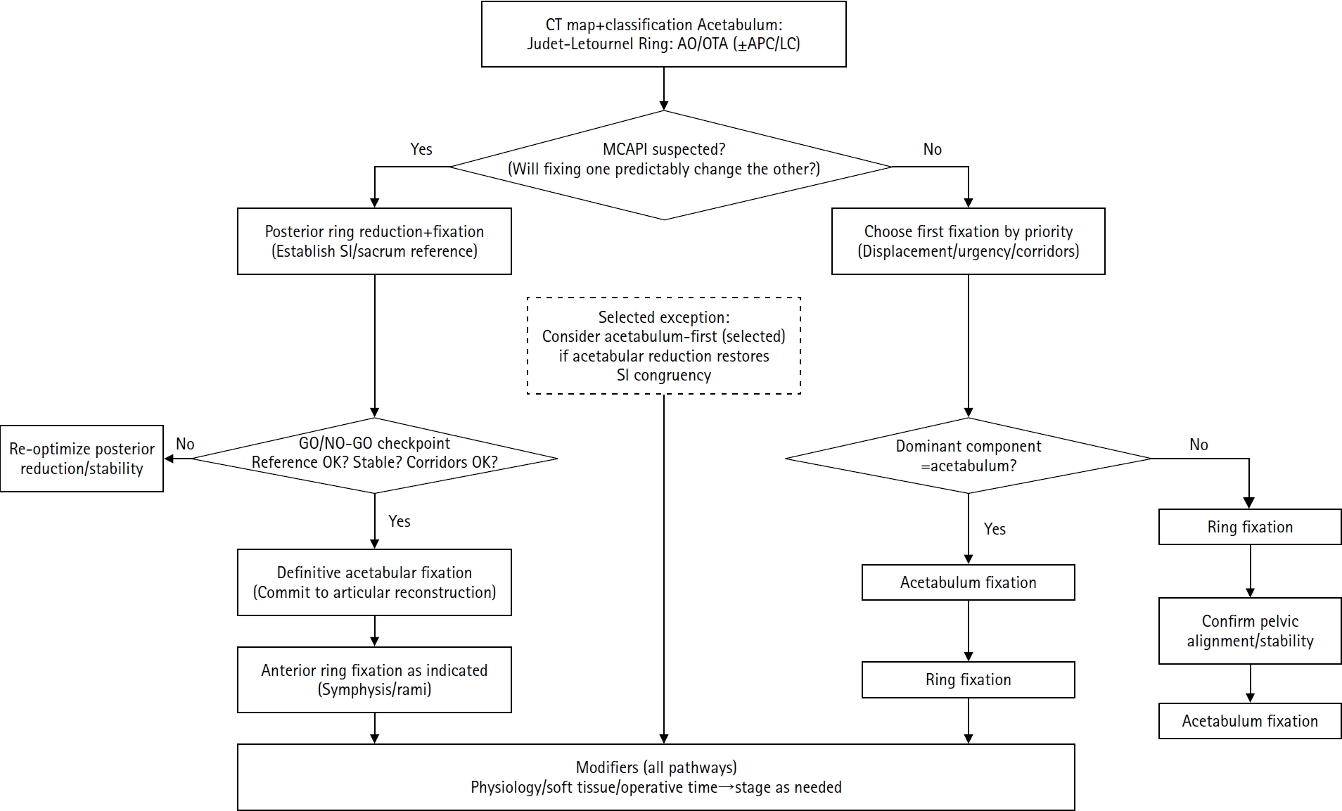
PDF - Combined acetabular and pelvic ring injuries are not simply “two fractures in one patient.” Reduction and fixation of one component can alter the alignment and reducibility of the other, rendering operative sequencing a primary decision variable rather than a secondary consideration. These injuries typically result from high-energy trauma, frequently occur in patients with polytrauma, and are further influenced by physiological tolerance and the feasibility of available operative corridors. The existing evidence base remains constrained by retrospective study designs, inconsistent definitions, variable classification systems, and heterogeneous outcome reporting, all of which limit the strength of comparative recommendations. This state-of-the-art review presents a surgeon-facing, algorithmic approach grounded in a reference-frame mindset. We emphasize computed tomography (CT)-based mapping and the use of consistent terminology to characterize acetabular morphology, pelvic ring instability, deformity vectors, suspicion of mechanical coupling, and feasible operative corridors. Mechanically connected acetabular and pelvic ring injuries (MCAPI) are introduced as a working framework for identifying patterns in which reduction or fixation of one injury predictably influences the other. In cases of suspected MCAPI, a posterior ring-based sequence is generally preferred, typically consisting of posterior ring reduction and fixation, definitive acetabular reconstruction, and subsequent anterior ring fixation. We propose an explicit intraoperative “GO/NO-GO” checkpoint (reference acceptable, stable, corridors feasible) to prevent acetabular reconstruction on a moving target. Acetabulum-first strategies may be appropriate only in selected anteroposterior compression- type configurations in which acetabular fixation plausibly restores sacroiliac congruency and posterior stabilization remains technically feasible. We summarize key outcome domains and complication patterns, highlighting hip dislocation as an important risk factor associated with both neurologic deficits and overall complications. Standardized CTbased definitions and outcome instruments, together with multicenter cohorts employing predefined decision pathways, are required to test sequencing strategies and to determine whether improved radiographic reduction translates into durable functional benefit.
- 602 View
- 19 Download

Case Reports
- Surgical Correction and Osteosynthesis for Cranial Displaced Pelvic Nonunion: Technical Note and Two Cases Report Regarding Anterior Correction and Osteosynthesis Following Posterior Release
- Kwang Cheon Choi, Ji Yoon Ha, Weon Yoo Kim
- J Korean Fract Soc 2014;27(2):151-156. Published online April 30, 2014
- DOI: https://doi.org/10.12671/jkfs.2014.27.2.151
-
Abstract
PDF
- Nonunion of an unstable pelvic fracture with cranial displacement pelvic surgery is technically difficult due to a large amount of bleeding and the risk of nerve damage. In addition, surgical correction of leg length discrepancy by reduction of a dislocated sacroiliac joint is in high demand. Nevertheless, when a patient is strongly disabled by a pelvic deformity, surgical correction may be necessary. Two patients with pelvic deformity were treated successfully by surgical correction and osteosynthesis.
- 593 View
- 0 Download
- Vertically Unstable Fracture of the Pelvis Combined with Anterior Dislocation of the Hip Joint: A Case Report
- Kap Jung Kim, Ha Yong Kim, Dae Suk Yang, Won Sik Choy
- J Korean Fract Soc 2007;20(3):272-276. Published online July 31, 2007
- DOI: https://doi.org/10.12671/jkfs.2007.20.3.272
-
Abstract
PDF
- Pelvic fractures result from high energy trauma and often associated with concomitant injuries. But, vertically unstable pelvic fractures combined with anterior dislocation of the hip is far less common. The traumatic dislocation of the hip is a true orthopedic emergency and it should be considered that a femoral head can be exposed to deteriorized vascularity. We report a case of vertically unstable pelvic fractures combined with traumatic anterior dislocation of the hip joint with the review of the literature.
- 645 View
- 1 Download
- Complete Rupture of Sciatic Nerve by Protruded Kuncher Nail in Pelvic Bone Fracture: A Case Report
- Yong Sik Kim, Nam Yong Choi, Suk Ku Han
- J Korean Fract Soc 2006;19(4):486-489. Published online October 31, 2006
- DOI: https://doi.org/10.12671/jkfs.2006.19.4.486
-
Abstract
- Rupture of sciatic nerve is a rare injury in minimally displaced pelvic bone fracture. We report one case of complete rupture of sciatic nerve that were resulted from the extremely protruded Kuncher nail inserted before accident and the preexisting heterotopic ossification with a review of the relevant literature.
- 540 View
- 0 Download
Original Articles
- Surgical Treatment of Unstable Pelvic Bone Fracture Involving Sacroiliac Joint
- Myung Ho Kim, Hee Gon Park, Moon jib Yoo, Jin Woo An
- J Korean Soc Fract 2003;16(4):433-440. Published online October 31, 2003
- DOI: https://doi.org/10.12671/jksf.2003.16.4.433
-
Abstract
PDF
- PURPOSE
To evaluate the results of surgical method using plate and screws in the treatment of unstable pelvic bone fracture involving Sacroiliac Joint.
MATERIALS AND METHOD
Authors reviewed 21 patients treated by surgical method from 1998 to 2002. Mean follow-up period was 15 months (12~24 month). Male were 16 and female were 5. We used plate and screws in 18 cases, just screws in 3 cases. We classified the type of fracture by Tile's classification and evaluated the results with Moon's criteria that based on reduction state in simple x-ray and patient's subjective satisfaction.
RESULTS
We got the bony union in all cases. By Moon's criteria, 10 cases were good, 7 cases were fair and 4 cases were poor. In 17 cases (80.9%), we got the results over fair. Mean weight bearing exercise periods were 6.4 weeks. There were 2 infection and 2 sacroiliac arthritis after operation.
CONCLUSION
As a method of surgical treatment on unstable pelvic bone fracture involving sacroiliac joint, we recommend open reduction and internal fixation with plate and screws and it may has particular advantages in early ambulation and satisfactory functional outcome.
- 672 View
- 1 Download
- Pelvic Bone Fractures in Children
- Byoung Suck Kim, Ye Yeon Won, Weon Ik Lee, Myeong Ryeol Song, Jae In Ahn
- J Korean Soc Fract 1998;11(1):107-114. Published online January 31, 1998
- DOI: https://doi.org/10.12671/jksf.1998.11.1.107
-
Abstract
PDF
- The pelvic bone fractures in children were uncommon, except for avulsion injuries in the literature and authors had 21 cases of children's pelvic bone fracture, ranging from 1 to 15 years. The mode of injury, type of fracture, associated injuries, morbidity and mortality, and out-come were retrospectively analyzed. The majority of injuries were from automobile-pedestrian collisions (81.0%). the Torode and Zieg type IV injury had the greatest morbidity, mortality, and complications. sixteen patients had non-orthopedic, associated injuries and fourteen required blood transfusions within initial 48 hours after injury. Two of them passed away due to hematologic unstableness. Twenty patients were managed by conservative method, except for one operative case by using of an external fixation device. This study included only 13 cases had average 1 year of follow-up. One acetabular dysplasia of 5 acetabular fractures was found at 12 months after injury. The nonoperative approach for the pelvic bone injury has had a satisfactory outcome in our hospital. so, authors think that if conservative methods will be properly applied, it may be one of the methods of treatment for the children's pelvic bone fracture. Even though there is no symptoms, long-term follow-up will be inevitable for checking more severe acetabular dysplasia and leg length discrepancy.
- 602 View
- 0 Download
- Operative treatment of the Unstable Pelvic Bone Fracture
- Byung Woo Min, Kwang Soon Song, Chul Hyung Kang, Young Soo Kim
- J Korean Soc Fract 1996;9(3):518-524. Published online July 31, 1996
- DOI: https://doi.org/10.12671/jksf.1996.9.3.518
-
Abstract
PDF
- Unstable pelvic bone fracture caused by high-energy trauma that can result in life-threatening situations in which intrapelvic hemorrhage and neurovascular injury. Long-term complications are frequently present, such as leg length discrepancy, gait disturbance and chronic low-back pain. Recently it is principle that it is mandatory to restore the anatomy of pelvic ring structure and to fixistably by means of internal fixation or extemal fixation for successful outcome after unstable pelvicring injury. 26 cases of unstable pelvic bone fracture were treated operatively at the authors hospital between 1992 and 1994. We analyse the clinical and the radiological result. The following results were obtained. 1. The incidence of the unstable pelvic bone fracture was 26 cases(18.4%) of all pelvic bone fractures(141 cases). 2. By the classification of modified Tile, type B1 were 8 cases, type B2(3 cases). type C1(7 cases) and type C3(8 cases). 3. Associated organ injury were found most commonly in the acetabular fracture(8 cases), and other extremity fracture(8 cases), genitourinary system(6 cases) and hemopenitoneum(4 cases). 4. The specific fracture pattern was classified according to various anatomical locations such as transsymphysis(7 cases), transpubic(7 cases), combination of the trassymphysis and traspubic(1 cases), trassacroiliac(7 cases), transiliac(9 cases), transsacral(1 case) and sacroiliac fracture dis location(1 case). 5. According to the fracture location, following methods of stabilization were applied. For the ante rior portion of pelvic ring, plates(13 cases), external fixators(3 cases) and wirings(3 cases) were used. For the posterior portion of pelvic ring, plates(9 cases), percutaneous iliosacral screws(3 cases) and lag screw(1 case) were used. 6. The results revealed as excellent in 20 cases, good in 5 case and fair in 1 case. 7. Postoperative complications were fixation failure(2 cases), metal failure(1 case) and nerve injury(1 case).
-
Citations
Citations to this article as recorded by
- Surgical Treatment of Malunion and Nonunion after Pelvic Bone Fracture
Byung-Woo Min, Kyung-Jae Lee
Journal of the Korean Fracture Society.2015; 28(4): 266. CrossRef - Clinical Results of Surgical Treatment of Acetabular Fractures according to Quality of Reduction
Sang-Hong Lee, Min-Kyu Shin, Sueng-Hwan Jo
The Journal of the Korean Orthopaedic Association.2007; 42(2): 153. CrossRef
- Surgical Treatment of Malunion and Nonunion after Pelvic Bone Fracture
- 822 View
- 0 Download
- 2 Crossref
 First
First Prev
Prev

