 E-submission
 TOTA
TOTA TOTS
TOTS
Search
- Page Path
- HOME > Search
Original Articles
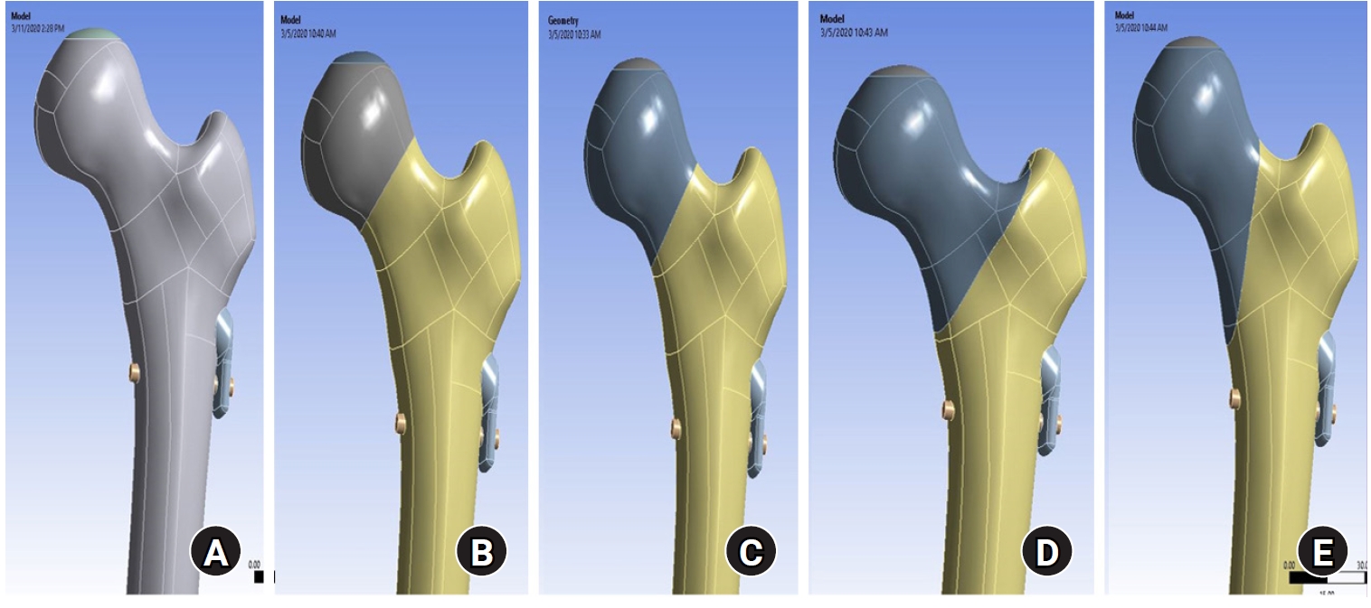
- Biomechanical finite element analysis of a femoral neck system fixation construct for femur neck fractures and clinical implications
- Hoon-Sang Sohn, Se-Lin Jeong, Gu-Hee Jung
- J Musculoskelet Trauma 2025;38(3):133-142. Published online July 22, 2025
- DOI: https://doi.org/10.12671/jmt.2025.00108
-
 Abstract
 PDF
PDF - Background
This study assessed the structural/mechanical stability of fixation constructs with a femoral neck system (FNS) via finite element analysis after simulating femoral neck fractures and explored the clinical implications.
Methods
We simulated subcapital, transcervical, basicervical, and vertical fracture models using a right femur (SAWBONES) and imported the implant model of FNS to Ansys (Ansys 19.0, Ansys Inc.) to place the implant in the optimal position. The distal end of the femur model was completely fixed and was abducted 7°. The force vector was set laterally at an angle of 3° and posteriorly at an angle of 15° in the vertical ground. The analysis was conducted using Ansys software with the von Mises stress (VMS) in megapascals (MPa).
Results
The maximum VMS of the fracture site was 67.01 MPa for a subcapital, 68.56 MPa for a transcervical, 344.54 MPa for a basicervical, and 130.59 MPa for a vertical model. The maximum VMS of FNS was 840.34 MPa for a subcapital, 637.37 MPa for a transcervical, 464.07 MPa for a basicervical, and 421.01 MPa for a vertical model. The stress distribution of basicervical and vertical fractures differed significantly, and the basicervical fracture had higher VMS at the bone, implant, and fracture sites.
Conclusions
FNS fixation should be performed with consideration the osseous anchorage in the femoral head, and this technique might be appropriate for vertical fractures. Regarding the VMS at the fracture site, FNS might be applied cautiously only to basicervical fractures with anatomical reduction without a gap or comminution. Level of evidence: IV. -
Citations
Citations to this article as recorded by
- Finite element analysis of screw thread geometry and titanium plate materials in internal fixation of the human femur
Abdessamed Bachiri, Mustapha Amine Arab, Nadia Kadouri
Computer Methods in Biomechanics and Biomedical Engineering.2026; : 1. CrossRef
- Finite element analysis of screw thread geometry and titanium plate materials in internal fixation of the human femur
- 2,812 View
- 99 Download
- 1 Crossref

- Comparison of Results between Minimally Invasive Plate Fixation and Antegrade Intramedullary Nailing of Recon-Type in Low-Energy Injury Distal Femoral Shaft Fractures
- Hong Moon Sohn, Gwangchul Lee, Ba Rom Kim, Jung Soo Oh
- J Korean Fract Soc 2024;37(2):87-94. Published online April 30, 2024
- DOI: https://doi.org/10.12671/jkfs.2024.37.2.87
-
Abstract
PDF
- Purpose
This study compared the outcomes of minimally invasive plate osteosynthesis and antegrade intramedullary nailing for low-energy fracture of the distal femoral shaft.
Materials and Methods
A study was conducted on 30 patients who underwent surgery for low-energy fractures of the distal femoral shaft between January 2016 and April 2022. The study compared 15patients who underwent minimally invasive plate osteosynthesis (Group P) with 15 patients who underwent recon-type antegrade intramedullary nailing (Group N). We evaluated intraoperative blood loss, operative time, C-arm exposure time, bone density, final union status, anatomical reduction, and clinical evaluation. The complications were also examined, and statistical analysis was conducted to compare the two groups.
Results
The blood loss, surgery time, and C-arm time were similar in the two groups. The radiographic assessments and clinical evaluations were also similar in the two groups. The clinical results showed no difference between the groups. Group N had one case of nonunion and one case of delayed union, while Group P had one case of nonunion and one case of peri-prosthetic fracture.
Conclusion
Antegrade intramedullary nailing of the recon-type demonstrated comparable results to minimally invasive plate osteosynthesis. Hence, antegrade intramedullary nailing of the recon-type, which enhances stability by fixing the entire femur and providing additional fixation in the distal portion, is deemed appropriate for treating distal femoral shaft fractures.
- 993 View
- 18 Download
- Effect of Additional Medial Locking Plate Fixation and Autogenous Bone Graft for Distal Femur Nonunion after Lateral Locking Plate Fixation
- Ho Min Lee, Jong Pil Kim, In Hwa Baek, Han Sol Moon, Sun Kyo Nam
- J Korean Fract Soc 2024;37(1):30-38. Published online January 31, 2024
- DOI: https://doi.org/10.12671/jkfs.2024.37.1.30
-
Abstract
PDF
- Purpose
This study examined the outcomes of additional medial locking plate fixation and autogenous bone grafting in the treatment of nonunions that occurred after initial fixation for distal femoral fractures using lateral locking plates.
Materials and Methods
The study involved eleven patients who initially underwent minimally invasive lateral locking plate fixation for distal femoral fractures between January 2008 and December 2020. The initial procedure was followed by additional medial locking plate fixation and autogenous bone grafting for clinically and radiographically confirmed nonunions, while leaving the stable lateral locking plate in situ. A clinical evaluation of the bone union time, knee joint range of motion, visual analog scale (VAS) pain scores, presence of postoperative complications, and functional evaluations using the lower extremity functional scale (LEFS) were performed.
Results
In all cases, bone union was achieved in an average of 6.1 months after the secondary surgery. The range of knee joint motion, weight-bearing ability, and VAS and LEFS scores improved at the final follow-up compared to the preoperative conditions. All patients could walk without walking assistive devices and did not experience pain at the fracture site. On the other hand, three patients complained of pain in the lateral knee joint caused by irritation by the lateral locking plate; hence, lateral hardware removal was performed. One patient complained of mild paresthesia at the anteromedial incision site. Severe complications, such as deep infection or metal failure, were not observed.
Conclusion
For nonunion with stable lateral locking plates after minimally invasive lateral locking plate fixation of distal femur fractures, additional medial locking plate fixation and autogenous bone grafting, while leaving the lateral locking plate intact, can achieve successful bone union.
- 651 View
- 7 Download
Case Report
- Recurrent Treatment Failure in Vancouver Classification Type C Periprosthetic Fractures around a Well Fixed Short Femoral Stem
- Byeong Yeol Choi, Hong-Man Cho, Jiyeon Park
- J Korean Fract Soc 2022;35(1):16-20. Published online January 31, 2022
- DOI: https://doi.org/10.12671/jkfs.2022.35.1.16
-
Abstract
PDF
- A short femoral stem (type 1 cementless stem) is being increasingly used to perform total hip arthroplasty; however, various types of intra- or postoperative periprosthetic fractures have been reported in recent times. A 66-year-old woman with a history of bilateral total hip arthroplasties using a type 1B femoral stem was admitted 2 months post-operation for a Vancouver type C periprosthetic fracture. She underwent open reduction and internal fixation; however, we observed recurrent non-union and plate breakage at the same site. In this case report, we discuss the factors associated with treatment failure in patients with a Vancouver type C periprosthetic fracture following type 1 femoral stem im-plantation.
- 606 View
- 1 Download
Original Articles
- Comparing Outcomes of Retrograde Intramedullary Nail and Locking Plate Fixation in Distal Femoral Fractures
- Byung-Ho Yoon, Bo Kwon Hwang, Hyoung-Keun Oh, Suk Kyu Choo, Jong Min Sohn, Yerl-Bo Sung
- J Korean Fract Soc 2021;34(4):131-136. Published online October 31, 2021
- DOI: https://doi.org/10.12671/jkfs.2021.34.4.131
-
Abstract
PDF
- Purpose
We compared the radiological and clinical results of fixation for distal femoral fracture (DFF) using a locking compression plate (LCP) or a retrograde intramedullary nail (RIN).
Materials and Methods
From October 2003 to February 2020, 52 cases of DFF with a minimum 1-year follow-up (with a mean follow-up of 19.1 months) were included: 31 were treated with LCP and 21 with RIN. The operation time, blood loss, and hospitalization period were compared, and the incidence of postoperative nonunion, malunion, delayed union and metal failure and other post-operative complications were evaluated and compared.
Results
There was no significant difference in the operating time between the two groups, but the mean blood loss was significantly higher in the LCP group (LCP 683.5 ml vs RIN; 134.9 ml; p=0.015). In 49 out of 52 cases, bone union was achieved without additional surgery in an average of 6.8 months, and a complete union was achieved after additional surgery in three cases of nonunion (LCP 2 cases vs RIN 1 case; p=0.065). One case of malunion and superficial infection was confirmed in each group.
Conclusion
Internal fixation using LCP and RIN give good outcomes with a low complication rate and can therefore be considered useful surgical treatments for DFF.
- 722 View
- 7 Download
- Treatment of Proximal Femur Fracture with a Newly Designed Nail: Trochanteric Fixation Nail-Advanced (TFNA)
- Jae Youn Yoon, Ji Wan Kim
- J Korean Fract Soc 2020;33(4):189-195. Published online October 31, 2020
- DOI: https://doi.org/10.12671/jkfs.2020.33.4.189
-
Abstract
PDF
- Purpose
This study evaluated the clinical results and implant safety of a newly developed implant, Trochanteric Fixation Nail-Advanced (TFNA; DePuy Synthes), in the treatment of proximal femur fractures.
Materials and Methods
This was a retrospective cohort study of 26 patients diagnosed with proximal femur fracture and treated surgically with TFNA. The patients’ demographic data, surgical data, radiologic findings, and functional outcomes, including complications, were evaluated.
Results
The mean age of the patients was 71.2 years (95% confidence interval [CI], 68.2-74.2); 65.4% were female. The mean Carlson comorbidity index score was 5.4, and the mean Koval grade before fracture was 2.1. Fracture classification included four cases of AO/OTA 31.A1, nine cases of A2, six cases of A3, and seven cases of 32A including six cases of atypical femoral fractures. The mean operating time was 53.3 minutes (95% CI, 43.6-63.1). There were no early postoperative complications, such as postoperative infection, deep vein thrombosis, pulmonary embolism, or in-hospital death, except one case of pneumonia. The mean Koval score at the postoperative six-month follow-up was 2.9. EuroQol-5 Dimension (EQ-5D) increased from 0.05 to 0.54 after three months and 0.72 at six months postoperatively. Bone union was observed in all cases with a mean union time of 12.9 weeks. No implant failure occurred, and no cases required secondary revision surgery.
Conclusion
A new intramedullary nail system, TFNA, showed excellent outcomes and safety in the surgical treatment of proximal femur fractures. -
Citations
Citations to this article as recorded by- Intermediate Length Cephalomedullary Nails in Proximal Femoral Fractures: Review of Indications and Outcomes
Daniel Scott Horwitz, Ahmed Nageeb Mahmoud, Michael Suk
Journal of the American Academy of Orthopaedic Surgeons.2025; 33(19): 1071. CrossRef - Outcomes of Intertrochanteric Fracture Fixation Using the Trochanteric Fixation Nail Advanced (TFNA): A Retrospective Analysis
Ramprasad Jasti, Prithvi Mohandas, Mahesh K Ragavan, Sunil D Magadam, Umesh Kannadasan
Cureus.2025;[Epub] CrossRef - Clinical and Radiological Outcomes of Unstable Intertrochanteric Fractures Treated with Trochanteric Fixation Nail-Advanced and Proximal Femoral Nail Antirotation-II: Correlation between Lateral Sliding of the Helical Blade and Lateral Trochanteric Pain
Sung Yoon Jung, Myoung Jin Lee, Lih Wang, Hyeon Jun Kim, Dong Hoon Sung, Jun Ha Park
Journal of the Korean Orthopaedic Association.2024; 59(3): 208. CrossRef - Prospective randomized multicenter noninferiority clinical trial evaluating the use of TFN-advancedTM proximal femoral nailing system (TFNA) for the treatment of proximal femur fracture in a Chinese population
Lidan Zhang, Zhijun Pan, Xiaohui Zheng, Qiugen Wang, Peifu Tang, Fang Zhou, Fan Liu, Bin Yu, Frankie K. L. Leung, Alex Wu, Suzanne Hughson, Zhuo Chen, Michael Blauth, Anthony Rosner, Charisse Sparks, Manyi Wang
European Journal of Trauma and Emergency Surgery.2023; 49(3): 1561. CrossRef - Risk of shortening in operatively treated proximal femur fractures with cephalomedullary nails with dynamically versus statically locked helical blades
Nathan Cherian, Lasun Oladeji, Cole Ohnoutka, Dan Touhey, Madeline Sauer, Kyle A. Schweser, Mauricio Kfuri, James L. Cook, Gregory J. Della Rocca, Brett D. Crist
Injury.2023; 54(2): 669. CrossRef - GS Hip Nail versus Affixus Hip Fracture Nail for the Intramedullary Nailing of Intertrochanteric Fractures
Seungcheol Kwon, Minjae Lee, Heeyeon Lee, Jihyo Hwang
Journal of Clinical Medicine.2023; 12(21): 6720. CrossRef - Comparison of the Clinical and Radiological Outcomes of TFNA (Trochanteric Fixation Nail-Advanced) and PFNA-II (Proximal Femoral Nail Antirotation-II) Treatment in Elderly Patients with Intertrochanteric Fractures
Min Sung Kwon, Young Bok Kim, Gyu Min Kong
Journal of the Korean Fracture Society.2022; 35(4): 162. CrossRef - Analysis of Clinical and Functional Outcomes according to the Blood Sugar Control Status at the Time of Ankle Fractures Resulting from Rotational Injuries
Jun Young Lee, Dong Seop Lim, Seung Hyun Lee, Seo Jin Park
Journal of the Korean Fracture Society.2022; 35(4): 135. CrossRef - Conventional versus helical blade screw insertion following the removal of the femoral head screw: a biomechanical evaluation using trochanteric gamma 3 locking nail versus PFN antirotation
Hong Man Cho, Kwang Min Park, Tae Gon Jung, Ji Yeon Park, Young Lee
BMC Musculoskeletal Disorders.2021;[Epub] CrossRef - Clinical and Radiologic Outcome of Intertrochanteric Fracture Treatment Using TFNA (Trochanteric Fixation Nail-Advanced)
Hyeon Joon Lee, Hyun Bai Choi, Ba Rom Kim, Seung Hwan Jo, Sang Hong Lee
Journal of the Korean Fracture Society.2021; 34(3): 105. CrossRef
- Intermediate Length Cephalomedullary Nails in Proximal Femoral Fractures: Review of Indications and Outcomes
- 3,149 View
- 30 Download
- 10 Crossref
- Retrospective Comparative Study of the Intraoperative Fracture Gap Compression in the Treatment of Intertrochanteric Fracture Using Proximal Femoral Nail Antirotation
- Se Jin Kim, Hong Man Cho, Jiyeon Park, Ki Yong An, Young Woo Chung, Woojin Shin
- J Korean Fract Soc 2020;33(4):179-188. Published online October 31, 2020
- DOI: https://doi.org/10.12671/jkfs.2020.33.4.179
-
Abstract
PDF
- Purpose
Intertrochanteric fractures can be treated using proximal femoral nail antirotation (PFNA). This study examined the clinical and radiological results of the intraoperative fracture compression.
Materials and Methods
Ninety-four patients underwent intraoperative compression (Group I), and 88 patients underwent natural sliding only (Group II). The patients were followed-up for more than two years. All patients met the following seven conditions: (1) AO/OTA 31-A1, A2 type intertrochanter fracture, (2) availability of compression of more than one cortical bone in the anterior or medial region of the fracture site under the preoperative imaging test, (3) Singh index grade ≥3, (4) blade position: center-center, center-inferior, (5) tip-apex distance <25 mm, (6) reduction status of good or very good, and (7) positive or neutral medial cortical support position with slightly valgus reduction.
Results
A slight tendency toward significant differences in acute phase pain between the two groups was observed at six weeks postoperatively (p=0.073). Twenty-four months after surgery, lateral extension of the PFNA helical blade between the two groups showed significant differences (p=0.017). Fracture gaps measured immediately after surgery showed significant differences (p=0.001), and a clear tendency for a significant difference in the average fracture union time was found (p=0.065).
Conclusion
Intraoperative fracture compression, intraoperative fracture compression appears beneficial to achieve a successful union of trochanteric fractures provided that all conditions are met to apply the method safely. -
Citations
Citations to this article as recorded by- Benefits of a Demineralized Bone Matrix in Osteoporotic Intertrochanteric Femoral Fracture Patients
Se Jin Kim, Hong-Man Cho, Myung Cheol Jung
Journal of the Korean Fracture Society.2022; 35(4): 151. CrossRef
- Benefits of a Demineralized Bone Matrix in Osteoporotic Intertrochanteric Femoral Fracture Patients
- 1,150 View
- 4 Download
- 1 Crossref
- Does the Use of a Silicone Ring Tourniquet Help Reduce Bleeding in the Minimally Invasive Internal Fixation with Locking Plate for Distal Femoral Fractures?
- Ki-Bong Park, Hong-Ki Jin, Il-Yeong Hwang, Sung-Who Chang, Sung-Cheon Na
- J Korean Fract Soc 2020;33(3):148-153. Published online July 31, 2020
- DOI: https://doi.org/10.12671/jkfs.2020.33.3.148
-
Abstract
PDF
- Purpose
This study evaluated the usefulness of a silicone ring tourniquet by analyzing the changes in the perioperative hemoglobin (Hb) levels or amount of perioperative bleeding compared to those of a pneumatic tourniquet or no usage during minimally invasive plate fixation for distal femoral fractures.
Materials and Methods
From January 2017 to December 2019, 30 patients who underwent minimally invasive plate fixation using a locking compression plate for distal femoral fractures were evaluated and classified as a silicone ring tourniquet (Group 1), a pneumatic tourniquet (Group 2), and no usage (Group 3). The variables for analysis were age, sex, preoperative Hb (preHb), postoperative 72-hour Hb (postHb), differences between preHb and postHb (preHb-postHb), amount of intraoperative and overall transfusion, estimated unit of transfusion corrected by preHb-postHb and total transfusion (Hb-lost), amount of intraoperative and postoperative and total bleeding. One-way ANOVA was used to identify the differences between the groups.
Results
The age, sex, operation time, preHb, preHb-postHb, amount of intraoperative and overall transfusion and Hb-lost were similar in the two groups. The amount of intraoperative bleeding was significantly lower in Group 1 than Group 3 (p=0.004), but there was no difference in the amount of postoperative and total bleeding between the two groups.
Conclusion
The use of a silicone ring tourniquet in the minimally invasive plate fixation for distal femoral fractures decreased the amount of intraoperative bleeding compared to no use of a tourniquet. -
Citations
Citations to this article as recorded by- Silicone ring tourniquet could be a substitute for a conventional tourniquet in total knee arthroplasty with a longer surgical field: a prospective comparative study in simultaneous total knee arthroplasty
Tae sung Lee, Kwan Kyu Park, Byung Woo Cho, Woo-Suk Lee, Hyuck Min Kwon
BMC Musculoskeletal Disorders.2023;[Epub] CrossRef
- Silicone ring tourniquet could be a substitute for a conventional tourniquet in total knee arthroplasty with a longer surgical field: a prospective comparative study in simultaneous total knee arthroplasty
- 1,391 View
- 6 Download
- 1 Crossref
- Failure of Intramedullary Nailing for Subtrochanteric Atypical Femoral Fractures Caused by Endosteal Cortical Thickening
- Young Ho Roh, Kimoon Kang, Hee Joong Kim, Kwang Woo Nam
- J Korean Fract Soc 2019;32(4):211-221. Published online October 31, 2019
- DOI: https://doi.org/10.12671/jkfs.2019.32.4.211
- Correction in: J Musculoskelet Trauma 2020;33(1):63
-
Abstract
PDF
- PURPOSE
Recent literature has noted incidences of subtrochanteric atypical femoral fractures (AFFs) in patients who have taken long-term bisphosphonates (BPs). Most cases of subtrochanteric AFFs have been treated with intramedullary nailing and cases of delayed union have been reported. On the other hand, there is no data available on the complications associated with endosteal thickening or cortical thickening. This study evaluated the results of surgical treatment according to the endosteal thickening of the lateral cortex in subtrochanteric AFFs.
MATERIALS AND METHODS
Investigation was performed at the Department of Orthopaedic Surgery, Jeju National University Hospital. The study consisted of patients with subtrochanteric AFFs, defined by the American Society for Bone and Mineral Research (ASBMR) major criteria, who underwent intramedullary nailing from March 2012 to October 2014. The cases were categorized into two groups based on the presence of endosteal thickening. The evaluation included the demographic data, radiographic data of initial reduction state, and duration of BPs.
RESULTS
The demographic data and duration of BPs were similar in the two groups. On the other hand, varus reduction (Group I: 12.5% vs. Group II: 78.9%; p=0.001), delayed union (Group I: 0% vs. Group II: 70.0%; p=0.003), nonunion (Group I: 0% vs. Group II: 47.4%; p=0.017), and union time (Group I: 5.5 months vs. Group II: 8.3 months; p<0.001) were significantly different in the two groups.
CONCLUSION
Endosteal thickening of the lateral cortex in subtrochanteric AFFs was identified as an independent factor that decides the reduction of the fracture and nonunion. The endosteal thickening should be removed to obtain anatomical alignment for successful surgical results. -
Citations
Citations to this article as recorded by- Controlled bending of proximal femoral nails used in fractures of bowed femurs: biomechanical study with clinical application
Hong Moon Sohn, Suenghwan Jo
Medical Biological Science and Engineering.2022; 5(2): 63. CrossRef
- Controlled bending of proximal femoral nails used in fractures of bowed femurs: biomechanical study with clinical application
- 1,756 View
- 7 Download
- 1 Crossref
- Treatment of the Proximal Femoral Fracture Using the New Design Cephalomedullary Nail: Prospective Outcomes Study
- Young Ho Roh, Joseph Rho, Kwang Woo Nam
- J Korean Fract Soc 2019;32(1):35-42. Published online January 31, 2019
- DOI: https://doi.org/10.12671/jkfs.2019.32.1.35
-
Abstract
PDF
- PURPOSE
The aim of this study is to investigate the clinical performance and safety of Zimmer® natural nail cephalomedullary nail (ZNN CM nail) in the treatment of proximal femur fractures.
MATERIALS AND METHODS
The following research was conducted as a prospective, non-comparative, single center outcome study. Upon providing written informed consent, enrolled patients' data were collected and analyzed. Postoperative follow-up visits were scheduled at 6 weeks, 3 months, 6 months, and 1 year. Follow-up evaluation included radiographic assessment, physical examination, and quality of life and adverse events reports.
RESULTS
Thirty-nine patients were available for evaluation at one year postoperative. The patients reported the mean EuroQol-5 Dimension score increased after surgery: from 0.4 points at discharge (n=49) to 0.6 points at 1-year post-surgery (n=39). The mean Harris hip score also increased after surgery: from 56.3 points at discharge (n=49) to 72.1 points at 1 year (n=12). Bone union was seen in 64% (n=16) in 6 months and 95% (n=37) in 1 year.
CONCLUSION
The results of this 1-year follow-up study affirmed the effectiveness and safety of the ZNN CM nail in the treatment of proximal femur fractures. -
Citations
Citations to this article as recorded by- Clinical and Radiologic Outcome of Intertrochanteric Fracture Treatment Using TFNA (Trochanteric Fixation Nail-Advanced)
Hyeon Joon Lee, Hyun Bai Choi, Ba Rom Kim, Seung Hwan Jo, Sang Hong Lee
Journal of the Korean Fracture Society.2021; 34(3): 105. CrossRef - Treatment of Proximal Femur Fracture with a Newly Designed Nail: Trochanteric Fixation Nail-Advanced (TFNA)
Jae Youn Yoon, Ji Wan Kim
Journal of the Korean Fracture Society.2020; 33(4): 189. CrossRef
- Clinical and Radiologic Outcome of Intertrochanteric Fracture Treatment Using TFNA (Trochanteric Fixation Nail-Advanced)
- 1,352 View
- 0 Download
- 2 Crossref
- Radiologic and Serologic Factors Associated with Bone Union at Femoral Atypical Fracture
- Suc Hyun Kweon, Byung Min Yoo
- J Korean Fract Soc 2019;32(1):27-34. Published online January 31, 2019
- DOI: https://doi.org/10.12671/jkfs.2019.32.1.27
-
Abstract
PDF
- PURPOSE
The purpose of this study was to investigate the radiologic and serologic factors related to postoperative union using intramedullary (IM) internal fixation in atypical femoral fractures (AFF), which are closely related to bisphosphonates (BPs) for osteoporosis.
MATERIALS AND METHODS
From February 2008 to December 2016, 65 patients (71 cases) who had undergone IM nail fixation after diagnosis of AFF were enrolled in this study. Patients were divided into group A, who experienced union within 6 months and group B, who did not experience union within 6 months. They were evaluated for duration of BPs use, radiologic factors and serological factors.
RESULTS
The mean duration of BPs use was 6.17 years in group A and 8.24 years in group B (p=0.039). In the subtrochanteric area, there were 14 cases (27.5%) in group A and 14 cases (70.0%) in group B. In the femoral shaft, there were 37 cases (72.5%) in group A and 6 cases (30.0%) in group B (p=0.001). On the preoperative, the flexion in the coronal plane was 5.9° (2.1°–9.2°) in group A and 8.0° (3.1°–12.1°) in group B (p=0.041). On the postoperative, conversion to valgus was 15 cases (29.4%), 8 cases (40.0%); conversion to neutral was 34 cases (66.7%) and 8 cases (40.0%); conversion to varus was 2 cases (3.9%) and 4 cases (20.0%), each (p=0.037). The fracture site gap was 1.5 mm (0–2.9 mm) on the front side and 1.2 mm (0–2.2 mm) on lateral side and 2.2 mm (0.9–4.7 mm) and 1.9 mm (0.5–3.5 mm), each (p=0.042, p=0.049). Among serological factors, there was no significant difference between the two groups.
CONCLUSION
Factors adversely affecting the union should be recognized before surgery, such as longterm BPs use or a severe degree of bending of the femur in the coronal plane. During surgery, proper reduction and spacing of the fracture site on the coronal plane should allow adequate reduction of the anterior and posterior surfaces. Obtaining anatomic reduction would be most beneficial for union, but if that is not possible, obtaining congenital valgus rather than varus on the coronal plane may be helpful for union. -
Citations
Citations to this article as recorded by- Subtrochanteric Fracture Reduction during Intramedullary Nailing: Technical Note
Gyu Min Kong
Journal of the Korean Fracture Society.2019; 32(2): 107. CrossRef
- Subtrochanteric Fracture Reduction during Intramedullary Nailing: Technical Note
- 1,231 View
- 1 Download
- 1 Crossref
- Risk Factors for Knee Stiffness in Distal Femoral Fractures
- Dong Wook Son, Hyoung Soo Kim, Woo Young Choi
- J Korean Fract Soc 2018;31(4):123-131. Published online October 31, 2018
- DOI: https://doi.org/10.12671/jkfs.2018.31.4.123
-
Abstract
PDF
- PURPOSE
The aims of this study were to evaluate risk factors for knee stiffness after the fixation of distal femoral fractures, and to analyze the clinical and radiologic outcomes.
MATERIALS AND METHODS
This is a retrospective case control study of 104 consecutive patients who have a distal femoral fracture and were treated with a submuscular locking plate. The case group comprised of patients with 12-month postoperative range of motion (ROM) ≤90° or a history of manipulation under anesthesia. The case group was compared with the control group of patients with a 12-month postoperative ROM >90°. The possible risk factors were evaluated by univariate and logistic regression analysis. The postoperative ROM and Knee Society clinical rating system was evaluated for the clinical assessment and the distal femoral angle on a whole-extremity scanogram was measured for radiologic assessments.
RESULTS
Fifty-four patients were included in the study (14 in the case group, 40 in the control group). Univariate analysis showed that comminuted fracture, intra-articular fracture, open fracture, temporary external fixation, severe osteoarthritis, and prolonged immobilization placed patients at an increased risk for knee stiffness. On the other hand, multivariate logistic regression showed that an extensor mechanism injury was the only significant predictor (p=0.001; odds ratio, 42.0; 95% confidence interval, 5.0–350.7). The ROM and Knee Society score were significantly lower in the case group; however, the coronal alignment was similar in the case and control group.
CONCLUSION
Various factors that delay postoperative knee motion place patients at increased risk of knee stiffness. Understanding these risk factors may help surgeons prevent postoperative knee stiffness after distal femoral fractures. In particular, extensor mechanism injury, such as patella fracture or open quadriceps injury, was found to be an independent predictable factor associated with knee stiffness. -
Citations
Citations to this article as recorded by- Post operative knee stiffness after surgical fixation of knee osseous injuries
Oitangor Arthur, Sekamatte Yasin, Mulepo Phillip
Adesh University Journal of Medical Sciences & Research.2026; 0: 1. CrossRef - The outcomes of patients with segmental long bone fractures treated with SIGN nail at Addis Ababa Burn, Emergency and Trauma Hospital
Cheru B. Tesso, Lelisa Merga, Samuel Kebede
OTA International.2026;[Epub] CrossRef - A Comprehensive Approach to Stiffness in Total Knee Arthroplasty
Brian P. Chalmers, Linda I. Suleiman, Peter K. Sculco, Matthew P. Abdel
The Journal of Arthroplasty.2025; 40(9): S59. CrossRef - Staged Management for Distal Femur Fractures: Impacts on Reoperation, Stiffness, and Overall Outcomes
Matthew T. Yeager, Robert W. Rutz, Alex Roszman, Gerald McGwin, James E. Darnley, Joseph P. Johnson, Clay A. Spitler
Journal of Orthopaedic Trauma.2024; 38(11): 577. CrossRef - Outcome of the Masquelet Technique for Complex Bilateral Distal Femoral Bone Defects
Ziad A Aljaafri, Abdullah Alzahrani, Ali Alshehri, Ahmed AlHussain, Faisal Alzahrani, Khalid Alsheikh
Cureus.2023;[Epub] CrossRef - Efficacy of non-operative treatment of patients with knee arthrofibrosis using high-intensity home mechanical therapy: a retrospective review of 11,000+ patients
Shaun K. Stinton, Samantha J. Beckley, Thomas P. Branch
Journal of Orthopaedic Surgery and Research.2022;[Epub] CrossRef - Distal Femoral Replacement and Extensor Mechanism Repair Reinforced With Synthetic Mesh for Distal Femur Fracture With Patellar Ligament Avulsion
Charles Powell, Kristopher Sanders, Neal Huang, Luis Felipe Colón, Colton Norton
Arthroplasty Today.2022; 16: 31. CrossRef - The fragility of statistical significance in distal femur fractures: systematic review of randomized controlled trials
Michael Megafu, Hassan Mian, Emmanuel Megafu, Sulabh Singhal, Alexander Lee, Richawna Cassie, Paul Tornetta, Robert Parisien
European Journal of Orthopaedic Surgery & Traumatology.2022; 33(6): 2411. CrossRef - Association Between Femoral “Spike” Size After Intramedullary Nailing and Subsequent Knee Motion Surgery
Michael G. Schloss, Nathan N. O'Hara, Syed M. R. Zaidi, Zachary D. Hannan, Dimitrius Marinos, Jared Atchison, Alexandra Mulliken, Jason W. Nascone, Robert V. O'Toole
Journal of Orthopaedic Trauma.2021; 35(2): 100. CrossRef - Distal Femur Replacement Versus Surgical Fixation for the Treatment of Geriatric Distal Femur Fractures: A Systematic Review
Brett P. Salazar, Aaron R. Babian, Malcolm R. DeBaun, Michael F. Githens, Gustavo A. Chavez, L. Henry Goodnough, Michael J. Gardner, Julius A. Bishop
Journal of Orthopaedic Trauma.2021; 35(1): 2. CrossRef
- Post operative knee stiffness after surgical fixation of knee osseous injuries
- 997 View
- 17 Download
- 10 Crossref
Case Reports
- Delayed Sciatic Nerve Palsy due to Hematoma Related with Anticoagulants Prophylaxis in the Femur Intramedullary Nailing: A Case Report
- Young Mo Kim, Yong Bum Joo, Seok Hwan Song
- J Korean Fract Soc 2017;30(4):198-202. Published online October 31, 2017
- DOI: https://doi.org/10.12671/jkfs.2017.30.4.198
-
Abstract
PDF
- Femur intramedullary nailing can be one of the most predictable procedures in orthopedic traumatology. The advantage of this method is that the fracture site does not have to be widely exposed for reduction, which can minimize soft tissue damage. For this reason, the incidence of complications related to hematoma has been rare. We experienced only one case of sciatic nerve palsy due to hematoma after intramedullary nailing; the patient was receiving an anticoagulant therapy. Therefore, we report this case with literature review.
-
Citations
Citations to this article as recorded by- Ipsilateral Foot Drop After Leg Traction on Fracture Table for Mid-Shaft Femur Fracture Nailing: A Rare Case Report
Jehad A Alzahrani, Ahmed A Alabdali, Mohammed O Albariqi
Cureus.2023;[Epub] CrossRef
- Ipsilateral Foot Drop After Leg Traction on Fracture Table for Mid-Shaft Femur Fracture Nailing: A Rare Case Report
- 1,241 View
- 8 Download
- 1 Crossref
- Insufficiency Fracture of the Femoral Neck after Intramedullary Nailing for the Treatment of Atypical Femoral Fracture - A Case Report -
- Nam Hoon Moon, Jae Hoon Jang, Tae Hyuk Hwang, Ki Young Park
- J Korean Fract Soc 2016;29(4):258-264. Published online October 31, 2016
- DOI: https://doi.org/10.12671/jkfs.2016.29.4.258
-
Abstract
PDF
- Although several publications have reported delayed or non-union, there is a consensus that the standard treatment for atypical femoral fracture (AFF) is an intramedullary nailing. However, no case of tensile insufficiency fracture of femoral neck associated with intramedullary nailing in patients with AFF have been reported. Here, we report an 82-year-old woman with tensile type of insufficiency fracture of the femoral neck after intramedullary nailing for the treatment of AFF.
- 739 View
- 4 Download
- Huge Pseudoaneurysm of Popliteal Artery Following Conservative Treatment of a Distal Femur Fracture: A Case Report
- Won Chul Cho, Chong Bin Park, Young Jun Choi, Hyun Il Lee, Hee Jae Won, Jae Kwang Hwang
- J Korean Fract Soc 2016;29(2):137-142. Published online April 30, 2016
- DOI: https://doi.org/10.12671/jkfs.2016.29.2.137
-
Abstract
PDF
- A pseudoaneurysm is a contained arterial disruption in the intimal and medial layers of an arterial wall. It may originate from a perforation caused by traumatic or iatrogenic injury or the dehiscence of a surgical anastomosis. Because of its insidious onset and delayed presentation, orthopaedic surgeons should be aware of the possibility of such a lesion after an initial trauma. We report on a case of a delayed huge pseudoaneurysm of the popliteal artery that occurred 11 months after conservative treatment of a supracondylar fracture of the femur in order to keep in mind the possibility of the delayed presentation of vascular injury after a distal femur fracture.
- 972 View
- 3 Download
Original Articles
- The Clinical and Radiological Results of Vancouver Type B1 and C Periprosthetic Fractures
- Bo Ram Na, Taek Rim Yoon, Kyung Soon Park
- J Korean Fract Soc 2016;29(1):26-33. Published online January 31, 2016
- DOI: https://doi.org/10.12671/jkfs.2016.29.1.26
-
Abstract
PDF
- PURPOSE
The purpose of this study is to evaluate the clinical and radiologic results of plate fixation in the Vancouver B1 and C periprosthetic femoral fracture (PFF).
MATERIALS AND METHODS
Twenty patients who had sustained a Vancouver type B1 and C periprosthetic fracture after hip arthroplasty (years 2002-2012) were identified. The mean age was 66.0 years (range, 43-85 years) and the mean follow-up duration of the group was 38 months (range, 12-102 months). The dynamic compression plate (DCP) group included 12 patients and the locking compression plate (LCP) group included eight patients. Harris hip score (HHS) and walking ability, knee joint range of motion (ROM) were compared before injury and last follow-up. Fracture union rate and period were compared.
RESULTS
The mean HHS score was 90.7 (64-96). There was no statistical difference between the two groups. At the last follow-up, knee joint ROM was 103.3degrees (105degrees-140degrees) in the DCP group and 118.4degrees (110degrees-140degrees) in the LCP group, showing good results in the LCP group (p=0.043). No significant difference in the fracture union rate and union periods was observed between the two groups.
CONCLUSION
A better result for the postoperative knee flexion exercise capacity was observed in the LCP group. Use of LCP plate fixation is a good option in management of Vancouver classification B1 and C PFF.
- 987 View
- 5 Download
- Steinmann Pin Assisted Reduction of Subtrochanteric Femoral Fracture
- Seung Wan Lim, Oog Jin Shon
- J Korean Fract Soc 2015;28(1):17-22. Published online January 31, 2015
- DOI: https://doi.org/10.12671/jkfs.2015.28.1.17
-
Abstract
PDF
- PURPOSE
Nail insertion is the treatment of choice for subtrochanteric femoral fracture, but displacement of proximal bone fragment makes it difficult to find an ideal entry point. Therefore, in this study we aimed to determine the usefulness of treatment of subtrochanteric femoral fracture using Steinmann pin assisted reduction, internal fixation, and insertion of intramedullary nails.
MATERIALS AND METHODS
We evaluated 33 patients who were followed-up more than a year with a displaced subtrochanteric femoral fracture treated with closed reduction and intramedullary nail fixation between January 2008 and March 2013. In addition, we studied postoperative bone union time, postoperative reduction status, change of the femur neck shaft angle, evaluation of hip joint function, return to daily life, and complications.
RESULTS
All fractures with Steinmann pin assisted reduction were united but they included three cases of delayed union. In Fogagnolo classification, all cases were up to acceptable states and the varus change of femur neck shaft angle was 0.94degrees+/-3.1degrees; no significant difference in Harris hip score was observed between preoperative and last follow-up (p>0.05).
CONCLUSION
There were satisfactory results in bone union and reduction state with Steinmann pin assisted reduction. Therefore, Steinmann pin assisted reduction is a useful surgical technique for subtrochanteric femoral fracture. -
Citations
Citations to this article as recorded by- Percutaneous acetabular anchoring pin-assisted cephalomedullary nailing for subtrochanteric and unstable intertrochanteric fractures
Keong-Hwan Kim, Youngsik Yoon, Eic Ju Lim
Injury.2020; 51(3): 769. CrossRef
- Percutaneous acetabular anchoring pin-assisted cephalomedullary nailing for subtrochanteric and unstable intertrochanteric fractures
- 1,331 View
- 22 Download
- 1 Crossref
- Results of Intramedullary Nailing of Femoral Shaft Fracture: Trochanteric Entry Portal (Sirus Nail) versus Piriformis Entry Portal (M/DN Nail)
- Sang Ho Ha, Woong Hee Kim, Gwang Chul Lee
- J Korean Fract Soc 2014;27(1):50-57. Published online January 31, 2014
- DOI: https://doi.org/10.12671/jkfs.2014.27.1.50
-
Abstract
PDF
- PURPOSE
To compare treatment results obtained using the trochanteric (Sirus nail) entry portal with those obtained using the Piriformis fossa (M/DN) entry portal during intramedullary (IM) nailing of femur shaft fractures.
MATERIALS AND METHODS
Four hundreds and thirty-two patients treated for femur shaft fracture using IM nails from February, 2001 to May, 2010 were divided into two groups. group 1 was composed of 180 patients treated through the trochanteric (Sirus nail; n=180) entry portal, while group 2 contained 170 patients treated through the piriformis fossa (M/DN nail; n=170) entry portal. We compared the clinical and radiographic findings of both groups to evaluate the treatment results.
RESULTS
Functional result, range of motion and union time (18, 20 weeks) were similar in both groups. The operation time of patients in the over-weighted group was 90 minutes in group 1 and 120 minutes in group 2 (p<0.05). Additionally, the blood loss was 280 ml in group 1 and 335 ml in group 2, and in case of over-weight patients, group 2 showed more blood loss (p<0.05). The duration of exposure to fluoroscopy differed slightly, with group 1 being less exposed than group 2; however, this difference was not significant (p>0.05). There were 18 iatrogenic fractures in group 1 and 4 in group 2 (p<0.05).
CONCLUSION
There was not much difference in complications based on clinical and radiographic findings of both groups. For groups using the trochanteric entry portal, the operation time was shorter and blood loss was lower than in groups using the piriformis entry portal. Iatrogenic fracture occurred more often in the group using the trochanteric entry portal than in the group using the piriformis entry portal. -
Citations
Citations to this article as recorded by- Analysis of different entry portals for femoral nail with two different nail designs-straight nail versus lateral angulated nail - Does it make a difference?
Sanjay Yadav, Saurabh Singh, Anil Kumar Rai
Journal of Clinical Orthopaedics and Trauma.2019; 10(5): 912. CrossRef - Comparing Entry Points for Antegrade Nailing of Femoral Shaft Fractures
Ujash Sheth, Chetan Gohal, Jaskarndip Chahal, Aaron Nauth, Tim Dwyer
Orthopedics.2016;[Epub] CrossRef - The Curative Effect Comparison Between Prolonged Third Generation of Gamma Nail and Prolonged Dynamic Hip Screw Internal Fixation in Treating Femoral Intertrochanteric Fracture and the Effect on Infection
Wenye He, Wei Zhang
Cell Biochemistry and Biophysics.2015; 71(2): 695. CrossRef - Treatment of Femur Subtrochanteric Fracture Using the Intramedullary Long Nail; Comparison of Closed Reduction and Minimal Open Reduction
Sang Joon Lee, Sang Hong Lee, Sang Soo Park, Hyung Seok Park
Journal of the Korean Orthopaedic Association.2015; 50(1): 18. CrossRef - Failure to Remove a Trochanteric Entry Femoral Nail and Its Cause in Adolescent Patients: Two Cases Report
Ji-Hwan Kim, Seung-Oh Nam, Young-Soo Byun, Han-Sang Kim
Journal of the Korean Fracture Society.2015; 28(1): 71. CrossRef - Treatment of the Femoral Fracture Using Sirus® Nail: A Comparison of Complication according to the Entry Potal
Young-Yool Chung, Dong-Hyuk Choi, Dae-Hyun Yoon, Jung-Ho Lee, Ji-Hun Park
Journal of the Korean Fracture Society.2015; 28(2): 103. CrossRef - Comparison of Greater Trochanter Versus Piriformis Entry Nail for Treatment of Femur Shaft Fracture
Jong-Hee Lee, Jong-Hoon Park, Si-Yeong Park, Seong-Cheol Park, Seung-Beom Han
Journal of the Korean Fracture Society.2014; 27(4): 287. CrossRef
- Analysis of different entry portals for femoral nail with two different nail designs-straight nail versus lateral angulated nail - Does it make a difference?
- 2,005 View
- 21 Download
- 7 Crossref
- The Comparison of Minimally Invasive Percutaneous Plate Osteosynthesis versus Open Plate Fixation in the Treatment of in the Distal Femur Fracture
- Seong Jun Ahn, Suk Woong Kang, Bu Hwan Kim, Moo Ho Song, Seong Ho Yoo, Kwan Taek Oh
- J Korean Fract Soc 2013;26(4):314-320. Published online October 31, 2013
- DOI: https://doi.org/10.12671/jkfs.2013.26.4.314
-
Abstract
PDF
- PURPOSE
To evaluate the efficacy of surgical treatment through retrospective comparison of minimally invasive percutaneous plate osteosynthesis (MIPPO) vs open plate fixation in the treatment of the distal femur fractures.
MATERIALS AND METHODS
Thirty-one patients with distal femur fractures from January 2002 to December 2010 were divided into two groups depending on the surgical method. Minimum follow up was 12 months. Group A consisted of 17 patients treated with MIPPO, and group B was comprised of 14 patients treated with open plate fixation. Clinical outcomes including operation time, transfusion rate, rehabilitation, range of motion, and interval change of postoperative C-reactive protein (CRP) were evaluated to assess postoperative inflammatory reaction, postoperative complications and clinical results with the use of Sanders criteria.
RESULTS
The operative time was 86/135 min and transfusion volume was 0.8/1.9 unit respectively. The postoperative 3-day and 7-day CRP were 7.4/1.5 mg% in group A and 10.3/2.4 mg% in group B, showing more minimal tissue injury and early recovery in group A. There were no significant differences in clinical results by Sanders criteria in both groups.
CONCLUSION
Both MIPPO and open plate fixation for the treatment of distal femur fractures showed comparably good results. However, the MIPPO technique is superior to group B in view of minimal tissue injury and operation time and was proven to lessen the transfusion rate. -
Citations
Citations to this article as recorded by- Usefulness of Reduction and Internal Fixation Using a 2.4 mm Hand Plating System in Type AO 33-A3 Distal Femur Fracture: Technical Note
Bong-Ju Lee, Ja-Yeong Yoon, Seungha Woo
Journal of the Korean Fracture Society.2023; 36(1): 25. CrossRef
- Usefulness of Reduction and Internal Fixation Using a 2.4 mm Hand Plating System in Type AO 33-A3 Distal Femur Fracture: Technical Note
- 1,106 View
- 5 Download
- 1 Crossref
- Comparison of Results of Minimally Invasive Plate Osteosynthesis according to Types of Locking Plate in Distal Femoral Fractures
- Oog Jin Shon, Moon Soo Kwon, Chul Hyun Park
- J Korean Fract Soc 2012;25(4):269-276. Published online October 31, 2012
- DOI: https://doi.org/10.12671/jkfs.2012.25.4.269
-
Abstract
PDF
- PURPOSE
To compare results of minimally invasive plate osteosynthesis using a locking compression plate and a periarticular locking plate in distal femur fractures.
MATERIALS AND METHODS
We retrospectively reviewed 31 consecutive femoral fractures who treated by minimally invasive plate osteosynthesis from April 2006 to May 2009. Sixteen patients were treated using a locking compression plate (group A) and 15 patients were treated using a periarticular locking plate (group B).
RESULTS
The mean operation time was 78 minutes and 76 minutes (p=0.273), and the mean radiation exposure time was 1.9 minutes and 2.3 minutes (p=0.001) in the group A and B, respectively. The plate bending during operation was performed in 4 cases of group A. The knee range of motion was 117.5degrees and 118.2degrees (p=0.825), and the Lysholm score was 81.3 and 81.8 (p=0.723) in the group A and B, respectively. Schazker criteria showed more than good grade in 93.8% of group A and in 93.3% of group B (p=1.0).
CONCLUSION
No significant differences in clinical results were observed between the two groups. However, a lower anatomical compliance was showed in the locking compression plate, and a higher risk of radiation exposure was showed in the periarticular locking plate. -
Citations
Citations to this article as recorded by- Incidence of nonunion after surgery of distal femoral fractures using contemporary fixation device: a meta‐analysis
Byung-Ho Yoon, In Keun Park, Youngwoo Kim, Hyoung-Keun Oh, Suk Kyu Choo, Yerl-Bo Sung
Archives of Orthopaedic and Trauma Surgery.2021; 141(2): 225. CrossRef - The Mid-Term Result after Osteosynthesis of Intra-Articular Fractures of Distal Femur
Sam Guk Park, Jeong Jae Moon, Oog Jin Shon
Journal of the Korean Fracture Society.2016; 29(4): 242. CrossRef
- Incidence of nonunion after surgery of distal femoral fractures using contemporary fixation device: a meta‐analysis
- 1,077 View
- 0 Download
- 2 Crossref
- Treatment of Distal Femoral Fractures Using Polyaxial Locking Plate
- Sang Eun Park, Hyun Taek Kang, Young Yul Kim, Jae Jung Jeong, Jung U Lee, Weon Yoo Kim
- J Korean Fract Soc 2011;24(4):321-327. Published online October 31, 2011
- DOI: https://doi.org/10.12671/jkfs.2011.24.4.321
-
Abstract
PDF
- PURPOSE
To report the clinical outcome of polyaxial locking plate (Noncontact bridging (NCB) plate (Zimmer, Warsaw, Indiana)) for the treatment of distal femur fracture with minimal invasive percutaneous periosteal osteosynthsis (MIPPO) technique.
MATERIALS AND METHODS
Between February 2008 to April 2010, twenty six patients (11 men, 15 women), twenty eight cases diagnosed as distal femoral fractures are enrolled in this retrospective study. The mean age of the patients was 63 years (34 to 85) and the mean follow-up was 20.3 months (12 to 32). According to the AO/ASIF classification, 15 fractures were type A, 1 type B and 9 type C. And there were 3 periprsthetic fractures around knee. The analysis of the clinical and radiologic outcome were performed by Sanders functional evaluation scale and radiologic follow up after operation, respectively.
RESULTS
Among 28 cases, 25 cases united without additional operation. According to Sanders functional evaluation scale, there were 11 excellent, 9 good, 4 fair, 2 poor. As complications, there were 1 knee stiffness, 1 delayed union, 1 implant failure with refracture, 1 implant loosening. Three patients except one knee stiffness, underwent a second LISS plating using NCB plate and and bone grafting, resulting in a satisfactory final outcome.
CONCLUSION
Internal fixation using polyaxial locking plate with MIPO technique may be one of the most effective methods for the treatment of distal femoral fractures. -
Citations
Citations to this article as recorded by- Usefulness of Reduction and Internal Fixation Using a 2.4 mm Hand Plating System in Type AO 33-A3 Distal Femur Fracture: Technical Note
Bong-Ju Lee, Ja-Yeong Yoon, Seungha Woo
Journal of the Korean Fracture Society.2023; 36(1): 25. CrossRef - Incidence of nonunion after surgery of distal femoral fractures using contemporary fixation device: a meta‐analysis
Byung-Ho Yoon, In Keun Park, Youngwoo Kim, Hyoung-Keun Oh, Suk Kyu Choo, Yerl-Bo Sung
Archives of Orthopaedic and Trauma Surgery.2021; 141(2): 225. CrossRef
- Usefulness of Reduction and Internal Fixation Using a 2.4 mm Hand Plating System in Type AO 33-A3 Distal Femur Fracture: Technical Note
- 1,537 View
- 11 Download
- 2 Crossref
- The Treatment of Subtrochanteric Fracture with Cephallomedually Nail: Minimal Incision and Lowman Clamp Assisted Reduction
- Jang Seok Choi, Do Hyun Moon, Young Tae Noh
- J Korean Fract Soc 2011;24(4):301-306. Published online October 31, 2011
- DOI: https://doi.org/10.12671/jkfs.2011.24.4.301
-
Abstract
PDF
- PURPOSE
To evaluate the radiographic results of patients with subtrochanteric femoral fracture using minimal incision and cephalomedullary nail technique.
MATERIALS AND METHODS
This study was performed on 54 patients, 54 cases of hip, recruited among patients who underwent minimal incision and Cephalomedullary nail from September 2005 to August 2008 and were available for 1-year or longer follow up. The gender ratio was 37 males and 17 females, and the mean age at the time of surgery was 57.4 years (range; 16~81 years). According to injury mechanism, traffic accident was 29 cases, fall down form high height was 18 cases, slip down was 7 cases. In classification by Seinsheimer, type II was 23 cases (m/c), type III was 18 cases, type IV was 13 cases. Average follow up period was 14 months (12~18). Radiographic evaluation was performed for time taking union, mal-union and complication.
RESULTS
53 of the 54 cases united. 39 of 54 reductions were anatomic. 19 fractures had a monir varus deformity of proximal fragment (between 2degrees and 5degrees). There was no varus deformity of more than 5degrees. 1 case that had been treated with PFN had nail breakage without trauma. There were no other complications.
CONCLUSION
Surgical treatment of subtrochanteric fractures with minimal incision and Cephalomedullary nail technique can reslut in excellent reduction without complications including inflammation & malunion. Careful attention to detail for using Lowman clamp is demanding to decrease soft tissue injury. -
Citations
Citations to this article as recorded by- The Treatment of Subtrochanteric Fractures with Proximal Femoral Nail Antirotation
Chi Hyoung Pak, Sang Hong Lee, Sang Ho Ha, Gwang Chul Lee, Kyoung Chul Song
Journal of the Korean Fracture Society.2013; 26(4): 284. CrossRef - Fixation of the Femoral Subtrochanteric Fracture with Minimally Invasive Reduction Techniques
Chul-Hyun Park, Chul-Wung Ha, Sang-Jin Park, Min-Su Ko, Oog-Jin Shon
Journal of the Korean Fracture Society.2013; 26(2): 112. CrossRef
- The Treatment of Subtrochanteric Fractures with Proximal Femoral Nail Antirotation
- 1,082 View
- 0 Download
- 2 Crossref
- Surgical Treatment of AO Type C Distal Femoral Fractures Using Locking Compression Plate (LCP-DF, Synthes(R))
- Kap Jung Kim, Sang Ki Lee, Won Sik Choy, Won Cho Kwon, Do Hyun Lee
- J Korean Fract Soc 2010;23(1):20-25. Published online January 31, 2010
- DOI: https://doi.org/10.12671/jkfs.2010.23.1.20
-
Abstract
PDF
- PURPOSE
To analyze the surgical results of AO type C distal femoral fractures using locking compression plate.
MATERIALS AND METHODS
From February 2006 to June 2008, 14 patients 15 cases were included. Injury mechanisms, combined injuries, radiologic and clinical results and postoperative complications were analyzed.
RESULTS
The mean age was 59.6 (30~77) years. The mean follow up period was 25 (12~40) months. AO types were 3 of C1, 5 of C2 and 7 of C3. Injury mechanisms were 9 of traffic accident, 5 of slip down and 1 of fall from a height. Four cases were combined with other extremity injuries or fractures. The mean radiologic union was obtained at postoperative 15 (13~20) weeks. The mean Neer's functional score was 74.2 (58~97); 3 of excellent, 5 of satisfactory and 7 of unsatisfactory. Postoperative complications were 2 of infection and 1 of nonunion. There were no mechanical failures or fixation loss with locking compression plate at the final follow up.
CONCLUSION
Internal fixation using locking compression plate for AO type C distal femoral fractures provided excellent fixations. At the final follow up, the clinical results were variable. The affecting factors on the final results seemed to be joint congruencies after anatomical reduction and active rehabilitation. -
Citations
Citations to this article as recorded by- Functional outcome of distal femoral fractures treated with distal femoral locking compression plate: a cross-sectional study
Sandeep Kumar Kumar Deep, Varun Phogat, Sankar Debroy
International Journal of Research in Orthopaedics.2025; 11(5): 1089. CrossRef - A STUDY OF SURGICAL MANAGEMENT OF DISTAL FEMORAL FRACTURES BY DISTAL FEMORAL LOCKING COMPRESSION PLATE OSTEOSYNTHESIS
Dema Rajaiah, Yerukala Ramana, Kuppa Srinivas, Venkateswar Reddy S
Journal of Evidence Based Medicine and Healthcare.2016; 3(66): 3584. CrossRef
- Functional outcome of distal femoral fractures treated with distal femoral locking compression plate: a cross-sectional study
- 1,189 View
- 4 Download
- 2 Crossref
- Unstable Intertrochanteric Femoral Fracture Treated with Mini-incision Reduction Technique and Intramedullary Nail
- Oog Jin Shon, Dae Sung Kim
- J Korean Fract Soc 2010;23(1):13-19. Published online January 31, 2010
- DOI: https://doi.org/10.12671/jkfs.2010.23.1.13
-
Abstract
PDF
- PURPOSE
To evaluate the efficacy of mini-incision reduction technique in unstable intertrochanteric femoral fracture treated with intramedullary nail.
MATERIALS AND METHODS
From January, 2005 to December, 2007, we selected 26 patients of unstable intertrochanteric femoral fracture which underwent anatomic reduction by mini-incision reduction technique using various instruments, and treated with intramedullary nail. We evaluated the radiological results with the union time, change of femoral neck-shaft angle and distance of lag screw sliding by follow-up radiography, and the clinical results with the mobility score of Parker and Palmer, Salvati and Wilson hip function scoring system and Jensen index.
RESULTS
The mean union time was 18.9 weeks. The mean changes of femoral neck-shaft angle was 4.1 degree. The mean distance of lag screw sliding was 4.4 mm. Decrease of mobility score of Parker and Palmer, Salvati and Wilson hip function score was showed, and social function score of Jensen maintained 54% compared with preoperative score.
CONCLUSION
Mini-incision reduction technique using various instruments showed satisfactory clinical and radiological results, and we believe that it is a recommendable method in unstable intertrochanteric femoral fracture which manual reduction is difficult.
- 1,037 View
- 8 Download
- Treatment of the Proximal Femoral Fractures with Proximal Femoral Nail Antirotation (PFNA)
- Myung Sik Park, Young Jin Lim, Young Sin Kim, Kyu Hyung Kim, Hong Man Cho
- J Korean Fract Soc 2009;22(2):91-97. Published online April 30, 2009
- DOI: https://doi.org/10.12671/jkfs.2009.22.2.91
-
Abstract
PDF
- PURPOSE
To analyze the clinical and radiologic results of treatments in proximal femoral fracture with Proximal Femoral Nail-Antirotation (PFNA).
MATERIALS AND METHODS
We retrospectively reviewed the results of 21 cases of proximal femoral fracture treated with PFNA from September 2006 to October 2007 which could be followed up for minimum of more than a year. The mean age was 61.5 (20~88) years old. Male were involved in 12 cases, female in 9 cases. The mean follow up was 14.3 (12~18) months. The Garden alignment index, Cleveland index, tip apex distance were evaluated by post-operative radiologic evaluation and complications of bone union, failure of internal fixation and deformity were evaluated by follow up radiologic findings. Clinical results were assessed by social function score of Jensen and mobility score of Parker and Palmer at last follow up.
RESULTS
All fractures were united and the mean time to bone union was 15.7 (13~18) weeks. Garden alignment index showed good results of above 'good' in 15 cases (71.4%), Cleveland index showed 14 cases (66.4%) positioning in zone 5 and tip apex distance showed 17.81 (+/-5.65~27.52) mm in radiologic findings. The mean sliding of blade was 1.32 (0.34~2.94) mm in follow up radiologic findings and fracture of distal locking screw area was found in 1 case as a complication. Among 21 cases, the function before injury was completely recovered in 15 cases (71.4%) which were assessed by social function score of Jensen and 13 cases (61.9%) by mobility score of Parker and Palmer.
CONCLUSION
We think that PFNA is effective osteosynthetic device for proximal femur fracture with satisfactory radiologic and clinical outcomes. -
Citations
Citations to this article as recorded by- Clinical and radiological outcome of the Chimaera short nailing system in inter- and subtrochanteric fractures
Aurélien Traverso, Trieu-Hoai-Nam Ngo, Guillem Fernandez Gil, Xavier Lannes, Sylvain Steinmetz, Kevin Moerenhout
Injury.2023; 54(3): 970. CrossRef - Comparative Study of Intertrochanteric Fracture Treated with the Proximal Femoral Nail Anti-Rotation and the Third Generation of Gamma Nail
Jae-Cheon Sim, Tae-Ho Kim, Ki-Do Hong, Sung-Sik Ha, Jong-Seong Lee
Journal of the Korean Fracture Society.2013; 26(1): 37. CrossRef - The Treatment of Intertrochanteric Femoral Fracture with Proximal Femoral Nail Antirotation
Jong Won Kim, Hyun Soo Park, Young Soo Jang, Jae Hyuk Choi, Sung Ju Bae, Chan Il Bae
Journal of the Korean Fracture Society.2012; 25(4): 257. CrossRef - Cementless Bipolar Hemiarthroplasty Using a Rectangular Cross-section Stem for Type A2 or above Intertrochanteric Fractures
Bong-Ju Park, Hong-Man Cho, Cheol Park, Hwang-Se Bong
Hip & Pelvis.2012; 24(3): 222. CrossRef - Hip Arthroplasty for Failed Internal Fixation of Intertrochanteric Fractures
Ju-Oh Kim, Hong-Man Cho, Cheol Park, Ju-Hyun Sim
Hip & Pelvis.2012; 24(2): 94. CrossRef - Anatomical Measurement of Normal Korean Proximal Femur Using Plain Radiography: A Problem when using Proximal Femoral Nail Anti-rotation
Jong-Seok Park, Woo-Jong Kim, Jae-Wan Soh, Byung-Woong Jang, Tae-Heon Kim, You-Sung Suh
Hip & Pelvis.2011; 23(4): 303. CrossRef - The PFNA Nail for Pertrochanteric Fracture of the Femur without Fracture Table
Jeoung Ho Kim, Sang Hong Lee, Kwang Chul Lee, Sung Won Cho
Journal of the Korean Fracture Society.2011; 24(3): 217. CrossRef - PFNA and PFN in Intertrochanteric Fractures - Comparison Study of Sliding -
Suk Kyu Choo, Hyoung Keun Oh, Jun Young Choi
Hip & Pelvis.2010; 22(1): 79. CrossRef - Proximal Femoral Nail Antirotation versus Compression Hip Screw with Trochanter Stabilizing Plate for Unstable Intertrochanteric Hip Fractures
Jae-Young Rho, Sang-Bum Kim, Youn-Moo Heo, Seong-Jin Cho, Dong-Sik Chae, Woo-Suk Lee
Journal of the Korean Fracture Society.2010; 23(2): 161. CrossRef - Treatment of the Unstable Intertrochanteric Fracture with Proximal Femoral Nail Antirotation: Comparison with Compression Hip Screw with Trochanteric Stabilizing Plate
Tae-Ho Kim, Jong-Oh Kim, Seung-Yup Lee, Geon-Ung Yun
Journal of the Korean Fracture Society.2010; 23(4): 353. CrossRef
- Clinical and radiological outcome of the Chimaera short nailing system in inter- and subtrochanteric fractures
- 1,637 View
- 5 Download
- 10 Crossref
- Minimally Invasive Plate Osteosynthesis for Comminuted Subtrochanteric Fracture of the Femur
- Chang Wug Oh, Jong Keon Oh, Sung Jung Kim, Shin Yoon Kim, Seung Hoon Baek, In Ho Jeon, Poong Taek Kim, Sang Won Lee
- J Korean Fract Soc 2006;19(4):407-411. Published online October 31, 2006
- DOI: https://doi.org/10.12671/jkfs.2006.19.4.407
-
Abstract
- PURPOSE
To evaluate the outcomes of patients with comminuted subtrochanteric femoral fractures using minimally invasive plate osteosynthesis (MIPO) technique.
MATERIALS AND METHODS
Twelve patients with a mean age of 38.2 years, who sustained comminuted subtrochanteric femoral fractures, were treated using MIPO technique. All patients suffered these fractures either from traffic accidents (6) or falls from height (6). Average follow-up was 4.3 years (range, 29~78 months). Patients were assessed radiographically and clinically with regards to time to union, malunion, and complications. According to the Seinsheimer's classification, there were 1 type III, 7 type IV, and 4 type V. Type C fractures were ten according to AO-OTA classification.
RESULTS
Union was achieved in 7 of 12 cases, in an average of 23.4 weeks (range, 12~42 weeks). Three definite non-unions with implant failures, needed the procedure of implant change and bone graft. In other two patients, early bone graft was performed for anticipated nonunion of comminuted area. The most common complication was metal failures (2 plate failures and 3 screw breakages). Limb length shortening of 1.5 cm occurred in one patient, and external rotation malunion of 15 degrees was noted in one patient. No patients developed infection.
CONCLUSION
Preserving biology of the fracture fragments, the use of MIPO technique using DCS has proven to be less successful in comminuted subtrochanteric fractures, comparing to fractures in other areas. To avoid mechanical failure, the careful and protective weight bearing is needed until the callus-bridging is seen in the commniuted area. -
Citations
Citations to this article as recorded by- Minimally Invasive Plate Osteosynthesis for Femoral Mid-Diaphyseal Fractures
Hyoung-Keun Oh, Suk-Kyoo Choo, Jong-In Kim, Sung-Jong Woo
Journal of the Korean Fracture Society.2013; 26(2): 140. CrossRef - Fixation of the Femoral Subtrochanteric Fracture with Minimally Invasive Reduction Techniques
Chul-Hyun Park, Chul-Wung Ha, Sang-Jin Park, Min-Su Ko, Oog-Jin Shon
Journal of the Korean Fracture Society.2013; 26(2): 112. CrossRef - Minimally Invasive Plate Osteosynthesis of Subtrochanteric Femoral Fractures
Chang-Wug Oh
Journal of the Korean Fracture Society.2009; 22(2): 123. CrossRef - What is an Ideal Treatment?
Chang-Wug Oh
Journal of the Korean Fracture Society.2008; 21(4): 347. CrossRef
- Minimally Invasive Plate Osteosynthesis for Femoral Mid-Diaphyseal Fractures
- 925 View
- 0 Download
- 4 Crossref
- Treatment of Periprosthetic Femoral Fractures with Cable Plate
- Hyung Sun Ahn, Ki Won Lee, Chung Hwan Kim, Jae Hun Lee, Ju Sik Jeon
- J Korean Fract Soc 2006;19(1):78-82. Published online January 31, 2006
- DOI: https://doi.org/10.12671/jkfs.2006.19.1.78
-
Abstract
- PURPOSE
To evaluate the results of Cable plate fixation for the treatment of periprosthetic femoral fracture after hip arthroplasty.
MATERIALS AND METHODS
We reviewed 10 cases of periprosthetic femoral fractures after hip arthroplasty between Nov. 2002 and May 2004. The mean follow up periods were 20 months. The fractures were classified according to Vancouver classification. Seven cases of type B1, one case of type B3 and two cases of type C were treated with open reduction and internal fixation with Cable plate. Evaluation of results was based on mean union time, postoperative complications and Harris hip score.
RESULTS
The mean time for bony union was 4.8 months in type B1, 6 months in type B3 and 8 months in type C fracture. As for complications, there were refracture, metal breakage and nonunion. The postoperative mean Harris hip score was 91.5 points for type B1, 85 points for type B3 and 72.5 points for type C fracure.
CONCLUSION
Cable plate can be useful for treatment of periprosthetic femoral fractures after hip arthroplasty, but the selection of treatment methods should be cautiously made according to the type of fracture and status of patients. -
Citations
Citations to this article as recorded by- Treatment of Periprosthetic Femoral Fracture according to the Vancouver Classification
Il-Yong Choi, Duk-Moon Jung, Seoung-Pyo Seo, Young-Ho Kim
The Journal of the Korean Orthopaedic Association.2007; 42(2): 147. CrossRef
- Treatment of Periprosthetic Femoral Fracture according to the Vancouver Classification
- 866 View
- 0 Download
- 1 Crossref
- Minimal Invasive Plate Osteosynthesis for Distal Femoral Fracture
- Seung Beom Han, In Chung Choi, Soon Hyuck Lee, Dong Hoon Suh, Hyung Joon Cho
- J Korean Fract Soc 2006;19(1):11-16. Published online January 31, 2006
- DOI: https://doi.org/10.12671/jkfs.2006.19.1.11
-
Abstract
- PURPOSE
To evaluate the clinical and radiologic results of minimally invasive plate osteosynthesis, We analyzed the cases of distal femoral fracture treated with this newly developed surgical technique.
MATERIALS AND METHODS
We reviewed 12 cases of distal femoral fracture which had been treated with minimally invasive plate osteosynthesis and each patients had been followed up for a minimum twelve months. Post-operative function was evaluated with checking the range of motion of knee joint and Knee Society Score. Union period and post-operative alignment was measured on radiograph.
RESULTS
In all cases, bony union was obtained in average fifteen weeks after operation without bone graft. The arc of motion of knee joint which was checked at the last follow up was 123.75 degrees on average. According to Knee Society Score, there were 9 excellent, 1 fair and 1 poor results. The post-operative complications were malunion in 1 case, soft tissue infection in 1 case and joint stiffness in 1 case.
CONCLUSION
The treatment of distal femoral fracture with minimally invasive plate osteosynthesis is one of the good surgical options for clinically preferable results with high union rate without bone graft and early joint motion. -
Citations
Citations to this article as recorded by- Surgical Treatment of AO/OTA 33-C Intra-Articular Distal Femoral Fractures through Parapatellar Approach
Suk Kyu Choo, Sung Tan Cho, Hyoung Keun Oh
Journal of the Korean Fracture Society.2022; 35(1): 1. CrossRef - Minimally Invasive Plate Osteosynthesis for Femoral Mid-Diaphyseal Fractures
Hyoung-Keun Oh, Suk-Kyoo Choo, Jong-In Kim, Sung-Jong Woo
Journal of the Korean Fracture Society.2013; 26(2): 140. CrossRef - Minimally Invasive Plate Osteosynthesis with Locking Compression Plate for Distal Femur Fracture
Sung Won Cho, Sang Ho Ha, Gwang Chul Lee, Woong Hee Kim
Journal of the Korean Fracture Society.2013; 26(3): 205. CrossRef - Treatment of Distal Femur Fracture with Minimally Invasive Locking Compression Plate Osteosynthesis
Ki-Chul Park, Kyu-Sung Chung, Joon-Ki Moon
Journal of the Korean Fracture Society.2012; 25(1): 13. CrossRef
- Surgical Treatment of AO/OTA 33-C Intra-Articular Distal Femoral Fractures through Parapatellar Approach
- 853 View
- 0 Download
- 4 Crossref
- Results of Operative Treatment of Distal Femoral Fracture
- Sung Soo Kim, Sung Keun Sohn, Kyung Taek Kim, Kyu Yeol Lee, Chul Hong Kim, Myung Jin Lee, Hyung Seo Jang, Il Kwon Jung
- J Korean Fract Soc 2005;18(3):232-237. Published online July 31, 2005
- DOI: https://doi.org/10.12671/jkfs.2005.18.3.232
-
Abstract
PDF
- PURPOSE
To evaluate the result of comparative study about the cases in the fracture of the distal femur treated with plate and screw, dynamic condylar screw, blade plate, retrograde intramedullary nail and external fixator.
MATERIALS AND METHODS
The AO classification system was used. 84 cases who were preformed operation during the period from March 1996 to May 2002, were included in this study. The mean duration of follow-up was 25 months. According to Sachatzker criteria, we classified the following results to excellent, good, fair and poor.
RESULTS
Type A were excellent or good result when treated with plate and screw, dynamic condylar screw and retrograde intramedullary nail. Type B were excellent or good result when treated with cannulated screw. Type C were excellent or good result when treated with plate and screw and blade plate.
CONCLUSION
We conclude that the most important thing in operation is firmly internal fixation and to obtain this, accurately anatomical reduction and the choice of suitable instrument for the type of the fracture are needed. cannulated screw. Type C were excellent or good result when treated with plate and screw and blade plate. -
Citations
Citations to this article as recorded by- Comparison of Results of Minimally Invasive Plate Osteosynthesis according to Types of Locking Plate in Distal Femoral Fractures
Oog Jin Shon, Moon Soo Kwon, Chul Hyun Park
Journal of the Korean Fracture Society.2012; 25(4): 269. CrossRef
- Comparison of Results of Minimally Invasive Plate Osteosynthesis according to Types of Locking Plate in Distal Femoral Fractures
- 853 View
- 1 Download
- 1 Crossref
- Effect of Alternative Splinting at Extension and 90degrees Flexion on Range of Motion after Open Reduction and Internal Fixation of Distal Femur Fracture
- Chong Kwan Kim, Jong Ho Yoon, Byung Woo Ahn, Chin Woo Jin, Dong Wook Kim, Young Il Kwan, Young Ho Lee
- J Korean Fract Soc 2005;18(2):144-148. Published online April 30, 2005
- DOI: https://doi.org/10.12671/jkfs.2005.18.2.144
-
Abstract
PDF
- PURPOSE
To evaluate the usefulness of early range of motion exercise by using 90degrees knee flexion splint after open reduction and internal fixation in fracture of distal femur.
MATERIALS AND METHODS
We reviewed twenty-six cases of distal femur fractures which were treated with open reduction and internal fixation from February 2002 to November 2003. One group (group A) were treated by using 30degrees knee flexion splint, the other group (group B) were treated by using 90degrees flexion and full extension splint alternativley by post-operative 1 week. The follow up period was minimally 12 months. The range of motion and Schatzker and Lambert criteria were evaluated.
RESULTS
The mean period to gain 90degrees knee flexion was 11.4 (7~14) weeks in group A, and 6.6 (3~8) weeks in group B. Mean range of motion was 94.7degrees (average flexion contracture 9.5degrees ) in A group and 108.7degrees (average flexion contracture 6.3degrees ) in B group at 12 weeks follow-up. According to Schatzker and Lambert criteria, excellent result was achieved in 10 cases (38%), good result in 13 cases (50%), fair result in 3 cases (12%).
CONCLUSION
This study demonstrates that alternative splinting at extension and 90degrees flexion contribute to early recovery of range of motion in distal femur fractures treated with internal fixation. -
Citations
Citations to this article as recorded by- Treatment of Femur Supracondylar Fracture with Locking Compression Plate
Seong Ho Bae, Seung Han Cha, Jeung Tak Suh
Journal of the Korean Fracture Society.2010; 23(3): 282. CrossRef
- Treatment of Femur Supracondylar Fracture with Locking Compression Plate
- 914 View
- 4 Download
- 1 Crossref
- Minimally Invasive Plate Osteosynthesis for Distal Femoral Fractures
- Sung Jung Kim, Chang Wug Oh, In Ho Jeon, Hee Soo Kim, Byung Chul Park, Hee Soo Kyung, Joo Chul Ihn, Ho Sung Jung
- J Korean Soc Fract 2003;16(4):474-481. Published online October 31, 2003
- DOI: https://doi.org/10.12671/jksf.2003.16.4.474
-
Abstract
PDF
- PURPOSE
We retrospectively reviewed the outcomes and advantages of minimal invasive plating osteosynthesis (MIPO) technique as a new treatment of distal femoral fractures.
MATERIALS AND METHODS
Sixteen supracondylar femoral fractures (15 patients) were treated by MIPO technique and evaluated radiologically and functionally after minimal 1 year follow-up (average; 22 months, range; 13~42 months). There were 9 women and 6 men with a mean age of 46 years old (range 35 to 64 years). Seven fractures were extended into knee joints (AO/OTA type C), and 9 were extraarticular (AO/OTA type A). Five cases were open fractures (type I; 2, type II; 3) according to the Gustilo-Anderson classification. After minimal lateral parapatellar incision and accurate reduction of intra-articular fractures, the supracondylar fractures were fixed by percuatneous plating method without exposure of fracture area. Neer scoring was used for functional evaluation of knee.
RESULT
At a mean of 17 weeks (range 14 to 22), most fractures united without secondary procedures. One case of nonunion had the procedure of bone graft, but there were no other complications including shortening over 1 cm, mal-alignment over 10 degrees, or deep infections. All the cases had good or excellent knee function, and the average range of knee motion was 120.6 degrees.
CONCLUSION
MIPO technique is a worthwhile method of managing distal femoral fractures with good unions and functional recovery. -
Citations
Citations to this article as recorded by- The Comparison of Minimally Invasive Percutaneous Plate Osteosynthesis versus Open Plate Fixation in the Treatment of in the Distal Femur Fracture
Seong-Jun Ahn, Suk-Woong Kang, Bu-Hwan Kim, Moo-Ho Song, Seong-Ho Yoo, Kwan-Taek Oh
Journal of the Korean Fracture Society.2013; 26(4): 314. CrossRef - Treatment of Distal Femur Fracture with Minimally Invasive Locking Compression Plate Osteosynthesis
Ki-Chul Park, Kyu-Sung Chung, Joon-Ki Moon
Journal of the Korean Fracture Society.2012; 25(1): 13. CrossRef - Axial Malalignment after Minimally Invasive Plate Osteosynthesis in Distal Femur Fractures with Metaphyseal Comminution
Jae-Ho Jang, Gu-Hee Jung, Jae-Do Kim, Cheung-Kue Kim
Journal of the Korean Orthopaedic Association.2011; 46(4): 326. CrossRef - Surgical Treatment of AO Type C Distal Femoral Fractures Using Locking Compression Plate (LCP-DF, Synthes®)
Kap-Jung Kim, Sang Ki Lee, Won-Sik Choy, Won-Cho Kwon, Do Hyun Lee
Journal of the Korean Fracture Society.2010; 23(1): 20. CrossRef - What is an Ideal Treatment?
Chang-Wug Oh
Journal of the Korean Fracture Society.2008; 21(4): 347. CrossRef
- The Comparison of Minimally Invasive Percutaneous Plate Osteosynthesis versus Open Plate Fixation in the Treatment of in the Distal Femur Fracture
- 914 View
- 3 Download
- 5 Crossref
- Treatment of Unstable Intertrochanteric Femoral Fracture with the AO/ASIF Proximal Femoral Nail (PFN)
- Do Hyun Moon, Jang Seok Choi, Geon Beom Kim, Jin Won Kim, Ki Tae Kim
- J Korean Soc Fract 2003;16(2):136-142. Published online April 30, 2003
- DOI: https://doi.org/10.12671/jksf.2003.16.2.136
-
Abstract
PDF
- PURPOSE
To review the clinical and radiographic results of the treatment of unstable intertrochanteric femoral fractures with a proximal femoral nail (PFN).
MATERIALS AND METHODS
We reviewed 47 unstable intertrochanteric femoral fracture cases that had been treated with a PFN operatively. The clinical and radiographic results and complications were analyzed. The mean age was 76.8 years old (62~96 years old) and the mean duration of follow-up was 15 months (12~24 months).
RESULTS
The postoperative walking ability was regarded as satisfactory when the patient could walk alone using an walking frame without others aids and satisfactory results was achieved in 43 cases (91.5%). In all cases the radiologic bone union was obtained. The average sliding of femoral neck screw was 3.0 mm and the average change of neck-shaft angle was 2.6 degree. There were three cases of postoperative complication which were including 1 case of cut-out of femoral neck screw, local superficial infection in 1 case and pain complaints over trochanteric area in 1 case.
CONCLUSION
The PFN is an useful implant for the treatment of unstable intertrochanteric femoral fracture because of the simplicity of the surgical technique and the low level of the complications encountered. -
Citations
Citations to this article as recorded by- Cementless Bipolar Hemiarthroplasty for Unstable Intertrochanteic Fractures in the Elderly
Byung-Hak Kim, Young-Yool Chung, Sung-Chang Ki, Dae-Hyun Yoon, Ji-Hoon Ryu
Journal of the Korean Orthopaedic Association.2011; 46(5): 399. CrossRef - Comparison Study of Intertrochanteric Fractures Treated with Intertrochanteric/subtrochanteric Fixation with a Standard vs a Mini-incision
Se Dong Kim, Oog Jin Sohn, Jae Ho Cho
Journal of the Korean Fracture Society.2008; 21(1): 1. CrossRef - Complications of Femoral Pertrochanteric Fractures Treated with Proximal Femoral Nail (PFN)
Kee-Byoung Lee, Byung-Taek Lee
Journal of the Korean Fracture Society.2007; 20(1): 33. CrossRef - Treatment of Intertrochanteric Fracture with Proximal Femoral Nail
Dae Joong Kim, Sung Chan Ki, Young Yool Chung
Journal of the Korean Fracture Society.2007; 20(1): 40. CrossRef
- Cementless Bipolar Hemiarthroplasty for Unstable Intertrochanteic Fractures in the Elderly
- 865 View
- 0 Download
- 4 Crossref
- Retrograde Intramedullary Nail for Femoral Shaft Fracture with Limited Indications
- Sung Jung Kim, Chang Wug Oh, Joo Chul Ihn, Hee Soo Kim, In Ho Jeon, Hee Soo Kyung, Il Hyung Park, Kyung Hoon Kim
- J Korean Soc Fract 2003;16(1):45-51. Published online January 31, 2003
- DOI: https://doi.org/10.12671/jksf.2003.16.1.45
-
Abstract
PDF
- PURPOSE
This is a retrospective study to analyze the results after retrograde intramedullary(IM) nailing in femoral shaft fractures with limited indications.
MATERIALS AND METHODS
Twenty-four femoral shaft fractures(21 patients) were operated with unreamed IM nail(Unreamed femoral nail, SynthesR) in a retrograde method and were followed for more than 1 years. There were 16 men and 5 women, and the mean age at index operation was 41 years (range 18-76 years). In Winquist-Hansen classifications, there were 10 of type I, five of type II, three of type III, and six of type IV. All the patients had associated fractures or injuries, and there were eight ipsilateral tibia fractures, five ipsilateral proximal femoral fractures(including neck and trochanter), four ipsilateral pelvic or acetabular fracture, three bilateral femoral fractures, and one ipsilateral knee injury according to the used indications. In radiological study, we evaluated the time for union, non-unions and malunion, and clinical evaluation with Neer 's criteria was done.
RESULTS
Most fractures(87.5%) were primarily united cases, and the mean time for union was 15.8 weeks(range 12-20 weeks). Three cases of delayed union or nonunion were developed, but a shortening over 1cm or malunion over 10 degrees angular deformity were not found. Evaluating the knee functions, the Neer score was 86.9 in average and all the cases were above satisfactory grade. The average range of knee motion was 120.2 degrees, and the mild knee pain was developed in three cases.
CONCLUSION
The retrograde IM nailing can be a useful option for femoral shaft fractures with limited indications, including ipsilateral fractures of other areas or multiple fractures. -
Citations
Citations to this article as recorded by- Comparison of Operative Methods between Retrograde and Antegrade Nailing for Ipsilateral Femoral Shaft and Neck Fracture
Chang-Wug Oh, Jong-Keon Oh, Woo-Kie Min, Shin-Yoon Kim, Seung-Hoon Baek, Byung-Chul Park, Hyung-Soo Ahn, Tae-Gong Kim
Journal of the Korean Fracture Society.2007; 20(2): 135. CrossRef - Retrograde Intramedullary Nailing for the Treatment of Ipsilateral Femoral Shaft and Neck Fracture
Chang-Wug Oh, Jong-Keon Oh, Shin-Yoon Kim, Ki-Bong Cha, In-Ho Jeon, Byung-Chul Park, Woo-Kie Min, Tae-Gong Kim
The Journal of the Korean Orthopaedic Association.2007; 42(3): 380. CrossRef
- Comparison of Operative Methods between Retrograde and Antegrade Nailing for Ipsilateral Femoral Shaft and Neck Fracture
- 760 View
- 0 Download
- 2 Crossref
- Treatment of the Femoral Shaft Fractures using Unreamed Interlocking Intramedullary Nail
- Sung Taek Jung, Eun Sun Moon, Moon Lee
- J Korean Soc Fract 1998;11(2):471-476. Published online April 30, 1998
- DOI: https://doi.org/10.12671/jksf.1998.11.2.471
-
Abstract
PDF
- The current trend in the use of the unreamed intramedullary nail to avoid the increased damage to the intramedullary blood supply may be theoretically attractive for femoral shaft fracture stabilization but little clinical and radiological attention was reported. We have evaluated the results of treatment of femoral shaft fracture with unreamed interlocking intramedullary nail. Thirty-eight femoral fractures have been followed for more than twelve months were included in this study. Most of the fractures were the result of moderate to high-energy trauma. Thirty-three cases were fresh closed fracture and five were open fractures. Winquist-Hansen type I fracture (16 cases) were most common and healing period was shorter than other type. Healing occurred in 35 cases and mean healing period was 18.7 weeks with a range of 11 to 32 weeks. Postoperative complications were delayed union in 3 cases. We concluded that unreamed interlocking nailing for femoral shaft fracture seems to be a useful method with low complication rate.
- 548 View
- 0 Download
- Structural Study of Proximal femur in the Elderly Femoral Neck & Trochanteric Fracture
- Byung Chul Park, Chang Wug Oh, Seung Hoon Oh
- J Korean Soc Fract 1998;11(1):175-180. Published online January 31, 1998
- DOI: https://doi.org/10.12671/jksf.1998.11.1.175
-
Abstract
PDF
- The factors that determine whether a proximal femoral fracture is neck or trochanteric area are a matter of controversy. So we studied the BMD(bone mineral density) and the morphology of the contralateral femur in subcapital fracture and intertrochanteric fracture(Boyd - Griffin Type I,II). The bone density of femoral neck, Ward's triangle and trochanteric region was measured by dual energy X-ray absortiometry(DEXA) in 41 patients with femoral neck fracture value and fracture type in same patients, we calculate the femoral neck length from the plain X-ray film. The results were as follows. 1. The ratio of BMD in the neck and trochanter area was higher in the trochanter fracture group. 2. The level of BMD of the trochanter fracture group was lower than the neck fracture group in all opints of measurement. 3. In the measurement of femoral neck length at plain X-ray film, the neck length of trochanter fracture group was longer than the neck fracture group. It may be that difference in BMD and femoral neck length is related to the site at which a proximal femoral fracture occurs.
- 554 View
- 0 Download
- Treatment of Pediatric Femoral Fractures Using External Fixator
- Sung Kwan Hwang, Seung Tae Jeong
- J Korean Soc Fract 1997;10(1):25-30. Published online January 31, 1997
- DOI: https://doi.org/10.12671/jksf.1997.10.1.25
-
Abstract
PDF
- Seven patients with femur fractures were treated with external fixators. The average age at fracture was 8 years 4 months ranging from 6 years to 10 years. Of 7 fractures, 5 were closed fractures and 2 were open fractures. The average time to healing of fractures was 7.3 weeks(ranging from 4 to 20 weeks). Duration of extemal Dxation averaged 12 weeks. There were 2 cases of pin tract infection; none resulted in osteomyelitis. Of all cases, angulation at the fracture site was less than 5 degrees. Two patients had leg length discrepancy less than 10mm. Conclusion ; External fxation is a well-proven technique for managing femoral fractures in the child with multiple injuries. It is also an effective means of treating isolated femoral fractures in the podiatric population.
- 639 View
- 0 Download
- Problems and Complications after Interlocking Intramedullary Nailing for Femoral Fracture
- Young Bae Pyo, Sang Hong Lee, Young Hyun Jeon
- J Korean Soc Fract 1995;8(3):505-512. Published online July 31, 1995
- DOI: https://doi.org/10.12671/jksf.1995.8.3.505
-
Abstract
PDF
- Interlocking nailing is the best method available in treatment of femoral shaft fracture dui to its many advantages compared with other method. However, we have encountered many problems during the operative procedure and postoperative periods due to technical complexities. After we have evaluated problems during and after the procedure of interlocking nailing for the femoral fractures in 56 cases, we suggest the means to avoid these errors and complications. 1. Among the 54 patients,39 complications and technical errors have been reported in 19 patients. 2. Intraoperative complications and technical errors were encountered in ; 9 cases of improper portal of entry, 3 cases of new fragmentation,3 cases of angulation,2 cases of new fracture line,2 cases of failure of distal locking screw insertion,2 cases of distraction of fracture site, 1 case of proximal protrusion of nail and 1 case of rotation of nail. 3. Postoperative complications were encountered ,6 cases of delayed union,2 cases of nonunion,2 cases of limb shortening,2 cases of deep infection,2 cases of loosening of distal locking screw, 1 case of breakage of distal locking screw and 1 case of failure of nail.
- 593 View
- 0 Download
- A Simplified Technique of Distal Locking Screw Fixation of Interlocking Nail
- Young Bok Jung, Ki Ser Kang, Boo Sup Kim, Han Jun Lee, Whui Jae Jin
- J Korean Soc Fract 1995;8(1):248-253. Published online January 31, 1995
- DOI: https://doi.org/10.12671/jksf.1995.8.1.248
-
Abstract
PDF
- Failure to determine distal femoral screw hole position can sometimes prolong operating time for placing interlocking screw and increased radiation hazard. We attempted to assess progress in the insertion of distal locking screw with target device. Insertion method of distal locking screw in femoral nail that uses target device improved the accuracy of distal screw placement and reduced the radiation exposure. The authors analyzed 30 patients(33 cases) of the femur shaft fractures that treated by interlocking IM nailing using target divice in the Department of Orthpaedic Surgery, Chung-Ang university from August 1990 to July 1994. Among these patients, except 6 cases, all of the distal femoral drill holes attempted were successfully made with the first pass of a dirll without image intensiner monitoring. Compared with the commonly used free-hand method, our target divice assisted screw placement offer a reduction in the amount of time and radiation exposure to insert distal locking femoral screws.
- 597 View
- 10 Download
- Treatment of Infected Nonunion of Femoral Fracture
- Chil Soo Kwon, Young Uck Kim, Byung Hyun Jung, Kyeong Seog Kong
- J Korean Soc Fract 1995;8(1):13-21. Published online January 31, 1995
- DOI: https://doi.org/10.12671/jksf.1995.8.1.13
-
Abstract
PDF
- There are many difficult problems in the treatment of inferted nonunions of the femur, e.g, eradication of infection and osseous union even in the presence of devastating infection. To solve these diffcult problems, various methods were tried including thorugh debridement and rigid internal nxation or external fixation, and bone graft. Recently, large bone defect due to resection of infected bone was treated excellently using llizarov external fixator. Authors treated 13 cases of infected nonunions of femur with Ilizarov external fixator from september 1989 to July 1994. The results were as follows. 1. Among 13 cases of infected nonunion of femoral fracture, 1 case was treated with internal fixation and others with external fixation, and obtaned bone union in all cases. 2. Mean number of previous surgery in other hospital was 3.2(1-5) times and mean number of surgery in our hospital was 2.5(1-5) times. 3. Lengthening(including length of internal bone transport) was performed from 2.5cm to 22cm. 4. Duration of treatment was from 4 months to 27 months(Mean : 11 months). In conclusion, infected femoral nonunion in very selective cases could be treated with retention of internal fixation (Exchange of nail, or plate and screws). But converison to external fixator should be well prepared in most cases. Infected nonunion could be managed in staged surgery with use of modular external fixator like llizarov external fixator. Management of infection, malposition, and shortening could be controlled more safely and more definitely with the ring external fixator.
- 658 View
- 2 Download
- Complications in the Use of Interlocking Intramedullary Nailing for the Femoral Fractures
- Sung Kwan Hwang, Jae Beum Han
- J Korean Soc Fract 1995;8(1):1-12. Published online January 31, 1995
- DOI: https://doi.org/10.12671/jksf.1995.8.1.1
-
Abstract
PDF
- A retrospective review was undertaken in 31 patient with femoral fracture which had complication after undergoing the closed reduction and internal fixation using interlocking intramedullay nail. The technical complexity associated with the locking nail introduces a new set of complications. This article discusses these problems and suggests means to avoid certain difficulties. The results were as follws; 1 Intraoperative complications were new fracture near the original fracture site(3 cases), Iatrogenic femur neck fracture(1 case), pudendal nerve neuropraxia(1 case). 2. Postoperative complications were delayed union(13 cases), limb shortening(4 cases), nonunion(3 cases), infection(3 cases), distal screw breakage(3 cases), nail breakage(1 case), proximal screw breakage(1 case), and angulation(1 case). 3. At final follow up, the major complications were 11 cases(13.4%) but, bone union was achived in all cases except 3 case, union rate was 96.4%. 4. Highly developed operative technique and postoperative management were necessary to diminish complications.
- 1,000 View
- 7 Download
- Angular Changes after Operative Treatment for the Supracondylar fractures of the Femur
- Hyoun Oh Cho, Kyoung Duek Kwak, Sung Do Cho, Cheol Soo Ryoo, Bub Jae Lee
- J Korean Soc Fract 1994;7(1):174-180. Published online May 31, 1994
- DOI: https://doi.org/10.12671/jksf.1994.7.1.174
-
Abstract
PDF
- A fractures of the distal femur is an extremely complex fracture with which to deal. Poor bone stock, proximity to the knee joint, and a truncated shape make this area of the femur less suitable for internal fixation than other areas. Ideal care still somewhat controversial. Major complications include nonunion, delayed union, infection, loss of fixation, Joint stiffness and angular deformity, etc. The authors reviewed postoperative tiblo-femoral angular changes from the twenty six cases of supracondylar and intercondylar fractures of the femur treated operatively from January, 1988 to December, 1991. Fractures of a single condyle were excluded. The fractures were fixed internally with dynamic condylar screw(DCS) in nine cases, anatomic plate in nine, dynamic compression plate(DCP) in six and others in two. During the periods of follow up there noted varus change of 3.7 degrees in average. We reviewed retrospectively these postoperative varus or valgus angular changes with reference to the degree of injury, type of fracture, time elapsed from injury to operation, kind of internal fixation device, initial bone graft and postoperative brace supplement. Less angular changes in the tiblo-femoral angle with better results were noted in cases with lower energy injuries, AO type Al, A2 or Cl fractures, operation within 1 week of injury, internal fixation with DCS, initial bone graft and postoperative corrective brace supplement.
- 637 View
- 0 Download
- Proximal Femral Fracture During Closed Intramedullary Interlocking Delta Nailing
- Phil Hyun Chung, Moon Jib Yoo, Suk Kang, Eung Nam Cha, Yong Min Kim, Jong Won Kim
- J Korean Soc Fract 1992;5(2):378-382. Published online November 30, 1992
- DOI: https://doi.org/10.12671/jksf.1992.5.2.378
-
Abstract
PDF
- Closed intramedullary nailing is a complex technique which usually requires fracture table and image intensifier, so that the patient and surgeon are exposed to the radiation. But this technique affords considerable advantages such as high rate of union, less infection rate and early weight bearing, etc. The main causes of failure or complication of this procedure are inapproprisate entry point and inadequate nail size. These are especially important problems in the patient who is femoral canal diameter is very small (8 or 9mm). The Delta femoral interlocking nails (diameter 10mm and 11mm)were devised for the femurs with narrow canal diameter. However, proximal portion of the Delta nail (about 7cm from the proximal end)is thick (diameter 13mm)to gain strength enough for holding the insertion device and fixation of the interlocking screws. If the insertion point is not correct or proximal reaming is inadequate, iatrogenic proximal femoral fracture may occur during final insertion of the nail. We experienced 2 cases of this complication during fixation of femoral shaft fractures using the Delta nails. We managed thls problem with hip spica cast immobilization in one case, and multiple pinning of femur neck in the other.
- 562 View
- 0 Download
 First
First Prev
Prev

