
 E-submission
 KOTA
KOTA TOTA
TOTA TOTS
TOTS
Search
- Page Path
- HOME > Search
Original Article
- Multiple blocking screws reduce peak stress in a smaller-diameter tibial nail to a level similar to that in a canal-filling nail: a comparative finite element analysis
- Jungtae Ahn, Se Lin Jeong, Gu-Hee Jung
- Received July 22, 2026 Accepted September 10, 2026 Published online October 13, 2026
- DOI: https://doi.org/10.12671/jmt.2026.00255 [Epub ahead of print]
-
 Abstract
 PDF
PDF - Background
Cortical contact at the medullary isthmus permits cortical load sharing and constrains nail motion, and is considered important for stable intramedullary nailing. Blocking screws may provide the latter function. This finite element analysis examined whether adding multiple blocking screws to a smaller-diameter nail would reduce peak nail stress to a level similar to that of a canal-filling construct.
Methods
Three configurations were modeled in a tibia with a 5-mm diaphyseal defect at the isthmus: an 11-mm canal-filling nail (model A), an 8-mm nail alone (model B), and an 8-mm nail with four 3.5-mm blocking screws positioned circumferentially (model C). An 800-N static axial load was applied at a medial-to-lateral ratio of 3:2 with the distal end fixed. Total deformation and von Mises stresses in bone, nail, and screws were analyzed using a linear elastic, small-deformation formulation.
Results
Maximum total deformation was 5.60, 33.33, and 18.82 mm in models A, B, and C, respectively, occurring at the proximal tibia in all models. Peak nail stress in model B was 891.9 MPa, slightly exceeding the assumed 880-MPa yield threshold, whereas peak stresses were similar in models A (272.9 MPa) and C (265.8 MPa). Screw stress was asymmetric: 57.9 and 58.2 MPa in the posterior and medial screws versus 3.2 and 6.8 MPa in the lateral and anterior screws, indicating differential mechanical engagement of the blocking screws.
Conclusions
Under the single static loading condition evaluated, four circumferential blocking screws reduced the peak nail stress of a smaller-diameter nail to a level similar to that of a canal-filling nail. Total deformation nevertheless remained approximately 3.4 times that of the canal-filling construct, and peak bone stress differed between constructs. The asymmetric screw-stress distribution was consistent with differential mechanical engagement of the blocking screws. Mechanical equivalence was not established for outcomes other than peak nail stress. Level of evidence: Not applicable.
- 120 View
- 2 Download

Case Report
- Fixation with two headless compression screws and a distal-radius bone graft for proximal scaphoid nonunion with dorsal intercalated segment instability deformity: a report of three cases
- Seonjeong Lee, Won Sun Lee, Jae Kwang Kim
- J Musculoskelet Trauma 2026;39(3):252-261. Published online July 7, 2026
- DOI: https://doi.org/10.12671/jmt.2026.00136
-
Abstract
PDF
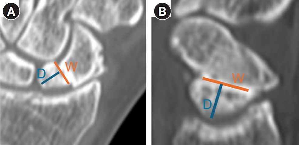
- Proximal scaphoid nonunion is difficult to treat because the proximal fragment is small and biologically compromised. Secure fixation can be especially challenging when dorsal intercalated segment instability (DISI) is present and requires correction. We report three male patients with proximal scaphoid nonunion and DISI deformity who underwent volar fixation with two 2.4-mm headless compression screws and cancellous bone graft harvested from the distal radius at the Lister tubercle. Preoperative computed tomography confirmed a proximal nonunion line and proximal fragment dimensions considered sufficient for dual-screw fixation, and magnetic resonance imaging showed no definite avascular necrosis. Early postoperative computed tomography showed maintained fixation and alignment in all three patients. Although CT-confirmed union at final follow-up was not uniformly available, radiographic union defined on plain radiographs was achieved in all cases without loss of alignment or carpal collapse. Carpal alignment improved in two patients, but DISI persisted in one. One patient underwent secondary removal of a preexisting loose body and one screw. This small case series suggests that dual-screw fixation with cancellous grafting may be technically feasible in selected proximal scaphoid nonunions with concomitant DISI. Level of evidence: V.
- 467 View
- 12 Download
Original Article
- Sex-specific bottlenecks and risk zones in the retrograde superior pubic ramus screw corridor: a 3D CT-based morphometric cadaver study
- Ji Won Jeong, Jung Tae Ahn, Gu Hee Jung, Kun Tae Kim
- J Musculoskelet Trauma 2026;39(2):103-116. Published online March 26, 2026
- DOI: https://doi.org/10.12671/jmt.2026.00066
-
Abstract
PDF
 Supplementary Material
 - Background
Superior ramus screw fixation is commonly used to stabilize anterior pelvic ring injuries but is constrained by a narrow, irregular, and curved intraosseous corridor. Trajectory-based morphometric analysis may assist in screw diameter selection and enable identification of reproducible anatomic constriction zones.
Methods
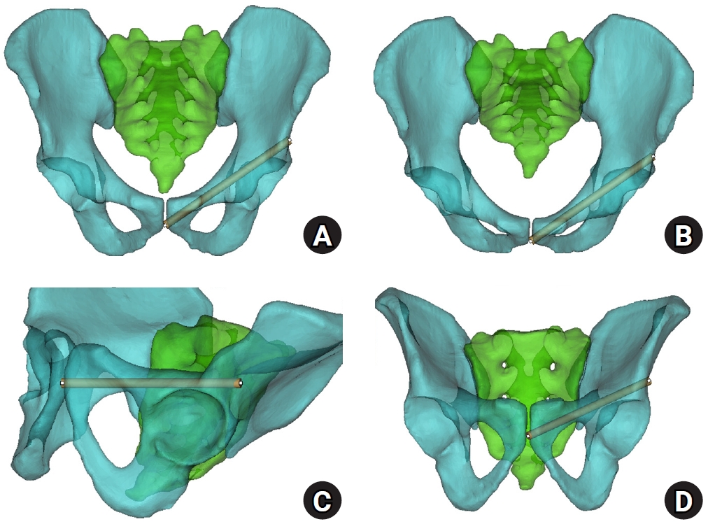
We conducted a cross-sectional computed tomography (CT)-based morphometric study of 82 cadaveric pelvises (42 males, 40 females). Bottleneck diameter was defined as the diameter of the largest fully contained virtual cylinder along the planned trajectory, and cylinder length was recorded. Orthogonal cross-sections at 9.5-mm intervals (up to 12 segments) were generated to measure segment-wise effective diameter (defined as twice the minimum centerline-to-cortex distance) and cortical clearance, which was used as a diameter-based safety margin. Segments were realigned to the acetabular start segment to define relative segment positions (Δ seg). Feasibility was assessed for prespecified screw diameters ranging from 3.5 to 7.3 mm.
Results
Mean bottleneck diameter was larger in males than in females (7.34±1.10 vs. 5.93±0.98 mm), whereas trajectory length was similar between sexes (127.85±8.54 vs. 128.85±8.20 mm). Δ seg realignment localized corridor constriction to two discrete zones: a preacetabular zone (Δ seg −6 to −4) and a periacetabular zone (Δ seg 1 to 2), where effective diameter and cortical clearance were most limited. Feasibility rates were 100% at 3.5–4.5 mm, 95.2% vs. 82.5% at 5.0 mm, 81.0% vs. 27.5% at 6.5 mm, and 59.5% vs. 10.0% at 7.3 mm in males and females, respectively.
Conclusions
Female models demonstrated smaller trajectory-wide bottleneck diameters and segment-wise effective diameters than male models. Acetabular-referenced Δ seg realignment identified two reproducible anatomic risk zones: a preacetabular zone adjacent to the obturator neurovascular bundle and a periacetabular zone near the external iliac vessels. At diameters ≥6.5 mm, cortical proximity increased more prominently in females than in males. Level of evidence: III.
- 1,615 View
- 35 Download
 First
First Prev
Prev


