 E-submission
 TOTA
TOTA TOTS
TOTS
Articles
- Page Path
- HOME > J Musculoskelet Trauma > Volume 23(1); 2010 > Article
-
Original Article
- Treatment of the Posterior Malleolar Fracture Using Posterior Approach
- Hyun Wook Chung, M.D., Dong Hwan Kim, M.D., Si Hoon Yoo, M.D., Jin Soo Suh, M.D.
-
Journal of the Korean Fracture Society 2010;23(1):50-56.
DOI: https://doi.org/10.12671/jkfs.2010.23.1.50
Published online: January 31, 2010
Department of Orthopedic Surgery, Inje University College of Medicine, Ilsan Paik Hospital, Goyang, Korea.
- Address reprint requests to: Jin Soo Suh, M.D. Department of Orthopedic Surgery, Inje University College of Medicine, Ilsan Paik Hospital, 2240, Daehwa-dong, Ilsan-seo-gu, Goyang 411-706, Korea. Tel: 82-31-910-7968, Fax: 82-31-910-7967, sjs0506@paik.ac.kr
• Received: June 7, 2009 • Revised: October 29, 2009 • Accepted: November 4, 2009
Copyright © 2010 The Korean Fracture Society
- 1,017 Views
- 3 Download
- 3 Crossref
Abstract
-
Purpose
- For fixation of the large posterior malleolar fracture fragment, indirect anterior fixation with cannulated screw has been widely used, but the anatomical reduction is not always obtained. The purpose of this article is to evaluate the clinical result of posterior malleolar fractures treated with anatomical reduction and internal fixation using posterior approach.
-
Materials and Methods
- We have analyzed the 15 patients with posterior malleolar fractures, treated with posterior approach from August 2005 to August 2008. The mean follow up period was 17.6 months, We have reviewed the perioperative joint integrity, method of operation, postoperative care, bony union and complication. A clinical outcome was evaluated by AOFAS (American orthopedic foot and ankle society) scaling system and Olerud & Molander scoring system.
-
Results
- Among 15 cases, posterolateral approach and posteromedial approach were chosen in 9 cases and 6 cases respectively. The radiologic unions were achieved at 12.4 (12~18) weeks. Mean AOFAS score was 90.3 (72~98), and Olerud & Molander score was "excellent" in 5 cases, "good" in 7 cases, "fair" in 1 case and "poor" in 2 cases. Postoperative complications in 2 cases revealed a posttraumatic arthritis and a scar band contracture respectively.
-
Conclusion
- In posterior malleolar fracture of ankle joint, the integrity of joint has closely affected clinical outcomes. We suggest that a posterior approach for posterior malleolar fracture with especially incarcerated fragments and comminuted fractures, can be a useful method for anatomical reduction and stable fixation, and satisfactory clinical results.
- 1. Bae SY, Shin DH. The role of posterior malleolar fragments in ankle pain after trimalleolar fractures. J Korean Soc Fract, 2003;16:59-66.Article
- 2. Fitzpatrick DC, Otto JK, McKinley TO, Marsh JL, Brown TD. Kinematic and contact stress analysis of posterior malleolus fracture of the ankle. J Orthop Trauma, 2004;18:271-278.
- 3. Hartford JM, Gorczyca JT, McNamara JL, Mayor MB. Tibiotalar contact area. Contribution of posterior malleolus and deltoid ligament. Clin Orthop Relat Res, 1995;320:182-187.
- 4. Huber M, Stutz P, Gerber C. Open reduction and internal fixation of the posterior malleolus with a posterior antiglide plate using a posterolateral approach: a preliminary report. Foot Ankle Surg, 1996;2:95-103.Article
- 5. Jaskulka RA, Ittner G, Schedl R. Fractures of the posterior tibial margin: their role in the prognosis of malleolar fractures. J Trauma, 1989;29:1565-1570.
- 6. Kang CN, Kim JO, Lee SB, Kang OY, Shin MS. Treatment of the posterior lip fracture of distal tibia using posteromedial approach. J Korean Soc Fract, 1995;8:594-599.Article
- 7. Kitaoka HB, Alexander IJ, Adelaar RS, Nunley JA, Myerson MS, Sanders M. Clinical rating systems for the ankle-hindfoot, midfoot, hallux, and lesser toes. Foot Ankle Int, 1994;15:349-353.ArticlePDF
- 8. Langenhuijsen JF, Heetveld MJ, Ultee JM, Steller EP, Butzelaar RM. Results of ankle fractures with involvement of the posterior tibial margin. J Trauma, 2002;53:55-60.Article
- 9. Lee CS, Suh JS, Yi JW. Comparative study for the results of ankle fracture depending on the extension of the posterior malleolus fracture. J Korean Orthop Assoc, 2007;42:470-474.Article
- 10. Lindsjö U. Operative treatment of ankle fractures. Acta Orthop Scand Suppl, 1981;189:1-131.
- 11. Lindsjö U. Operative treatment of ankle fracture-dislocations. A follow-up study of 306/321 consecutive cases. Clin Orthop Relat Res, 1985;199:28-38.
- 12. Macko VW, Matthews LS, Zwirkoski P, Goldstein SA. The Joint-contact area of the ankle. The contribution of the posterior malleolus. J Bone Joint Surg Am, 1991;73:347-351.
- 13. McDaniel WJ, Wilson FC. Trimalleolar fractures of the ankle. An end result study. Clin Orthop Relat Res, 1977;122:37-45.
- 14. Olerud C, Molander H. A scoring scale for symptom evaluation after ankle fracture. Arch Orthop Trauma Surg, 1984;103:190-194.PDF
- 15. Papachristou G, Efstathopoulos N, Levidiotis C, Chronopoulos E. Early weight bearing after posterior malleolar fractures: an experimental and prospective clinical study. J Foot Ankle Surg, 2003;42:99-104.
- 16. Talbot M, Steenblock TR, Cole PA. Posterolateral approach for open reduction and internal fixation of trimalleolar ankle fractures. Can J Surg, 2005;48:487-490.
- 17. Weber M. Trimallolar fractures with impaction of the posteromedial tibial plafond: implications for talar stability. Foot Ankle Int, 2004;25:716-727.PDF
REFERENCES
Fig. 1Posterolateral approach to the posterior malleolar fracture. Intraoperative finding showing fixation of posterior malleolus using plate and screws.


Fig. 3
(A) Preoperative CT image showing posterior malleolar fracture with displacement and incarcerated bony fragment.
(B) Postoperative lateral radiograph showing anatomical reduction of posterior malleolus.

Fig. 4
(A) Preoperative sagittal CT image showing posterior malleolar fracture with comminution.
(B) Postoperative lateral radiograph showing reduction and stabilization with plate and screws (C) 1 year follow-up sagittal CT image showing incongruity of joint.
(D) 2 year follow-up lateral radiograph with arthritic changes.


Figure & Data
REFERENCES
Citations
Citations to this article as recorded by 
- Single lateral approach for open reduction and internal fixation of posterior malleolar fragment in Weber B rotational ankle fracture
Jaehyung Lee, Hwan Ryu, Jae Yong Park
Medicine.2023; 102(3): e32725. CrossRef - Posterior Malleolus Fractures in Trimalleolar Ankle Fractures: Malleolus versus Transyndesmal Fixation
Bilgehan Tosun, Ozgur Selek, Umit Gok, Halil Ceylan
Indian Journal of Orthopaedics.2018; 52(3): 309. CrossRef - Single Oblique Posterolateral Approach for Open Reduction and Internal Fixation of Posterior Malleolar Fractures With an Associated Lateral Malleolar Fracture
Jun Young Choi, Ji Hoon Kim, Hyeong Tak Ko, Jin Soo Suh
The Journal of Foot and Ankle Surgery.2015; 54(4): 559. CrossRef
 Cite
CiteTreatment of the Posterior Malleolar Fracture Using Posterior Approach
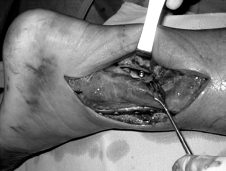


Fig. 1
Posterolateral approach to the posterior malleolar fracture. Intraoperative finding showing fixation of posterior malleolus using plate and screws.
Fig. 2
A schematic picture showing the posterolateral approach.
Fig. 3
(A) Preoperative CT image showing posterior malleolar fracture with displacement and incarcerated bony fragment.
(B) Postoperative lateral radiograph showing anatomical reduction of posterior malleolus.
Fig. 4
(A) Preoperative sagittal CT image showing posterior malleolar fracture with comminution.
(B) Postoperative lateral radiograph showing reduction and stabilization with plate and screws (C) 1 year follow-up sagittal CT image showing incongruity of joint.
(D) 2 year follow-up lateral radiograph with arthritic changes.
Fig. 1
Fig. 2
Fig. 3
Fig. 4
Treatment of the Posterior Malleolar Fracture Using Posterior Approach
Summary of cases
*PM: Posteromedial, †PL: Posterolateral.
Table 1
Summary of cases
*PM: Posteromedial, †PL: Posterolateral.

