 E-submission
 TOTA
TOTA TOTS
TOTS
Articles
- Page Path
- HOME > J Musculoskelet Trauma > Volume 26(2); 2013 > Article
-
Case Report
- Extensive Metallosis Caused by Plate and Screw Construct for Distal Fibular Fracture - A Case Report -
- Ki-Tae Park, M.D., Kwang-Bok Lee, M.D.
-
Journal of the Korean Fracture Society 2013;26(2):147-150.
DOI: https://doi.org/10.12671/jkfs.2013.26.2.147
Published online: April 22, 2013
Department of Orthopedic Surgery, Chonbuk National University Hospital, Chonbuk National University Medical School, Gwangju, Korea.
- Address reprint requests to: Kwang-Bok Lee, M.D. Department of Orthopedic Surgery, Chonbuk National University Hospital, Chonbuk National University Medical School, 20 Geonji-ro, Deokjin-gu, Jeonju 561-712, Korea. Tel: 82-63-250-2586, Fax: 82-63-271-6538, osdr2815@naver.com
• Received: June 4, 2012 • Revised: September 1, 2012 • Accepted: December 31, 2012
Copyright © 2013 The Korean Fracture Society
- 697 Views
- 3 Download
- 1 Crossref
Abstract
- Metallosis has been reported in the setting of weight-bearing joint arthroplasties, like the hip and knee joints. However, the prevalence of metallosis in non-articular portions is very uncommon. We report a rare case of a patient who had metallosis secondary by fibular nonunion after fixation with plate and screw. In addition, we discuss the clinical and the operative findings, as well as the outcome of this uncommon complication.
- 1. Berry DJ, Barnes CL, Scott RD, Cabanela ME, Poss R. Catastrophic failure of the polyethylene liner of uncemented acetabular components. J Bone Joint Surg Br, 1994;76:575-578.ArticlePDF
- 2. Bullough PG. Metallosis. J Bone Joint Surg Br, 1994;76:687-688.ArticlePDF
- 3. Case CP, Langkamer VG, James C, et al. Widespread dissemination of metal debris from implants. J Bone Joint Surg Br, 1994;76:701-712.ArticlePDF
- 4. Chang JD, Lee SS, Hur M, Seo EM, Chung YK, Lee CJ. Revision total hip arthroplasty in hip joints with metallosis: a single-center experience with 31 cases. J Arthroplasty, 2005;20:568-573.
- 5. Danzig LA, Woo SL, Akeson WH, Jemmott GF, Wickham MG. Internal fixation plates after fifty-six years of implantation: report of a case. Clin Orthop Relat Res, 1980;149:201-206.
- 6. De Smet L. Metallosis mimicking osteomyelitis from a forearm plate retained for 50 years. Acta Orthop Belg, 2000;66:289-291.
- 7. Huo MH, Romness DW, Huo SM. Metallosis mimicking infection in a cemented total knee replacement. Orthopedics, 1997;20:466-470.Article
- 8. Khan RJ, Wimhurst J, Foroughi S, Toms A. The natural history of metallosis from catastrophic failure of a polyethylene liner in a total hip. J Arthroplasty, 2009;24:1144.e1-1144.e4.
- 9. McGovern TF, Moskal JT. Radiographic evaluation of periprosthetic metallosis after total knee arthroplasty. J South Orthop Assoc, 2002;11:18-24.
- 10. Tan GM, Lynne G, Sarbjit S. Osteolysis and wear debris after total knee arthroplasty presenting with extra-articular metallosis in the calf. J Arthroplasty, 2008;23:775-780.
REFERENCES
Fig. 1Preoperative left tibia anteroposterior (A) lateral (B) images show the nonunion, osteolytic bone defect and hardware the failure on the fibular fracture site.

Fig. 2Operative photos (A-D) show the broken plate and extensive gray-black colored tissue around the plate, and clean bony tissue after the removal of the plate and gray-black colored tissue.

Fig. 3Postoperative left tibia anteroposterior (A) lateral (B) images show the internal fixation with rush pin and autogenous bone graft for bone defect and nonunion site.
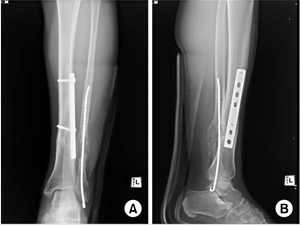

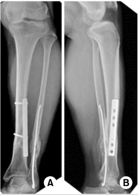
Figure & Data
REFERENCES
Citations
Citations to this article as recorded by 
- Plate on Plate Osteosynthesis for the Treatment of Nonhealed Periplate Fractures
Georgios Arealis, Vassilios S. Nikolaou, Andrew Lacon, Neil Ashwood, Mark Hamlet
ISRN Orthopedics.2014; 2014: 1. CrossRef
 Cite
CiteExtensive Metallosis Caused by Plate and Screw Construct for Distal Fibular Fracture - A Case Report -
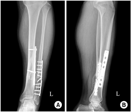
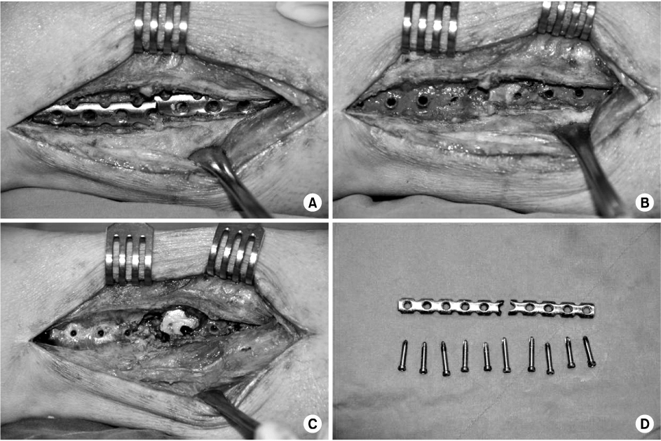

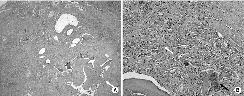

Fig. 1
Preoperative left tibia anteroposterior (A) lateral (B) images show the nonunion, osteolytic bone defect and hardware the failure on the fibular fracture site.
Fig. 2
Operative photos (A-D) show the broken plate and extensive gray-black colored tissue around the plate, and clean bony tissue after the removal of the plate and gray-black colored tissue.
Fig. 3
Postoperative left tibia anteroposterior (A) lateral (B) images show the internal fixation with rush pin and autogenous bone graft for bone defect and nonunion site.
Fig. 4
Histologic photos demonstrate the fibrous stroma admixed with bone and necrotic debris (white arrow) (A: H&E, ×40) and inflammatory cells (white arrow) and foreign-body giant cell (black arrow) (nuclei arranged haphazardly) in the fibrous stroma (B: H&E, ×200).
Fig. 5
Final follow-up left tibia anteroposterior (A) lateral (B) images show bony consolidation of fibular nonunion and bony defect area.
Fig. 1
Fig. 2
Fig. 3
Fig. 4
Fig. 5
Extensive Metallosis Caused by Plate and Screw Construct for Distal Fibular Fracture - A Case Report -

