 E-submission
 TOTA
TOTA TOTS
TOTS
Articles
- Page Path
- HOME > J Musculoskelet Trauma > Volume 27(1); 2014 > Article
-
Original Article
- Fixation of the Greater Trochanter in Arthroplasty for Unstable Intertrochnateric Fracture
- Dong-Hyeok Choi, M.D., Ju-Yeong Heo, M.D., Young-Jae Jang, M.D., Young-Yool Chung, M.D.
-
Journal of the Korean Fracture Society 2014;27(1):58-64.
DOI: https://doi.org/10.12671/jkfs.2014.27.1.58
Published online: January 17, 2014
Department of Orthopaedic Surgery, Kwangju Christian Hospital, Gwangju, Korea.
- Address reprint requests to: Young-Yool Chung, M.D. Department of Orthopaedic Surgery, Kwangju Christian Hospital, 37 Yangrim-ro, Nam-gu, Gwangju 503-715, Korea. Tel: 82-62-650-5064, Fax: 82-62-650-5066, paedic@chol.com
• Received: September 28, 2013 • Revised: November 4, 2013 • Accepted: December 18, 2013
Copyright © 2014 The Korean Fracture Society. All rights reserved.
This is an Open Access article distributed under the terms of the Creative Commons Attribution Non-Commercial License (http://creativecommons.org/licenses/by-nc/3.0/) which permits unrestricted non-commercial use, distribution, and reproduction in any medium, provided the original work is properly cited.
- 1,685 Views
- 38 Download
- 4 Crossref
Abstract
-
Purpose
- We classified fractures of the greater trochanter (GT) and evaluated fracture fragment stability according to GT type.
-
Materials and Methods
- A total of 43 patients with an unstable intertrochanteric fracture treated between January 2007 and July 2009 with bipolar hemiarthroplasty were included in this study. GT fractures were classified as type A, B, C, or D and fixed using either cerclage wiring alone, cerclage wiring and non-absorbable suture or a greater trochanteric reattachment (GTR) plate.
-
Results
- Type A fractures were fixed using cerclage wiring with non-absorbable suture in two cases, cerclage wiring in six cases and GTR plate in seven cases. Failure occurred in three cases of type A fractures treated with cerclage wiring alone. A total of 11 type B fractures were fixed with cerclage wiring (7), cerclage wiring and non-absorbable suture (3) and GTR plate (1). There was no failure of type B fractures. Type C fractures were fixed using cerclage wiring with non-absorbable suture in one case and GTR plate in three. There was no fixation in three cases. Of 10 type D fractures, six were treated with cerclage wiring and one with GTR plate. Fixation was not performed in three patients. There was no failure in type C and D type fractures.
-
Conclusion
- Fracture fragment stability differed according to fracture types. Cerclage wiring alone was insufficient to fix type A fractures, so type A fracture required a stronger fixation method.
- 1. Anglen JO, Weinstein JN. American Board of Orthopaedic Surgery Research Committee. Nail or plate fixation of intertrochanteric hip fractures: changing pattern of practice. A review of the American Board of Orthopaedic Surgery Database. J Bone Joint Surg Am, 2008;90:700-707.
- 2. Aros B, Tosteson AN, Gottlieb DJ, Koval KJ. Is a sliding hip screw or im nail the preferred implant for intertrochanteric fracture fixation? Clin Orthop Relat Res, 2008;466:2827-2832.Article
- 3. Audigé L, Hanson B, Swiontkowski MF. Implant-related complications in the treatment of unstable intertrochanteric fractures: meta-analysis of dynamic screw-plate versus dynamic screw-intramedullary nail devices. Int Orthop, 2003;27:197-203.ArticlePDF
- 4. Barrack RL, Butler RA. Current status of trochanteric reattachment in complex total hip arthroplasty. Clin Orthop Relat Res, 2005;441:237-242.Article
- 5. Broos PL, Rommens PM, Geens VR, Stappaerts KH. Pertrochanteric fractures in the elderly. Is the Belgian VDP prosthesis the best treatment for unstable fractures with severe comminution? Acta Chir Belg, 1991;91:242-249.
- 6. Geiger F, Zimmermann-Stenzel M, Heisel C, Lehner B, Daecke W. Trochanteric fractures in the elderly: the influence of primary hip arthroplasty on 1-year mortality. Arch Orthop Trauma Surg, 2007;127:959-966.ArticlePDF
- 7. Hamadouche M, Zniber B, Dumaine V, Kerboull M, Courpied JP. Reattachment of the ununited greater trochanter following total hip arthroplasty. The use of a trochanteric claw plate. J Bone Joint Surg Am, 2003;85:1330-1337.
- 8. Hamadouche M, Zniber B, Dumaine V, Kerboull M, Courpied JP. Reattachment of the ununited greater trochanter following total hip arthroplasty. J Bone Joint Surg Am, 2004;86-A:Suppl 1. 112-118.Article
- 9. Hardy DC, Descamps PY, Krallis P, et al. Use of an intramedullary hip-screw compared with a compression hip-screw with a plate for intertrochanteric femoral fractures. A prospective, randomized study of one hundred patients. J Bone Joint Surg Am, 1998;80:618-630.Article
- 10. Harwin SF, Stern RE, Kulick R. Primary Bateman-Leinbach bipolar prosthetic replacement of the hip in the treatment of unstable intertrochanteric fractures in the elderly. Orthopedics, 1990;13:1131-1136.Article
- 11. Hendel D, Yasin M, Garti A, Weisbort M, Beloosesky Y. Fracture of the greater trochanter during hip replacement: a retrospective analysis of 21/372 cases. Acta Orthop Scand, 2002;73:295-297.
- 12. Hersh CK, Williams RP, Trick LW, Lanctot D, Athanasiou K. Comparison of the mechanical performance of trochanteric fixation devices. Clin Orthop Relat Res, 1996;(329):317-325.Article
- 13. Kim JH, Park JH, Kim HS, et al. Methods to increase the effectiveness of trochanteric stabilizing plate for unstable femoral intertrochanteric fractures with gtreater trochanteric fracture: fixation of greater trochanter with wire and screw. J Korean Hip Soc, 2007;19:58-63.Article
- 14. Kim SY, Kim YG, Hwang JK. Cementless calcar-replacement hemiarthroplasty compared with intramedullary fixation of unstable intertrochanteric fractures. A prospective, randomized study. J Bone Joint Surg Am, 2005;87:2186-2192.Article
- 15. Kim WY, Han CH, Park JI, Kim JY. Failure of intertrochanteric fracture fixation with a dynamic hip screw in relation to pre-operative fracture stability and osteoporosis. Int Orthop, 2001;25:360-362.ArticlePDF
- 16. Koyama K, Higuchi F, Kubo M, Okawa T, Inoue A. Reattachment of the greater trochanter using the Dall-Miles cable grip system in revision hip arthroplasty. J Orthop Sci, 2001;6:22-27.Article
- 17. Kyle RF, Gustilo RB, Premer RF. Analysis of six hundred and twenty-two intertrochanteric hip fractures. J Bone Joint Surg Am, 1979;61:216-221.Article
- 18. Little NJ, Verma V, Fernando C, Elliott DS, Khaleel A. A prospective trial comparing the Holland nail with the dynamic hip screw in the treatment of intertrochanteric fractures of the hip. J Bone Joint Surg Br, 2008;90:1073-1078.ArticlePDF
- 19. Mariani EM, Rand JA. Nonunion of intertrochanteric fractures of the femur following open reduction and internal fixation. Results of second attempts to gain union. Clin Orthop Relat Res, 1987;(218):81-89.
- 20. McCarthy JC, Bono JV, Turner RH, Kremchek T, Lee J. The outcome of trochanteric reattachment in revision total hip arthroplasty with a Cable Grip System: mean 6-year follow-up. J Arthroplasty, 1999;14:810-814.Article
- 21. Moran CG, Wenn RT, Sikand M, Taylor AM. Early mortality after hip fracture: is delay before surgery important? J Bone Joint Surg Am, 2005;87:483-489.
- 22. Nam HJ, Sun DH, Jang SW. Fixation of greater trochanteric fracture using double strands and double loops with figure of 8 wiring in non-cement total hip arthroplasty for unstable intertrochanteric fracture. Hip Pelvis, 2012;24:316-321.Article
- 23. Pervez H, Parker MJ, Vowler S. Prediction of fixation failure after sliding hip screw fixation. Injury, 2004;35:994-998.Article
- 24. Pritchett JW. Fracture of the greater trochanter after hip replacement. Clin Orthop Relat Res, 2001;(390):221-226.Article
- 25. Ritter MA, Eizember LE, Keating EM, Faris PM. Trochanteric fixation by cable grip in hip replacement. J Bone Joint Surg Br, 1991;73:580-581.ArticlePDF
- 26. Rothman RH, Cohn JC. Cemented versus cementless total hip arthroplasty. A critical review. Clin Orthop Relat Res, 1990;(254):153-169.
- 27. Saudan M, Lübbeke A, Sadowski C, Riand N, Stern R, Hoffmeyer P. Pertrochanteric fractures: is there an advantage to an intramedullary nail?: a randomized, prospective study of 206 patients comparing the dynamic hip screw and proximal femoral nail. J Orthop Trauma, 2002;16:386-393.
- 28. Schwab JH, Camacho J, Kaufman K, Chen Q, Berry DJ, Trousdale RT. Optimal fixation for the extended trochanteric osteotomy: a pilot study comparing 3 cables vs 2 cables. J Arthroplasty, 2008;23:534-538.
- 29. Suh YS, Choi SW, Park JS, Yim SJ, Shin BJ. Comparison between the methods for fixation of greater trochanteric fragment in cemented bipolar hemiarthroplasty for unstable intertrochanteric fracture. J Korean Hip Soc, 2008;20:104-109.Article
- 30. Thakur NA, Crisco JJ, Moore DC, Froehlich JA, Limbird RS, Bliss JM. An improved method for cable grip fixation of the greater trochanter after trochanteric slide osteotomy. J Arthroplasty, 2010;25:319-324.Article
REFERENCES
Fig. 1Classification of greater trochanteric fracture. (A) Type A is a transverse fracture above the inferior border of greater trochanter (GT). The fracture fragment was displaced superiorly by the pulling force of the hip abductors. (B) Type B is a vertical fracture that is not extended to the inferior border of GT. Type B was similar in stability of fracture fragment to type A. (C) Type C is the transverse fracture below the inferior border of GT. Type B fracture was more stable than type A because of the balanced pulling forces of abductors proximally and vastus lateralis distally. (D) Type D is a vertical fracture that extended to below to inferior border of GT. Type D was similar in stability of fracture fragment to type C. The muscles were attached to the fracture fragment proximally and distally, which stabilized the fracture.

Fig. 2
(A) Preoperative radiograph of type A greater trochater (GT) fracture in a 78-year-old male. (B) Postoperative radiograph of a type A GT fracture fixed with a greater trochanteric reattachment plate.

Fig. 3
(A) Preoperative radiograph of a type D greater trochater (GT) fracture in an 85-year-old woman. (B) The displaced GT fragment was reduced and fixed with cerclage wiring and non-absorbable suture. (C) Postoperative radiograph after bipolar hip arthroplasty and fixation of GT fracture with cerclage wiring and non-absorbable suture.
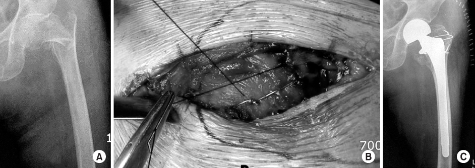
Figure & Data
REFERENCES
Citations
Citations to this article as recorded by 
- Primary Arthroplasty for Unstable and Failed Intertrochanteric Fractures: Role of Multi-Planar Trochanteric Wiring Technique
Javahir A. Pachore, Vikram Indrajit Shah, Sachin Upadhyay, Shrikunj Babulal Patel
Hip & Pelvis.2023; 35(2): 108. CrossRef - The Efficacy of Suture Fixation of the Greater Trochanter in Unstable Intertrochanteric Fractures
Ki-Choul Kim, Hee-Gon Park, Jae-Wook Park
Clinics in Orthopedic Surgery.2021; 13(4): 468. CrossRef - Is rigid fixation of the greater trochanter necessary for arthroplasty of intertrochanteric fractures?
Kee Haeng Lee, Dong Hun Lee, Jong Ho Noh, Yoon Vin Kim
Orthopaedics & Traumatology: Surgery & Research.2019; 105(1): 41. CrossRef - Selecting Arthroplasty Fixation Approach Based on Greater Trochanter Fracture Type in Unstable Intertrochanteric Fractures
Min-Wook Kim, Young-Yool Chung, Sung-an Lim, Seung-Woo Shim
Hip & Pelvis.2019; 31(3): 144. CrossRef
 Cite
CiteFixation of the Greater Trochanter in Arthroplasty for Unstable Intertrochnateric Fracture
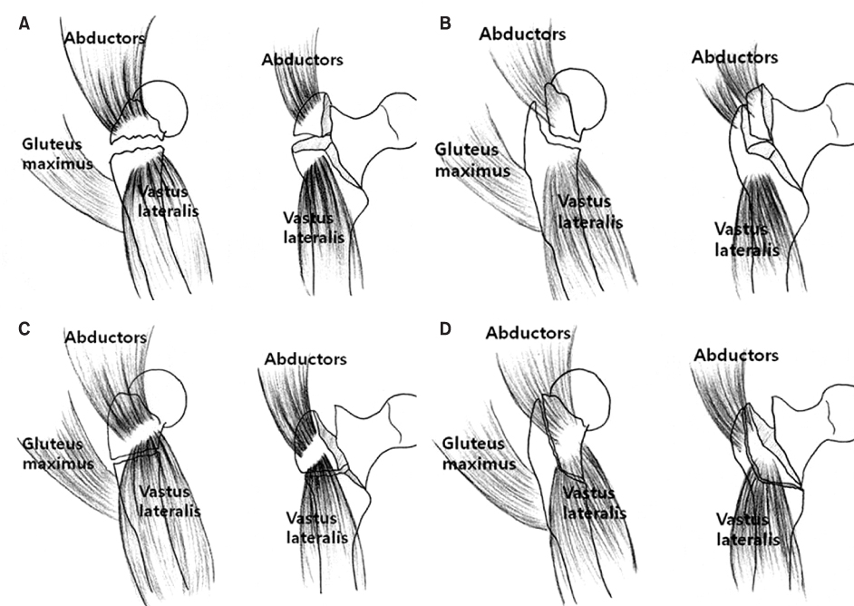
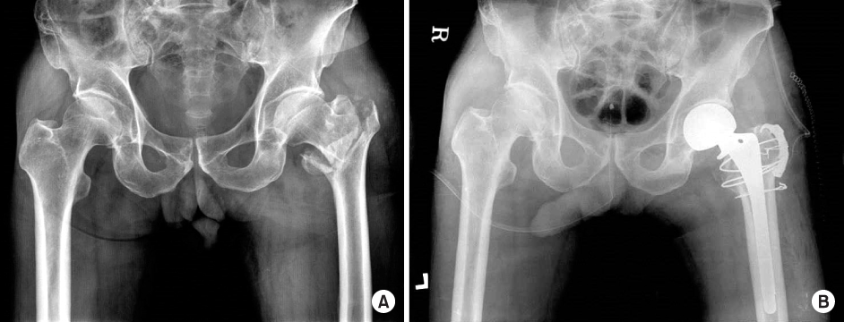

Fig. 1
Classification of greater trochanteric fracture. (A) Type A is a transverse fracture above the inferior border of greater trochanter (GT). The fracture fragment was displaced superiorly by the pulling force of the hip abductors. (B) Type B is a vertical fracture that is not extended to the inferior border of GT. Type B was similar in stability of fracture fragment to type A. (C) Type C is the transverse fracture below the inferior border of GT. Type B fracture was more stable than type A because of the balanced pulling forces of abductors proximally and vastus lateralis distally. (D) Type D is a vertical fracture that extended to below to inferior border of GT. Type D was similar in stability of fracture fragment to type C. The muscles were attached to the fracture fragment proximally and distally, which stabilized the fracture.
Fig. 2
(A) Preoperative radiograph of type A greater trochater (GT) fracture in a 78-year-old male. (B) Postoperative radiograph of a type A GT fracture fixed with a greater trochanteric reattachment plate.
Fig. 3
(A) Preoperative radiograph of a type D greater trochater (GT) fracture in an 85-year-old woman. (B) The displaced GT fragment was reduced and fixed with cerclage wiring and non-absorbable suture. (C) Postoperative radiograph after bipolar hip arthroplasty and fixation of GT fracture with cerclage wiring and non-absorbable suture.
Fig. 1
Fig. 2
Fig. 3
Fixation of the Greater Trochanter in Arthroplasty for Unstable Intertrochnateric Fracture

