 E-submission
 TOTA
TOTA TOTS
TOTS
Articles
- Page Path
- HOME > J Musculoskelet Trauma > Volume 28(1); 2015 > Article
-
Case Report
- Infected Nonunion of Clavicle Shaft after Operation: A Case Report
- Ho-Su Jang, M.D., Suk-Hwan Jang, M.D.
-
Journal of the Korean Fracture Society 2015;28(1):77-81.
DOI: https://doi.org/10.12671/jkfs.2015.28.1.77
Published online: January 20, 2015
Department of Orthopedic Surgery, Seoul Paik Hospital, Inje University College of Medicine, Seoul, Korea.
- Address reprint requests to: Suk-Hwan Jang, M.D. Department of Orthopedic Surgery, Seoul Paik Hospital, Inje University College of Medicine, 9 Mareunnae-ro, Jung-gu, Seoul 100-032, Korea. Tel: 82-2-2270-0028, Fax: 82-2-2270-0023, orthopodjang@gmail.com
• Received: August 16, 2014 • Revised: October 7, 2014 • Accepted: December 18, 2014
Copyright © 2015 The Korean Fracture Society. All rights reserved.
This is an Open Access article distributed under the terms of the Creative Commons Attribution Non-Commercial License (http://creativecommons.org/licenses/by-nc/3.0/) which permits unrestricted non-commercial use, distribution, and reproduction in any medium, provided the original work is properly cited.
- 810 Views
- 7 Download
Abstract
- The infected nonunion of clavicle with bone defect is an uncommon complication following clavicle shaft fracture. There were a few reports regarding treatment of the infected nonunion after clavicle fracture. We report on a case of infected clavicle nonunion successfully treated with autologous bone graft and dual plate fixation.
- 1. Cho CH, Jang HG. Intercalary tricortical iliac bone graft in the surgical treatment of nonunion of midshaft clavicular fractures. Clin Should Elbow, 2012;15:32-36.ArticlePDF
- 2. Lazarides S, Zafiropoulos G. Conservative treatment of fractures at the middle third of the clavicle: the relevance of shortening and clinical outcome. J Shoulder Elbow Surg, 2006;15:191-194.Article
- 3. Fuchs B, Steinmann SP, Bishop AT. Free vascularized corticoperiosteal bone graft for the treatment of persistent nonunion of the clavicle. J Shoulder Elbow Surg, 2005;14:264-268.Article
- 4. Min HJ, Kim KW, Cho KH, et al. Infected nonunion of long bones treated with dual plate. J Korean Orthop Assoc, 1997;32:1701-1709.ArticlePDF
- 5. Duncan SF, Sperling JW, Steinmann S. Infection after clavicle fractures. Clin Orthop Relat Res, 2005;439:74-78.Article
- 6. Huang HK, Chiang CC, Su YP, et al. Role of autologous bone graft in the surgical treatment of atrophic nonunion of midshaft clavicular fractures. Orthopedics, 2012;35:e197-e201.Article
- 7. Ko SH, Cho SD, Park MS, et al. Internal fixation with plate and bone graft of mid-shaft clavicle nonunion. J Korean Shoulder Elbow Soc, 2005;8:19-22.Article
- 8. van der Meijden OA, Gaskill TR, Millett PJ. Treatment of clavicle fractures: current concepts review. J Shoulder Elbow Surg, 2012;21:423-429.Article
- 9. Kang HJ, Yoon HS, Hahn SB, Kim SJ. Operative Treatment of distal clavicle fracture nonunion. J Korean Shoulder Elbow Soc, 2007;10:220-226.Article
- 10. Wu CC. Single-stage surgical treatment of infected nonunion of the distal tibia. J Orthop Trauma, 2011;25:156-161.Article
REFERENCES
Fig. 1
(A) Preoperative radiograph taken 4 months after the initial operation shows nonunion of the right clavicle shaft with intervening sequestered bone fragment. (B) Gross photograph of the right shoulder showing the draining sinus with suppurative discharge over the clavicle shaft.


Figure & Data
REFERENCES
Citations
Citations to this article as recorded by 
 Cite
CiteInfected Nonunion of Clavicle Shaft after Operation: A Case Report
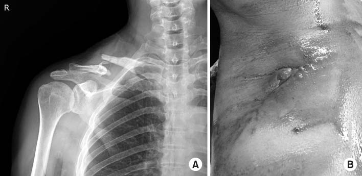

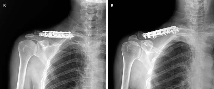
Fig. 1
(A) Preoperative radiograph taken 4 months after the initial operation shows nonunion of the right clavicle shaft with intervening sequestered bone fragment. (B) Gross photograph of the right shoulder showing the draining sinus with suppurative discharge over the clavicle shaft.
Fig. 2
(A) Postoperative radiograph after the initial operation at our hospital showing inserted antibiotic-impregnated cement beads with bone defect (B) Reconstructed images using 3-dimensional computed tomography of both clavicles were used to determine the difference between clavicle lengths.
Fig. 3
Radiograph at 2 years after final surgery shows complete bony union.
Fig. 1
Fig. 2
Fig. 3
Infected Nonunion of Clavicle Shaft after Operation: A Case Report

