
 E-submission
 KOTA
KOTA TOTA
TOTA TOTS
TOTS
Articles
- Page Path
- HOME > J Musculoskelet Trauma > Volume 39(3); 2026 > Article
-
Original Article
- Postoperative sagittal alignment and reoperation predict late outcomes after plate fixation of AO/OTA 33C distal femur fractures: a retrospective cohort study
- Hee Gon Park
-
Journal of Musculoskeletal Trauma 2026;39(3):216-226.
DOI: https://doi.org/10.12671/jmt.2026.00073
Published online: July 24, 2026
Department of Orthopedic Surgery, Dankook University College of Medicine, Cheonan, Korea
- Correspondence to: Hee Gon Park Department of Orthopedic Surgery, Dankook University College of Medicine, 119 Dandae-ro, Dongnam-gu, Cheonan 31116, Korea Tel: +82-41-550-6290 Email: osdku@dankook.ac.kr
• Received: January 27, 2026 • Revised: May 4, 2026 • Accepted: May 11, 2026
© 2026 The Korean Orthopaedic Trauma Association
This is an Open Access article distributed under the terms of the Creative Commons Attribution Non-Commercial License (http://creativecommons.org/licenses/by-nc/4.0/) which permits unrestricted noncommercial use, distribution, and reproduction in any medium, provided the original work is properly cited.
- 862 Views
- 24 Download


Abstract
-
Background Intra-articular distal femoral fractures are associated with high complication rates and variable late outcomes. Although the AO Foundation/Orthopaedic Trauma Association (AO/OTA) classification system is useful for describing injury severity and anticipating complications, whether fracture subtype alone determines clinically relevant late outcomes remains unclear. This study aimed to evaluate postoperative complications and late outcomes following plate fixation of intra-articular distal femoral fractures and to identify factors independently associated with final clinical outcomes.
-
Methods This retrospective cohort study was conducted at a regional Level I trauma center. Patients with AO/OTA 33C intra-articular distal femoral fractures who underwent plate fixation and were followed for at least 12 months were included. Radiographic parameters reflecting coronal and sagittal alignment were assessed using standardized measurement methods. Postoperative complications and reoperation were recorded as intermediate events. Clinically relevant late outcomes included final knee range of motion (ROM), leg length discrepancy, and radiographic osteoarthritis severity, assessed as the difference in Kellgren-Lawrence grade. Multivariable linear regression analyses were performed to identify factors independently associated with late outcomes.
-
Results The 33C3 group (n=33) had significantly higher rates of open fracture (54.5%), nonunion (39.4%), and reoperation (45.5%) than the 33C1–2 group (n=49). At the final follow-up, 33C3 fractures were associated with lower mean ROM (P<0.001) and greater osteoarthritis progression (P<0.001). However, multivariable analysis showed that sagittal malalignment (Δ anatomical posterior distal femoral angle: β=−2.35, P=0.001) and reoperation (β=−17.5, P=0.001), rather than AO/OTA subtype itself, were independent predictors of final ROM.
-
Conclusions Although fracture subtype according to the AO/OTA classification was associated with predictable complication patterns, clinically relevant late outcomes following intra-articular distal femoral fractures were more closely associated with postoperative alignment quality and the treatment course than with fracture classification alone. These findings highlight the importance of optimizing radiographic alignment and preventing complications that lead to reoperation to improve long-term outcomes after plate fixation.
-
Level of evidence III.
Introduction
Intra-articular distal femur fractures are complex injuries associated with substantial morbidity and functional impairment. Although relatively uncommon, these fractures present significant treatment challenges due to articular surface involvement, metaphyseal comminution, and the frequent coexistence of patient- and injury-related risk factors. Despite advances in fixation techniques, postoperative complications and inferior long-term outcomes remain common [1-3].
The AO Foundation/Orthopaedic Trauma Association (AO/OTA) classification system is widely used to describe fracture morphology and severity in distal femur fractures. Intra-articular fractures classified as AO/OTA type 33C encompass a broad spectrum of injury patterns, ranging from relatively simple articular fractures (C1 and C2) to highly complex multifragmentary fractures (C3) [4]. Previous studies have demonstrated that more complex fracture patterns are associated with higher rates of nonunion, fixation failure, and reoperation [5-7]. However, it remains unclear whether fracture subtype alone determines clinically relevant late outcomes.
In clinical practice, patients with intra-articular distal femur fractures frequently experience late problems such as limitation of motion (LOM), leg length discrepancy (LLD), and progressive post-traumatic osteoarthritis, even in the absence of overt fixation failure [8-10]. These observations raise an important question: are clinically relevant late outcomes primarily dictated by fracture classification, or are they more closely associated with modifiable factors such as postoperative alignment, reduction quality, and subsequent treatment course?
Therefore, this study aimed to evaluate postoperative complications and clinically relevant late outcomes following plate fixation of intra-articular distal femur fractures, investigating their associations with fracture subtype, radiographic parameters, and postoperative events. We hypothesized that while fracture subtype would be associated with predictable complication patterns, final clinical outcomes would be more closely related to radiographic alignment and the occurrence of reoperation than to fracture classification alone.
Methods
This study was approved by the Institutional Review Board (IRB) of Dankook University Hospital (IRB No. DKUH 2025-12-003). The requirement for informed consent was waived by the IRB due to its retrospective design.
This was a retrospective cohort study conducted in accordance with the Strengthening the Reporting of Observational Studies in Epidemiology (STROBE) guidelines for observational studies.
This retrospective cohort study was conducted at the Department of Orthopedic Surgery of Dankook University Hospital, a regional Level I trauma center in Cheonan, Korea. Consecutive adult patients who underwent plate fixation for AO/OTA 33C intra-articular distal femur fractures between 2004 and 2024 were identified. Outcomes were assessed using postoperative and follow-up clinical and radiographic data, with a minimum follow-up of 12 months.
We identified all consecutive patients who underwent operative treatment for intra-articular distal femur fractures at our regional Level I trauma center between 2004 and 2024. By including every patient who met the predefined inclusion criteria during this 20-year period, we aimed to minimize selection bias and ensure that the cohort reflects the true clinical spectrum of AO/OTA 33C fractures treated at our institution. Eligible patients were adults with unilateral AO/OTA type 33C intra-articular distal femur fractures treated with plate fixation, including cases in which accessory screws were used to facilitate articular reduction. Patients with a minimum follow-up of 12 months were included. Patients with pathologic, periprosthetic, or stress fractures, those requiring additional soft-tissue reconstructive procedures, and those treated without plate fixation were excluded. Given the institutional role as a Level I trauma center, patients with polytrauma were not excluded. A total of 242 patients were screened, of whom 174 had AO/OTA type 33C fractures. After excluding patients who did not meet the eligibility criteria (n=55), those treated without plate fixation (n=26), and those with a follow-up of less than 12 months (n=11), 82 patients were included in the final analysis (Fig. 1).
Study outcomes included postoperative complications and clinically relevant late outcomes. Postoperative complications were nonunion, deep infection, and implant failure, and reoperation was defined as any subsequent surgical intervention performed due to these complications. Late outcomes assessed at the final follow-up were final knee range of motion (ROM, degrees), LOM (ROM <90°), LLD (measured on standing scanograms), and radiographic osteoarthritis severity, quantified as the difference in Kellgren-Lawrence grade between the injured and contralateral knee (ΔK-L) at final follow-up. Conversion to total knee replacement arthroplasty (TKRA) was also recorded as a late clinical endpoint.
Data sources were obtained retrospectively from institutional electronic medical records and the radiology archive at Dankook University Hospital. Baseline demographics, injury characteristics, operative details, postoperative complications, reoperations, and clinical follow-up data (including knee ROM) were abstracted from medical charts.
Fractures were classified by two or more orthopedic surgeons using preoperative radiographs and computed tomography (CT) scans according to the AO/OTA classification system. Representative preoperative and postoperative radiographs of each AO/OTA 33C subtype are shown in Fig. 2. Injury mechanisms were classified into high-energy and low-energy injuries. High-energy injuries were defined as motor vehicle accidents (including car, motorcycle, and pedestrian accidents) and falls from a height of >3 m. Conversely, low-energy injuries were defined as falls from a standing height (slip down). In our cohort, slip downs were the only mechanism classified as low-energy injuries.
Radiographic assessment
Radiographic parameters were assessed using immediate postoperative and follow-up radiographs. To ensure the reliability of radiologic assessments, inter-observer reliability was evaluated. A subset of cases was randomly selected, and fracture classification and radiographic parameters—including anatomical lateral distal femoral angle (aLDFA), anatomical posterior distal femoral angle (aPDFA), hip-knee-ankle (HKA) angle, mechanical lateral distal femoral angle (mLDFA), LLD, and Kellgren-Lawrence (K-L) grades—were independently measured by two board-certified orthopedic surgeons who were blinded to the clinical outcomes. Reliability was assessed using the intraclass correlation coefficient (ICC) for continuous variables. The ICC was 0.71 (95% confidence interval [CI], 0.65–0.78), indicating substantial inter-observer agreement. Measurement methods are illustrated in Fig. 3. On anteroposterior knee radiographs of both limbs, the aLDFA was measured as the lateral angle between the anatomical axis of the femur and the distal femoral joint line (Fig. 3A). In the presence of fracture-related angulation, the femoral anatomical axis was determined using the proximal shaft segment (Fig. 3B). On standing full-length anteroposterior radiographs (whole lower extremity scanograms), overall lower-limb alignment was evaluated by measuring the HKA angle. The mLDFA and LLD were also measured according to standard definitions (Fig. 3C) [11-13].
Outcome measures
Postoperative articular reduction quality was evaluated on immediate postoperative radiographs and CT scans according to established criteria for complex intra-articular fractures. Reduction was classified as anatomical (articular step-off or gap <2 mm), good (step-off 2–5 mm), or poor (step-off >5 mm). This classification system is widely accepted for highly comminuted AO/OTA 33C3 fractures, where a 2-mm threshold represents a clinically acceptable limit for joint congruity. Postoperative complications included nonunion, infection, and implant failure. Reoperation was defined as any subsequent surgical intervention performed due to postoperative complications. Clinically relevant late outcomes included final knee ROM, LOM (ROM<90°), LLD, and conversion to TKRA. To account for individual anatomical variations, radiographic parameters were assessed by comparing the injured limb to the uninjured contralateral limb, which served as the patient-specific anatomical reference. Rather than relying on population-based normal ranges, we focused on the restoration of symmetry. Coronal alignment was expressed as ΔaLDFA, defined as the absolute difference between the affected and contralateral sides. Similarly, sagittal plane alignment was evaluated using the difference in aPDFA. A deviation of greater than 5° from the contralateral side was defined as malalignment, a threshold previously identified as clinically significant for joint loading and functional outcomes. Radiographic osteoarthritis severity was assessed using the K-L grading system. To ensure an accurate evaluation of joint space narrowing and degenerative changes, all follow-up radiographs, including standing anteroposterior views and full-length scanograms, were obtained in a weight-bearing position. The progression of osteoarthritis was defined as the ΔK-L between the injured knee and the uninjured contralateral knee at the final follow-up. Using the contralateral limb as a patient-specific control allowed us to account for preexisting degenerative changes. Conversion to TKRA was performed in patients who met both radiographic and clinical criteria for end-stage post-traumatic osteoarthritis. Radiographic indication was defined as progression to K-L grade 3 or higher. Clinical indications included refractory pain with a visual analog scale score of ≥7 despite more than six months of conservative management and significant functional impairment hindering activities of daily living.
To minimize selection bias, we included all consecutive eligible patients treated during the study period using predefined inclusion/exclusion criteria. To reduce measurement bias, radiographic outcomes were assessed using standardized measurement protocols; key imaging variables were independently measured by two board-certified orthopedic surgeons blinded to clinical outcomes, and inter-observer reliability was quantified using the ICC. Potential confounding was addressed by multivariable regression including clinically relevant covariates, although residual confounding inherent to retrospective studies may remain.
The sample size was justified by an observations-per-variable ratio of 20.5:1, which is adequate for multivariate regression analysis. Additionally, a post-hoc power analysis confirmed a statistical power of >0.80.
Continuous variables are presented as mean±standard deviation, and categorical variables as number (%). Comparisons between groups were performed using Student t-test or Mann-Whitney U test for continuous variables and chi-square or Fisher exact test for categorical variables, as appropriate. Multivariable linear regression analyses were conducted to identify factors associated with clinically relevant late outcomes, including final ROM, LLD, and ΔK-L. Fracture subtype, radiographic alignment parameters, and postoperative events were included as explanatory variables based on clinical relevance. To control for potential confounding factors, all multivariable models were adjusted for baseline clinical covariates, including injury mechanism (high- vs. low-energy), open fracture status, and treatment era. Missing data were addressed using complete-case analysis, and standard regression diagnostics were performed using IBM SPSS ver. 20.0 (IBM Corp.). A P<0.05 was considered statistically significant.
Results
A total of 82 patients were included (23 with type 33C1, 26 with 33C2, and 33 with 33C3). Baseline demographic and injury-related characteristics according to AO/OTA 33C subtype are summarized in Table 1. Compared to the 33C1–2 group, patients with 33C3 fractures significantly more often sustained high-energy injury mechanisms and open fractures (54.5% vs. 21.7%–42.3%). Regarding radiographic outcomes, the 33C3 group demonstrated inferior immediate postoperative articular reduction quality (anatomical reduction: 9.1% vs. 73.5%, P<0.001) and greater deviations in sagittal (ΔaPDFA, P=0.014) and coronal (ΔaLDFA, P=0.006) alignment. Complication rates were significantly higher in the 33C3 group, including nonunion (39.4% vs. 14.3%, P=0.020), infection (15.2% vs. 0%, P=0.009), and reoperation (45.5% vs. 20.4%, P=0.030).
Postoperative complications and subsequent interventions, and clinically relevant late outcomes are summarized in Tables 2–4. Radiographic parameters reflecting reduction quality and alignment are presented in Table 2. Immediate postoperative articular reduction quality differed according to fracture subtype, with inferior reduction quality observed more frequently in AO/OTA 33C3 fractures. Sagittal plane alignment, assessed using the aPDFA, and coronal alignment, assessed using ΔaLDFA, also demonstrated greater deviation in 33C3 fractures compared with 33C1–2 fractures. In contrast, differences in overall lower-limb alignment assessed at 1 year were less pronounced between groups. Postoperative complications and subsequent interventions are summarized in Table 3. Patients with AO/OTA 33C3 fractures experienced higher rates of nonunion and reoperation compared with those with 33C1–2 fractures. All infections occurred in the 33C3 group. Among patients who developed infection, four of five cases (80.0%) occurred in the setting of open fractures, while 15 of 20 nonunion cases (75.0%) were associated with open injuries. Clinically relevant late outcomes are summarized in Table 4. Patients with AO/OTA 33C3 fractures demonstrated lower final knee ROM, a higher incidence of LOM, greater radiographic osteoarthritis severity, and larger LLD compared with those with 33C1–2 fractures. Conversion to TKRA occurred in seven patients (8.5%).
At the final follow-up, the 33C3 group showed significantly worse late outcomes than the 33C1–2 group: lower mean ROM (89.7 vs. 119.5, P<0.001), greater radiographic osteoarthritis progression (ΔK-L grade: 0.9 vs. 0.1, P<0.001), and larger LLD (1.3 cm vs. 0.4 cm, P<0.001). Factors independently associated with clinically relevant late outcomes are summarized in Table 5. Multivariable linear regression analysis revealed that the independent predictors of late outcomes were specific surgical and clinical parameters rather than the fracture subtype itself. Specifically, final knee ROM was independently associated with sagittal malalignment (ΔaPDFA: β=–2.35, P=0.001) and the occurrence of reoperation (β=–17.5, P=0.001). LLD was also independently predicted by sagittal malalignment (ΔaPDFA: β=0.08, P<0.001).
Discussion
The primary finding of this study is that AO/OTA 33C3 fractures are associated with significantly higher complication rates and poorer functional outcomes compared to 33C1–2 subtypes. This is largely attributable to the high prevalence of open injuries (54.5%) and high-energy trauma mechanisms observed in the 33C3 group, which frequently lead to compromised soft-tissue envelopes and precarious vascularity at the fracture site.
Consistent with previous reports [2,3,5-7], more complex fracture subtypes (AO/OTA 33C3) in our cohort were associated with inferior immediate postoperative radiographic findings and higher complication rates, including a marked increase in nonunion (39.4%), infection (15.2%), and reoperation. This highlights the inherent biological and mechanical challenges of treating comminuted intra-articular fractures, while also supporting the clinical utility of fracture classification for early risk stratification and predicting postoperative complications. However, the present study demonstrates that clinically relevant late outcomes following intra-articular distal femur fractures are not solely determined by the initial fracture subtype; rather, they are more closely associated with radiographic alignment parameters and the postoperative treatment course.
Crucially, when fracture subtype was adjusted for as an explanatory variable in our multivariable regression models, it did not emerge as a significant independent predictor of late functional outcomes. This approach allowed identification of factors that are independently associated with late outcomes after accounting for fracture-related risk stratification. In these analyses, sagittal plane alignment of the distal femur, assessed using the aPDFA, demonstrated a consistent association with both final knee ROM and LLD. Additionally, the occurrence of reoperation was independently associated with inferior functional outcomes, reflecting the cumulative impact of postoperative complications and alterations in the treatment course.
Radiographic osteoarthritis severity was also associated with postoperative alignment parameters, reinforcing the concept that coronal and sagittal alignment influence joint loading and degenerative changes beyond the fracture healing period [11-13]. Taken together, these results support the notion that postoperative alignment does not represent a static parameter, but rather a key determinant of long-term joint health.
Intriguingly, our multivariable analysis revealed that sagittal malalignment (ΔaPDFA), rather than the AO/OTA classification itself, was an independent predictor of both final ROM and LLD. While sagittal alignment did not reach statistical significance as an independent predictor in the final ΔK-L model, it demonstrated a clear trend toward greater radiographic changes at final follow-up. A deviation in the aPDFA alters the knee's axis of rotation and the quadriceps moment arm, leading to abnormal contact stresses across the patellofemoral and tibiofemoral joints [10]. Our results, showing a β-coefficient of –2.35 for ROM and 0.08 for LLD, suggest that even minor sagittal deviations can lead to cumulative joint degeneration and functional impairment over time. Although sagittal malalignment was not identified as a definitive independent predictor of the severity of joint degeneration (ΔK-L grade) in our model, these altered biomechanics may still potentially predispose the joint to early degenerative changes. This emphasizes that surgeons should prioritize restoring the sagittal profile as meticulously as coronal alignment. Another critical finding was the profound negative impact of reoperation on final knee ROM. The occurrence of a reoperation was associated with an average decrease of 17.5° in ROM (β=–17.5, P=0.001), which is a clinically significant loss that often hinders activities of daily living. Repeated surgical trauma likely exacerbates intra-articular adhesions and periarticular fibrosis, creating a cycle of stiffness that is difficult to reverse. Therefore, achieving definitive stability during the index surgery to minimize the need for subsequent interventions is paramount for optimizing patient prognosis.
Reoperation represents a major alteration in the treatment course and often reflects the cumulative impact of complications such as nonunion, implant failure, or infection. While fracture subtype was associated with higher rates of reoperation, the present analyses suggest that the occurrence of reoperation itself is more closely related to inferior functional and structural outcomes than fracture classification alone. This highlights the clinical significance of preventing complications that necessitate reoperation, particularly in patients with complex fracture patterns. To address the potential confounding effect of reoperations on clinical outcomes, we performed a thorough analysis incorporating 'reoperation' as a key independent variable in our multivariable models. Rather than excluding these cases—which represent a significant clinical reality in complex AO/OTA 33C fractures—including them allowed us to quantify their impact directly. Our analysis identified that undergoing a reoperation was a significant independent predictor of poorer functional outcomes (P<0.05). The inclusion of these cases ensures that our findings reflect the true clinical burden and challenges associated with these high-energy injuries, rather than presenting an idealized outcome. From a clinical perspective, these results emphasize that efforts to improve outcomes after intra-articular distal femur fractures should focus on modifiable surgical and postoperative factors. While fracture classification provides valuable information regarding injury severity and anticipated complications, optimizing reduction quality, maintaining appropriate alignment, and preventing complications that lead to reoperation may have a greater impact on long-term functional and structural outcomes.
Several limitations of this study should be acknowledged. The retrospective design introduces inherent limitations, including potential selection and information bias. Follow-up duration varied among patients, although this variability was explicitly examined when evaluating radiographic osteoarthritis progression. The study was conducted at a single Level I trauma center, which may limit generalizability to other practice settings. In addition, this study did not perform a detailed subgroup analysis based on the specific anatomical locations of concomitant fractures, such as the ipsilateral hip, tibial plateau, or ankle. Concomitant fractures in the same limb—often described as 'floating knee' injuries—are associated with significantly higher morbidity and poorer functional recovery compared to isolated fractures. Demirtas et al. [14] emphasized that the presence of multi-level fractures complicates the early rehabilitation protocol and negatively impacts the final clinical scores. Similarly, Hierholzer et al. [6] highlighted that complex fracture patterns involving adjacent joints present unique biological and mechanical challenges that can compromise both reduction quality and ROM. Given the heterogeneity of these associated injuries, future research with larger cohorts is warranted to categorize how specific concomitant fracture patterns independently influence long-term outcomes.
The precise mechanism of LLD could not be definitively categorized into structural bone shortening, sagittal bowing, or functional shortening due to joint contracture. Because our radiographic assessment of LLD relied on standing anteroposterior (AP) scanograms without corresponding lateral views, we could not fully account for the influence of sagittal plane deformities or knee flexion contractures on the final limb length. Although our multivariable analysis suggested an association between sagittal malalignment (ΔaPDFA) and LLD, these findings are limited by the two-dimensional nature of the imaging. Moreover, it is worth noting that the mean LLD in our cohort was relatively small (approximately 1.3 cm). While statistically significant within our regression model, this magnitude of limb shortening remains below the threshold typically considered clinically symptomatic or disabling. Future studies utilizing three-dimensional CT reconstruction or bilateral lateral scanograms are warranted to differentiate these complex mechanisms of limb shortening following AO/OTA 33C fractures.
Finally, the minimum follow-up duration of 12 months is a clear limitation of this study, particularly for the definitive assessment of post-traumatic osteoarthritis. While our data captured significant early degenerative changes (ΔK-L grade), which may be attributed to the rapid progression of osteoarthritis following high-energy intra-articular trauma in AO/OTA 33C fractures, this duration is likely insufficient to determine the true long-term incidence of symptomatic osteoarthritis or late conversion to TKRA. Consequently, our findings should be interpreted as early to mid-term outcomes, and further studies with longer follow-up are required to evaluate the final structural and functional status of the joint.
Despite these limitations, the present study provides clinically relevant insights by integrating fracture classification, radiographic parameters, postoperative complications, and late outcomes within a unified analytical framework. By shifting the focus from fracture subtype alone to modifiable factors and treatment course, this study offers a practical perspective for improving long-term outcomes following intra-articular distal femur fractures.
Postoperative complications following intra-articular distal femur fractures demonstrated predictable patterns according to AO/OTA fracture subtype, confirming the relevance of fracture classification in anticipating complication risk. However, final outcomes such as knee ROM, LLD, and radiographic osteoarthritis were associated not only with fracture pattern but also with radiographic alignment parameters and the occurrence of reoperation. These findings emphasize that while fracture classification provides an important framework, active efforts to prevent complications and optimize alignment during treatment are critical to improving long-term outcomes after plate fixation.
-
Author contributions
All the work was done by Hee Gon Park.
-
Conflicts of interest
No potential conflict of interest relevant to this article was reported.
-
Funding
This research was supported by the Dankook University College of Medicine Research Fund in 2025 (Grant No. R202501235).
-
Data availability
Contact the corresponding author for data availability.
-
Acknowledgments
None.
-
Supplementary materials
None.
Article Information
Fig. 1.
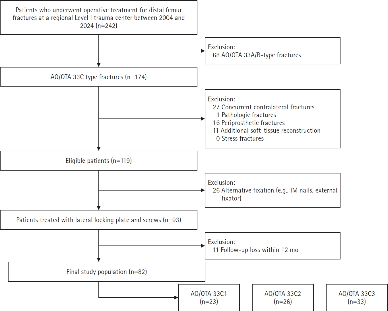
Study enrollment and patient selection flowchart. AO/OTA, AO Foundation/Orthopaedic Trauma Association; IM, intramedullary.
Fig. 2.
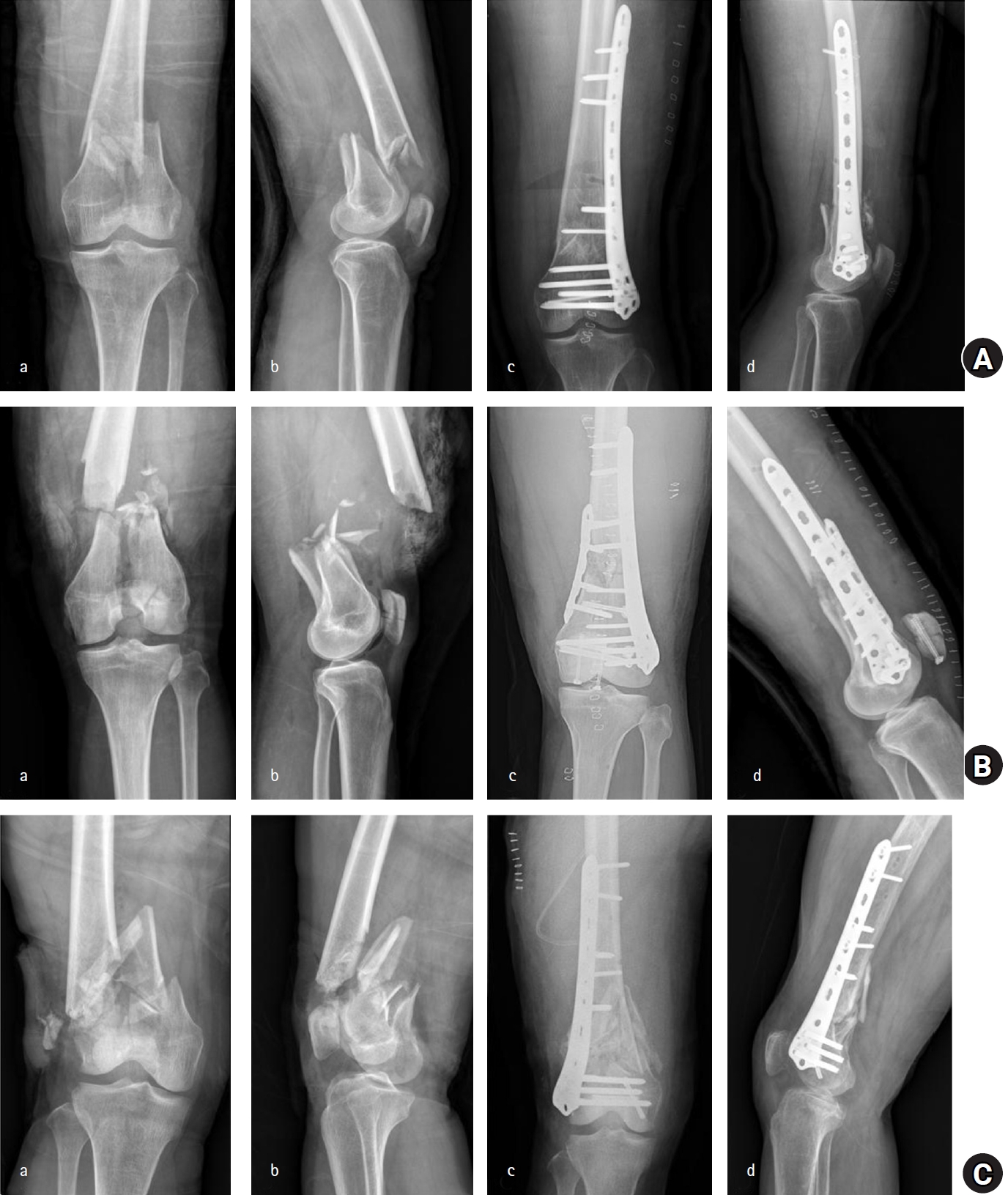
Representative preoperative and postoperative radiographs of AO Foundation/Orthopaedic Trauma Association (AO/OTA) 33C fracture subtypes. For each subtype, preoperative (a) anteroposterior and (b) lateral radiographs are followed by immediate postoperative (c) anteroposterior and (d) lateral radiographs after plate fixation. (A) AO/OTA 33C1 type fracture. (B) AO/OTA 33C2 type fracture. (C) AO/OTA 33C3 type fracture.
Fig. 3.
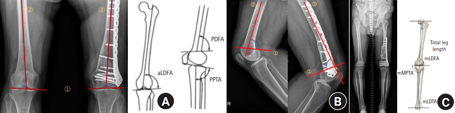
(A) The aLDFA was measured as the lateral angle between the distal joint line, defined as a line tangent to the most distal points of the medial and lateral condyles (①), and the anatomical axis of the femoral shaft (②). Coronal alignment was assessed as the angle between the anatomical axis of the proximal shaft fragment (③) and the distal joint line of the condylar fragment (①). (B) The aPDFA was measured between the reference line connecting the anterior and posterior junctions where the femoral metaphysis meets the condyles (①) and the anatomical axis of the femoral shaft (②). Sagittal angulation was assessed as the angle between the anatomical axis of the proximal shaft fragment (③) and the distal junctional line of the condylar fragment (①). The degree of deformity was calculated as the angular difference between the fractured side and the intact contralateral side. (C) Limb length discrepancy was measured as the absolute difference in total leg length between the two sides. Total leg length was defined as the distance from the highest point of the femoral head to the center of the ankle joint, corresponding to the midpoint of the tibial plafond. The hip-knee-ankle angle was defined as the angle formed by the mechanical axis of the femur, extending from the center of the femoral head to the center of the knee, and the mechanical axis of the tibia, extending from the center of the knee to the center of the ankle. aLDFA, anatomical lateral distal femoral angle; PDFA, posterior distal femoral angle; PPTA, proximal posterior tibial angle; mLDFA, mechanical lateral distal femoral angle; mMPTA, mechanical medial proximal tibial angle; mLDTA, mechanical lateral distal tibial angle.
Table 1.
Baseline characteristics according to the AO/OTA 33C subtype
| Variable | Total | C1 (n=23) | C2 (n=26) | C3 (n=33) |
|---|---|---|---|---|
| Age (yr) | 55.4±16.3 | 55.1±16.4 | 59.8±15.0 | 52.1±16.8 |
| Male sex | 50 (61.0) | 10 (43.5) | 17 (65.4) | 23 (69.7) |
| Follow-up (mo) | 28.2±18.1 | 30.7±20.4 | 25.1±14.3 | 28.8±19.2 |
| Mechanism of injury | ||||
| In-car accident | 30 (36.6) | 4 (17.4) | 12 (46.2) | 14 (42.4) |
| Motorbike accident | 12 (14.6) | 0 (0.0) | 5 (19.2) | 7 (21.2) |
| Pedestrian accident | 9 (11.0) | 4 (17.4) | 2 (7.7) | 3 (9.1) |
| Fall down | 10 (12.2) | 5 (21.7) | 2 (7.7) | 3 (9.1) |
| Slip downa) | 21 (25.6) | 10 (43.5) | 5 (19.2) | 6 (18.2) |
| Open fracture | 34 (41.5) | 5 (21.7) | 11 (42.3) | 18 (54.5) |
| Diabetes mellitus | 13 (15.9) | 7 (30.4) | 1 (3.8) | 5 (15.2) |
| Osteoporosis | 27 (32.9) | 8 (34.8) | 11 (42.3) | 8 (24.2) |
Table 2.
Radiographic reduction quality and alignment parameters
Values are presented as number (%) or mean± standard deviation. Δ indicates the difference between the affected and contralateral limb.
aPDFA, anatomical posterior distal femoral angle; aLDFA, anatomical lateral distal femoral angle; HKA, hip-knee-ankle angle; mLDFA, mechanical lateral distal femoral angle.
Table 3.
Postoperative complications and subsequent interventions according to the AO/OTA 33C subtype
| Variable | Total (n=82) | C1‒2 (n=49) | C3 (n=33) | P-value |
|---|---|---|---|---|
| Nonunion | 20 (24.4) | 7 (14.3) | 13 (39.4) | 0.020 |
| Infection | 5 (6.1) | 0 (0.0) | 5 (15.2) | 0.009 |
| Implant failure | 6 (7.3) | 3 (6.1) | 3 (9.1) | 0.681 |
| Reoperationa) | 25 (30.5) | 10 (20.4) | 15 (45.5) | 0.030 |
Table 4.
Clinically relevant late outcomes according to the AO/OTA 33C subtype
Table 5.
Multivariable linear regression analysis for clinical and radiographic outcomes
| Variable | βa) (95% CI) | P-value |
|---|---|---|
| Factors associated with final knee ROM | ||
| Age (yr) | −0.24 (−0.52 to 0.03) | 0.084 |
| Sagittal malalignment (ΔaPDFA, °) | −2.35 (−3.73 to −0.98) | 0.001 |
| Coronal malalignment (ΔaLDFA, °) | 0.08 (−2.01 to 2.18) | 0.938 |
| Reoperation (yes) | −17.5 (−27.8 to −7.1) | 0.001 |
| Factors associated with LLD | ||
| Sagittal malalignment (ΔaPDFA, °) | 0.08 (0.04 to 0.12) | <0.001 |
| Coronal malalignment (ΔaLDFA, °) | −0.01 (−0.08 to 0.05) | 0.670 |
| HKA (°) | 0.00 (−0.04 to 0.05) | 0.913 |
| Factors associated with radiographic osteoarthritis severity (ΔK-L)b) | ||
| Age (yr) | −0.008 (−0.016 to −0.001) | 0.027 |
| HKA (°) | −0.024 (−0.059 to 0.012) | 0.185 |
| ΔmLDFA (°) | 0.039 (−0.017 to 0.095) | 0.169 |
| Follow-up (mo) | 0.001 (−0.006 to 0.008) | 0.757 |
CI, confidence interval; ROM, range of motion; aPDFA, anatomical posterior distal femoral angle; aLDFA, anatomical lateral distal femoral angle; LLD, leg length discrepancy; HKA, hip-knee-ankle angle; K-L, Kellgren-Lawrence grade; mLDFA, mechanical lateral distal femoral angle.
a)Negative β indicates decreased ROM; positive β for LLD indicates increased LLD. b)ΔK-L represents injured minus contralateral knee.
- 1. Ehlinger M, Ducrot G, Adam P, Bonnomet F. Distal femur fractures. Orthop Traumatol Surg Res 2013;99:353-60.ArticlePubMed
- 2. Henderson CE, Lujan TJ, Kuhl LL, Bottlang M, Fitzpatrick DC, Marsh JL. Healing complications are common after locked plating for distal femur fractures. Clin Orthop Relat Res 2011;469:1757-65.ArticlePubMedPMC
- 3. Ricci WM, Loftus T, Cox C, Borrelli J. Locked plates combined with minimally invasive insertion technique for the treatment of periarticular fractures of the knee. J Orthop Trauma 2004;18:659-66.Article
- 4. Meinberg EG, Agel J, Roberts CS, Karam MD, Kellam JF. Fracture and dislocation classification compendium-2018. J Orthop Trauma 2018;32 Suppl 1:S1-170.Article
- 5. Zlowodzki M, Williamson S, Cole PA, Zardiackas LD, Kregor PJ. Biomechanical evaluation of fixation of distal femur fractures. J Orthop Trauma 2004;18:494-502.ArticlePubMed
- 6. Hierholzer C, von Ruden C, Potzel T, Woltmann A, Buhren V. Outcome analysis of retrograde nailing and less invasive stabilization system in distal femoral fractures: a retrospective analysis. Indian J Orthop 2011;45:243-50.ArticlePubMedPMCPDF
- 7. Elsoe R, Ceccotti AA, Larsen P. Population-based epidemiology of distal femur fractures. Int Orthop 2018;42:191-6.ArticlePubMed
- 8. Martinet O, Cordey J, Harder Y, Maier A, Buhler M, Barraud GE. The epidemiology of fractures of the distal femur. Injury 2000;31(Suppl 3):C62-3.ArticlePubMed
- 9. Pritchett JW. Supracondylar fractures of the femur. Clin Orthop Relat Res 1984;(184):173-7.Article
- 10. Kook I, Kim KY, Hwang KT. The impact of medial-first dual plating for reduction of distal femoral fractures: a retrospective comparative cohort study. Sci Rep 2025;15:15454.ArticlePubMedPMCPDF
- 11. Sharma L, Song J, Felson DT, Cahue S, Shamiyeh E, Dunlop DD. The role of knee alignment in disease progression and functional decline in knee osteoarthritis. JAMA 2001;286:188-95.ArticlePubMed
- 12. Brouwer GM, van Tol AW, Bergink AP, et al. Association between knee alignment and osteoarthritis progression. Arthritis Rheum 2007;56:1204-11.ArticlePubMed
- 13. Kellgren JH, Lawrence JS. Radiological assessment of osteo-arthrosis. Ann Rheum Dis 1957;16:494-502.ArticlePubMedPMC
- 14. Demirtas A, Azboy I, Alemdar C, et al. Functional outcomes and quality of life in adult ipsilateral femur and tibia fractures. J Orthop Transl 2018;16:53-61.Article
References
Figure & Data
REFERENCES
Citations
Citations to this article as recorded by 
 ePub Link
 Cite
Cite XML Download
Postoperative sagittal alignment and reoperation predict late outcomes after plate fixation of AO/OTA 33C distal femur fractures: a retrospective cohort study
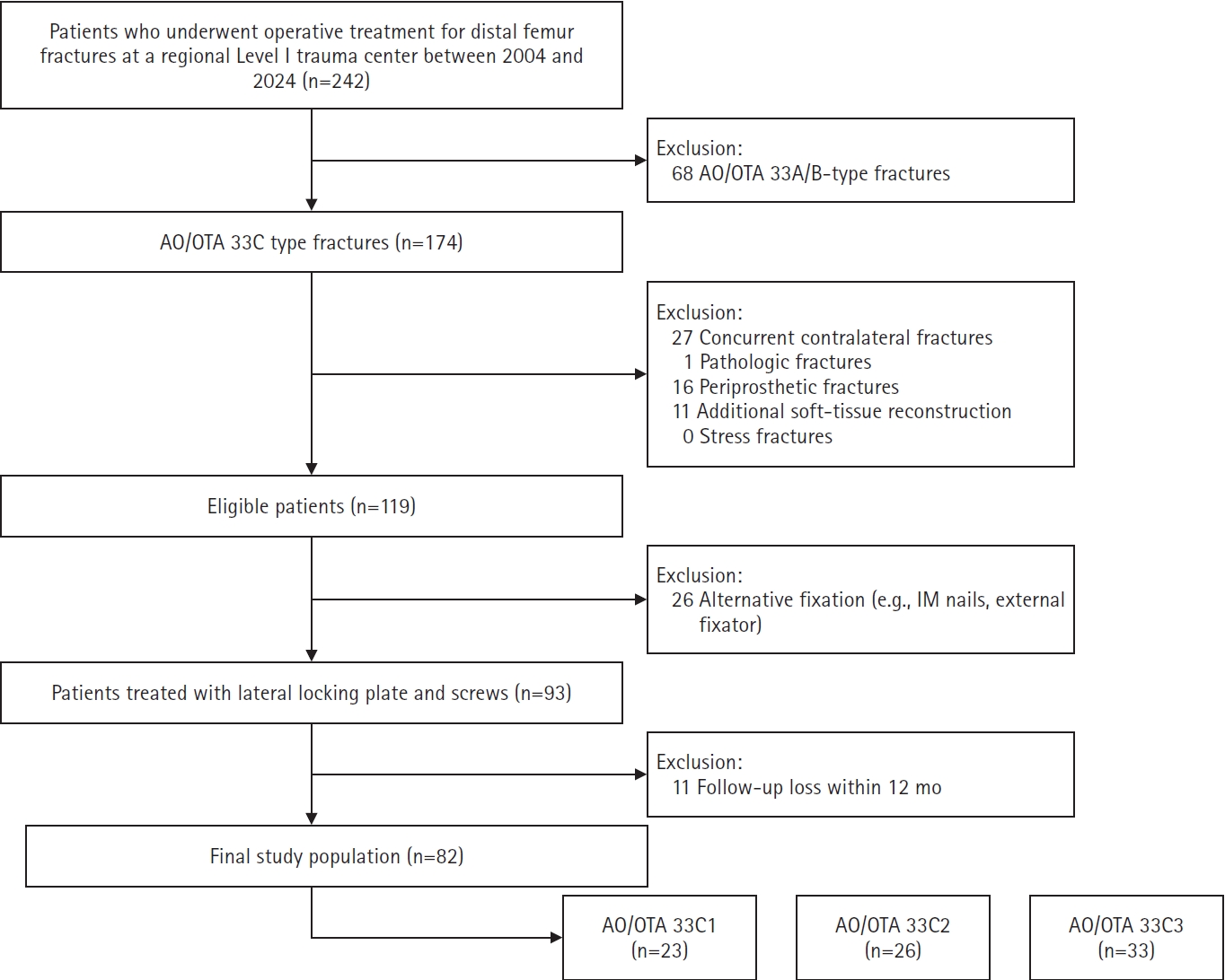
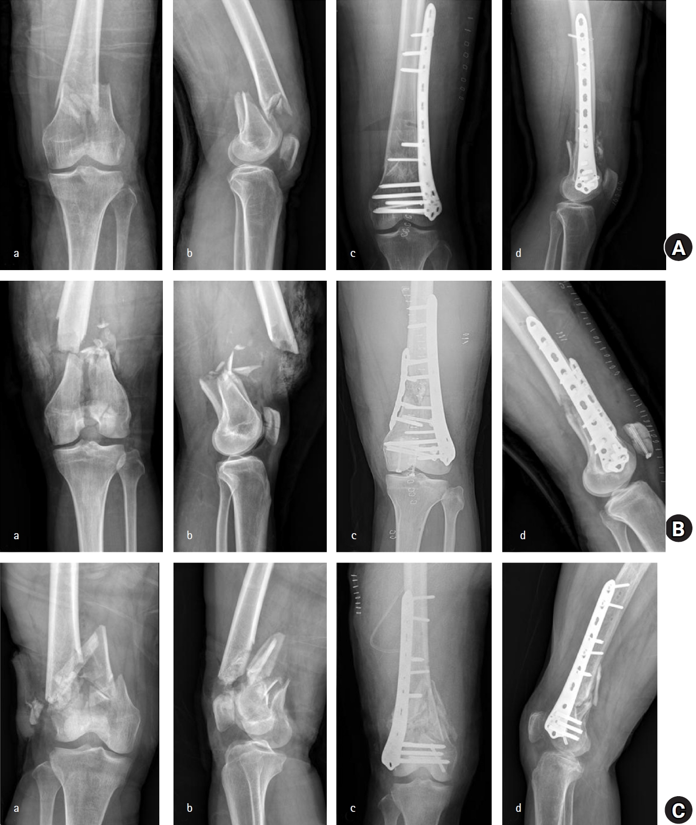
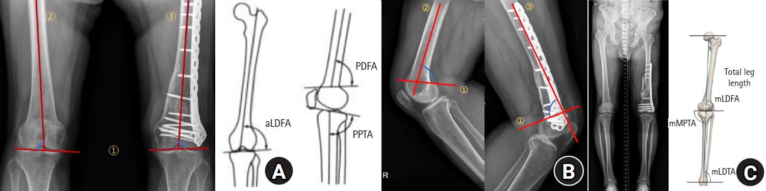
Fig. 1. Study enrollment and patient selection flowchart. AO/OTA, AO Foundation/Orthopaedic Trauma Association; IM, intramedullary.
Fig. 2. Representative preoperative and postoperative radiographs of AO Foundation/Orthopaedic Trauma Association (AO/OTA) 33C fracture subtypes. For each subtype, preoperative (a) anteroposterior and (b) lateral radiographs are followed by immediate postoperative (c) anteroposterior and (d) lateral radiographs after plate fixation. (A) AO/OTA 33C1 type fracture. (B) AO/OTA 33C2 type fracture. (C) AO/OTA 33C3 type fracture.
Fig. 3. (A) The aLDFA was measured as the lateral angle between the distal joint line, defined as a line tangent to the most distal points of the medial and lateral condyles (①), and the anatomical axis of the femoral shaft (②). Coronal alignment was assessed as the angle between the anatomical axis of the proximal shaft fragment (③) and the distal joint line of the condylar fragment (①). (B) The aPDFA was measured between the reference line connecting the anterior and posterior junctions where the femoral metaphysis meets the condyles (①) and the anatomical axis of the femoral shaft (②). Sagittal angulation was assessed as the angle between the anatomical axis of the proximal shaft fragment (③) and the distal junctional line of the condylar fragment (①). The degree of deformity was calculated as the angular difference between the fractured side and the intact contralateral side. (C) Limb length discrepancy was measured as the absolute difference in total leg length between the two sides. Total leg length was defined as the distance from the highest point of the femoral head to the center of the ankle joint, corresponding to the midpoint of the tibial plafond. The hip-knee-ankle angle was defined as the angle formed by the mechanical axis of the femur, extending from the center of the femoral head to the center of the knee, and the mechanical axis of the tibia, extending from the center of the knee to the center of the ankle. aLDFA, anatomical lateral distal femoral angle; PDFA, posterior distal femoral angle; PPTA, proximal posterior tibial angle; mLDFA, mechanical lateral distal femoral angle; mMPTA, mechanical medial proximal tibial angle; mLDTA, mechanical lateral distal tibial angle.
Fig. 1.
Fig. 2.
Fig. 3.
Postoperative sagittal alignment and reoperation predict late outcomes after plate fixation of AO/OTA 33C distal femur fractures: a retrospective cohort study
| Variable | Total | C1 (n=23) | C2 (n=26) | C3 (n=33) |
|---|---|---|---|---|
| Age (yr) | 55.4±16.3 | 55.1±16.4 | 59.8±15.0 | 52.1±16.8 |
| Male sex | 50 (61.0) | 10 (43.5) | 17 (65.4) | 23 (69.7) |
| Follow-up (mo) | 28.2±18.1 | 30.7±20.4 | 25.1±14.3 | 28.8±19.2 |
| Mechanism of injury | ||||
| In-car accident | 30 (36.6) | 4 (17.4) | 12 (46.2) | 14 (42.4) |
| Motorbike accident | 12 (14.6) | 0 (0.0) | 5 (19.2) | 7 (21.2) |
| Pedestrian accident | 9 (11.0) | 4 (17.4) | 2 (7.7) | 3 (9.1) |
| Fall down | 10 (12.2) | 5 (21.7) | 2 (7.7) | 3 (9.1) |
| Slip down |
21 (25.6) | 10 (43.5) | 5 (19.2) | 6 (18.2) |
| Open fracture | 34 (41.5) | 5 (21.7) | 11 (42.3) | 18 (54.5) |
| Diabetes mellitus | 13 (15.9) | 7 (30.4) | 1 (3.8) | 5 (15.2) |
| Osteoporosis | 27 (32.9) | 8 (34.8) | 11 (42.3) | 8 (24.2) |
| Variable | Total (n=82) | C1‒2 (n=49) | C3 (n=33) | P-value |
|---|---|---|---|---|
| Articular reduction quality | <0.001 | |||
| Anatomical | 39 (47.6) | 36 (73.5) | 3 (9.1) | |
| Good | 35 (42.7) | 13 (26.5) | 22 (66.7) | |
| Poor | 8 (9.8) | 0 (0.0) | 8 (24.2) | |
| Sagittal alignment (ΔaPDFA, °) | 4.0±3.6 | 3.2±2.8 | 5.3±4.2 | 0.014 |
| Coronal alignment (ΔaLDFA, °) | 1.3±3.0 | 0.5±2.8 | 2.5±3.1 | 0.006 |
| HKA (°) | 1.5±4.4 | 1.0±4.3 | 2.3±4.5 | 0.170 |
| mLDFA (°) | 88.9±2.7 | 88.5±2.7 | 89.5±2.7 | 0.091 |
| ΔmLDFA (°) | 1.9±2.7 | 1.5±2.7 | 2.5±2.7 | 0.091 |
| Variable | Total (n=82) | C1‒2 (n=49) | C3 (n=33) | P-value |
|---|---|---|---|---|
| Nonunion | 20 (24.4) | 7 (14.3) | 13 (39.4) | 0.020 |
| Infection | 5 (6.1) | 0 (0.0) | 5 (15.2) | 0.009 |
| Implant failure | 6 (7.3) | 3 (6.1) | 3 (9.1) | 0.681 |
| Reoperation |
25 (30.5) | 10 (20.4) | 15 (45.5) | 0.030 |
| Variable | Total (n=82) | C1‒2 (n=49) | C3 (n=33) | P-value |
|---|---|---|---|---|
| Final ROM (°) | 107.7±27.6 | 119.5±24.2 | 89.7±22.3 | <0.001 |
| LOM (ROM <90°) | 13 (15.9) | 4 (8.2) | 9 (27.3) | 0.029 |
| ΔK-L grade | 0.5±0.7 | 0.1±0.4 | 0.9±0.7 | <0.001 |
| LLD (cm) | 0.7±0.8 | 0.4±0.5 | 1.3±0.9 | <0.001 |
| TKRA | 7 (8.5) | 3 (6.1) | 4 (12.1) | - |
| Variable | β |
P-value |
|---|---|---|
| Factors associated with final knee ROM | ||
| Age (yr) | −0.24 (−0.52 to 0.03) | 0.084 |
| Sagittal malalignment (ΔaPDFA, °) | −2.35 (−3.73 to −0.98) | 0.001 |
| Coronal malalignment (ΔaLDFA, °) | 0.08 (−2.01 to 2.18) | 0.938 |
| Reoperation (yes) | −17.5 (−27.8 to −7.1) | 0.001 |
| Factors associated with LLD | ||
| Sagittal malalignment (ΔaPDFA, °) | 0.08 (0.04 to 0.12) | <0.001 |
| Coronal malalignment (ΔaLDFA, °) | −0.01 (−0.08 to 0.05) | 0.670 |
| HKA (°) | 0.00 (−0.04 to 0.05) | 0.913 |
| Factors associated with radiographic osteoarthritis severity (ΔK-L) |
||
| Age (yr) | −0.008 (−0.016 to −0.001) | 0.027 |
| HKA (°) | −0.024 (−0.059 to 0.012) | 0.185 |
| ΔmLDFA (°) | 0.039 (−0.017 to 0.095) | 0.169 |
| Follow-up (mo) | 0.001 (−0.006 to 0.008) | 0.757 |
Table 1. Baseline characteristics according to the AO/OTA 33C subtype
Values are presented as mean± standard deviation or number (%). AO/OTA, AO Foundation/Orthopaedic Trauma Association. a)Slip down was the only low-energy injury mechanism.
Table 2. Radiographic reduction quality and alignment parameters
Values are presented as number (%) or mean± standard deviation. Δ indicates the difference between the affected and contralateral limb. aPDFA, anatomical posterior distal femoral angle; aLDFA, anatomical lateral distal femoral angle; HKA, hip-knee-ankle angle; mLDFA, mechanical lateral distal femoral angle.
Table 3. Postoperative complications and subsequent interventions according to the AO/OTA 33C subtype
Values are presented as number (%). AO/OTA, AO Foundation/Orthopaedic Trauma Association. a)Reoperation was defined as a subsequent surgical intervention performed due to postoperative complications.
Table 4. Clinically relevant late outcomes according to the AO/OTA 33C subtype
Values are presented as mean± standard deviation or number (%). AO/OTA, AO Foundation/Orthopaedic Trauma Association; ROM, range of motion; LOM, limitation of motion; K-L, Kellgren-Lawrence; LLD, leg length discrepancy; TKRA, total knee replacement arthroplasty.
Table 5. Multivariable linear regression analysis for clinical and radiographic outcomes
CI, confidence interval; ROM, range of motion; aPDFA, anatomical posterior distal femoral angle; aLDFA, anatomical lateral distal femoral angle; LLD, leg length discrepancy; HKA, hip-knee-ankle angle; K-L, Kellgren-Lawrence grade; mLDFA, mechanical lateral distal femoral angle. a)Negative β indicates decreased ROM; positive β for LLD indicates increased LLD. b)ΔK-L represents injured minus contralateral knee.


