
 E-submission
 KOTA
KOTA TOTA
TOTA TOTS
TOTS
Articles
- Page Path
- HOME > J Musculoskelet Trauma > Volume 39(3); 2026 > Article
-
Review Article
- Spine OpenSim models: a narrative review of the literature
- Mohammad Taghi Karimi
-
Journal of Musculoskeletal Trauma 2026;39(3):195-204.
DOI: https://doi.org/10.12671/jmt.2026.00171
Published online: July 7, 2026
Orthotics and Prosthetics Department, Rehabilitation Faculty of Shiraz University of Medical Sciences, Shiraz, Iran
- Correspondence to: Mohammad Taghi Karimi Orthotics and Prosthetics Department, Rehabilitation Faculty of Shiraz University of Medical Sciences, Sadra City Road, Shahid Doran Campus, Shiraz 7198754361, Iran Tel: +98-71-3626-5489 Email: Mt_Karimi@Sums.ac.ir
• Received: April 13, 2026 • Revised: May 2, 2026 • Accepted: May 4, 2026
© 2026 The Korean Orthopaedic Trauma Association
This is an Open Access article distributed under the terms of the Creative Commons Attribution Non-Commercial License (http://creativecommons.org/licenses/by-nc/4.0/) which permits unrestricted noncommercial use, distribution, and reproduction in any medium, provided the original work is properly cited.
- 864 Views
- 18 Download


Abstract
- The human spine is affected by various traumatic and pathological conditions that can impair function during daily activities. Several software programs have been developed to assess the risk of spinal injury, and OpenSim is one platform used for this purpose. Various OpenSim spine models have been developed to analyze parameters such as range of motion, moments applied to the vertebrae, muscle forces, and joint contact forces. This review aimed to compile the available OpenSim models that can be used to study the spine under normal and pathological conditions. Published studies using spine models in OpenSim were reviewed, and information on model outputs, availability, and validity was extracted. Keywords such as “spine models,” “lumbar models,” “musculoskeletal modeling,” and “trunk models” were used in combination with “OpenSim.” Eighty papers on spine models in OpenSim were identified, of which 23 were selected for final analysis. The available OpenSim models were classified as specific spine models (17 models) or full-body models (six models) that include the spine as a component. The available models can be used to assess injury risk, muscle forces, and joint contact forces in the cervical and lumbar spine. Full-body models allow examination of kinematics and joint contact forces at L5/S1, although only a limited number of muscles are represented in these models. Output validity has been evaluated in only a few models.
Introduction
The performance of the musculoskeletal system influences individuals' abilities to carry out daily activities. Various diseases can impact this system’s performance, ultimately limiting daily activity. The treatment of these diseases depends on understanding their effects on kinematics, kinetics, joint contact forces, and muscle forces. While there are various methods available to determine and monitor these parameters, some can only be used in cadaveric studies. The moments and range of motion of the joints can be assessed using a motion analysis system and various approaches like inverse dynamics and inverse kinematics [1,2]. However, parameters such as joint contact forces must be evaluated based on sensor outputs inserted in the joints. These studies are often conducted on cadavers due to ethical concerns.
Now it is possible to determine joint contact forces and muscle forces using modeling approaches, which can be done with advanced software such as AnyBody and OpenSim [3]. AnyBody is commercial software, while OpenSim is free open-source software with a variety of models available.
Various models of OpenSim have been developed to analyse the motions of spine, muscle forces and also joint contact forces [4-6]. These models have been used for both normal and pathological conditions. Now use of these models is recommended to design various assistive devices, evaluation of sport activities and also to design implants. The models of the spine may include only articulated spine and also models with fully articulated thoracolumbar spine and rib cages. Depending on the models, they may include the trunk and cervical muscles. Although various models of the spine have been developed for use with OpenSim, it is not well determined which ones can be used for the cervical, lumbar, and total spine. Information regarding the validity and accuracy of the outputs of these models is not well-documented. Furthermore, it is not clear which types of these models can be used for designing assistive devices. The aim of this review was to introduce the available models of the spine that can be used in OpenSim software and to determine their validity based on available literature. Additionally, the goal was to determine the feasibility of using spine models depending on the research conducted.
This study was a narrative review and did not require institutional review board approval or informed consent.
Methods
A comprehensive search was conducted in the existing literature to gather all studies on the use of OpenSim software in the analysis of spine motions in both normal and pathological conditions. The search was done in some databases including PubMed, Scopus, Web of Science, and Google Scholar. The search was done for a time period up to the end of 2025. Some keywords such as spine model, cervical model, lumbar model, musculoskeletal modeling, and trunk model were used in combination with OpenSim. The main inclusion criteria for selecting the papers were that only studies focusing on the use of spine models were included. The selection of the papers was done based on titles and abstracts. We attempted to categorize the available models of the spine based on their outputs, validity, and availability.
Results
There were 80 papers on spinal models in OpenSim software. However, only 23 papers were selected for final analysis. The models of the spine in OpenSim can be categorized into two main groups, which include (1) specific models of the spine; (2) full-body models that also include the spine.
Musculoskeletal model of the lumbar spine
This model consists of a rigid pelvis and sacrum, five lumbar vertebrae, and torso (thoracic spine and ribcage). Each lumbar vertebra has 3 degrees of freedom (DOFs; flexion/extension, lateral bending, and axial rotation). The model has been developed to include 238 muscle fascicles which create eight muscle groups of the spine. In the newly developed model, each lumbar vertebra has 6 DOFs for both rotatory and translatory motions. The main limitation associated with this model is that it cannot be used for complex motions of the spine that involve motion across two or more planes [7]. Fig. 1 shows this model.
The thoracolumbar spine and rib cage model
This is fully articulated, including vertebrae from T1 to L5. It has 3 degrees of motion for rotational movement. This model also includes the head, neck, upper extremities, and most major muscles of the lumbar spine, abdomen, and intercostal muscles. There are 552 musculotendon actuators for trunk muscles and 620 actuators for the entire model, with a total of 93 DOFs in the spine. Static optimization can be used to determine muscle forces in this model, but computer muscle control cannot. The validity of the muscle forces and joint contact forces in this model was evaluated using data from the literature. This model can be used for designing exoskeletons and determining spinal loads in various lifting conditions [8]. Fig. 2 shows the thoracolumbar spine and rib cage model.
Full-body spine models with detailed thoracolumbar spine for children and adolescents
This model is a combination of articulated thoracolumbar spine and rib cage, combined head and neck with the upper and lower limb extremities. In this project, the data of 26 children and adolescents were used with ages of 6‒18 years. It should be emphasized that model 2354 was used in this project [9]. The validity of the outputs of this model was evaluated with literature.
A model for estimating spinal loading during lifting and lowering tasks
A specific model (Fig. 3) was developed in this project. A fully articulated thoracolumbar model was created and validated using nine dynamic lifting tasks. Passive structures and kinematic constraints were incorporated into the model to enhance its accuracy in predicting spinal loads during dynamic tasks. The results of this study demonstrated that the model can effectively estimate spinal loads during lifting and lowering tasks, as well as perform inverse dynamics [10].
Articulated kinematic model of the thoracic and lumbar spine
This dynamic articulated model of the lumbar and thoracic spine, Fig. 4, is designed for kinematic analysis. It is a modification of Christophy's model, with the following enhancements [7].
The thoracic vertebrae in this model have been separated to increase the DOF. The pelvic section now features a custom joint that allows for translation in three-dimensional space. The rib cage has been removed from the previous model. This modification allows for the addition of a greater number of markers to scale the model effectively.
Lumbar spine with passive elements
In this model, the structure of the lumbar spine has been enhanced to include passive elements, particularly between L3 and S1. This model allows for the simulation of the rotational moment/force generated by the intervertebral disc, facet articulations, and ligaments [11].
London lumbar spine model
This model includes the lumbar spine and lower limbs, and was created to analyse muscle force and joint contact force during daily activities, particularly lifting. The model, Fig. 5, was constructed using magnetic resonance imaging scans of the subject. Its accuracy was verified by comparing the momentum arm of the erector spinae during forward flexion to measurements from Jorgensen's method. Various parameters, such as joint contact force and muscle forces, were assessed in this study. The results of this study based on the outputs of the model aligned well with existing literature [4].
University of Waterloo cervical spine model
This model was specifically developed to study the forces and movements of the cervical spine. It includes 218 actuators representing 58 cervical muscles. Motion data of the cervical spine and electromyography (EMG) activities of the muscles were collected from eight healthy subjects. The compressive force at C7‒T1 was calculated in this model and compared to values from other models. The results revealed discrepancies between the forces in this model and those reported in existing literature, ranging from 25.5 to 368.1 N [12].
Personalized scoliotic spine models
This project aimed to create specific model of spine for scoliotic subjects. This project is a simplified model of spine adapted from Bruno et al. [13]. The model was scaled and changed based on the markers attached on the spine.
Model to determine muscle forces during sit-to-stand transfer
In this project a model was developed to analyse sit-to-stand transfer motion. The muscle forces can be determined in this task. Actually, this model consists of lower limb model 2010, musculoskeletal model of the lumbar spine, MoBL-ARMS Upper Limb Model, and head and neck musculoskeletal biomechanics model. This model has 46 DOFs with 194 actuators [14].
A model of cervical spine for injury mechanisms analysis and simulation in sport
Evaluation of the motion of the cervical spine in normal and pathological conditions can be done using advanced models of the cervical spine. There are some specific cervical models developed for this purpose, including the MASI (Fig. 6) and Rugby models. The MASI model is based on data from a healthy cadaver, which also includes the scapula and clavicle, allowing for scapular and clavicular coupled motion with respect to humeral elevation.
Rugby model
The Rugby model is another version of the MASI model used to estimate cervical spine loads in rugby and other sports activities, especially those with upper limb contacts. In the older version of this model, the internal loading of the cervical spine in non-injured activities can be evaluated. The new version of the model was developed to simulate the motions from C2‒C3 to C5‒C6 to simulate axial impact. Using this model, joint contact force and muscle forces can be analysed in impact activities [5,15].
The validity of both the Rugby and MASI models was established based on dynamic verification, kinematic validation, and dynamic validation. The muscle force of the cervical spine was determined using a computer muscle control approach. The results of studies based on these models support the reliability of the results. These models can be used to investigate the risks of injuries in various sports activities [15].
Full-body lifting model
This model is actually based on the full-body lumbar spine (FBLS) and was developed to estimate spinal loads during lifting. The validity of the model was assessed through various procedures, including comparisons with EMG muscle data, evaluation of intradiscal pressure, and comparison of contact forces with vertebral body implant measurements. It is important to note that this model is primarily used to determine joint loading of the lumbar spine. It appears that this model has acceptable validity for evaluating changes in lumbar loading during lifting [16].
A model to determine the role of the neck during impact loading
In this project a model of head and neck was used to determine the role of muscles and passive elements in the activities with impact loads. This model is based on works of Vasavada et al. [6] and Mortensen et al. [17]. The kinematics of head movement was evaluated in this study in various loading conditions [18,19].
Head and neck musculoskeletal biomechanics
This model of the head and neck was created based on data from five cadavers. The model includes the head, neck, scapula, and clavicle. In the first version of the model, the head had 3 DOFs (flexion/extension, abduction/adduction, and rotation) relative to the trunk. In the newer version, there were 6 DOFs (3 DOF for the motion of the skull relative to C2 and 3 DOF for the motion of C2 relative to T1). It appears that this model has a high level of validity to determine the range of motion [6].
A model to study infant head kinematics during abusive head trauma
This model was developed to determine the kinematics of head movement in infants during shaking. It is a combination of a 4.5-month-old infant model developed from computed tomography scans, and an upper body adult model [20].
Musculoskeletal model of head and neck suitable for dynamic simulations
The primary issue with previous head and neck models was their inability to calculate absolute values of moments in all directions. This was mainly due to the multi-directional moments of the cervical spine muscles. The accuracy of cervical moments has been improved by incorporating hyoid muscles into the models. This new model, known as the hyoid model, Fig. 7, can be utilized to calculate muscle forces using computer muscle control and joint contact forces using forward dynamics [17].
The second group of models that can be used to analyse the kinematics and kinetics of the spine is full-body models. The available full-body models can be divided into two main groups: those with detailed motions of the spine (especially for the lumbar vertebrae) and those with a combined spine (head, neck, and trunk). The second group includes models with only a link with 3 DOFs at L5/S1.
Model 1018
It is a simplified model with 10 DOFs and 18 actuators that primarily focuses on the lower extremities. This model includes a linkage with 3 DOFs between the pelvis and trunk (a combination of the head and spine). It showcases the utilization of the new Millard muscles in OpenSim. The purpose of this model is for educational, demonstrative, and initial prototyping purposes when quick simulation times are required. It can be utilized to analyse the kinematics of the trunk in relation to the pelvis. Additionally, this model can help to determine the joint contact force at L5/S1.
Model 2354
This model combines legs and trunk without upper limbs. There is a linkage between the trunk and pelvis with 3 rotational DOFs. This model consists of 23 DOFs with 54 actuators for 46 muscles. There are several trunk muscles associated with this model, including abdominal and spinal extensors. This model is mostly used for demonstration and education. It appears that this model can be used for analysing trunk kinematics relative to the pelvis [21].
Model 2392
It is a well-developed model of the lower extremity combined with a model of the trunk. While this model primarily focuses on leg muscles, it also includes a few trunk muscles. It is designed to determine the kinematics of the trunk and joint contact force at L5/S1. The model has 23 DOFs and utilizes 92 actuators for 76 muscles. Additionally, there is a link between L5/S1 with 3 DOFs in this model, allowing for the analysis of joint contact force at that specific location.
Full-body model Hamner
This is a well-developed full-body model primarily used for walking and running. It includes legs, upper limbs, and the spine. The model is also utilized to study the range of motion and moments of the joints. It is important to note that the trunk is constructed as a unit consisting of the head, neck, and spine, connecting to the pelvis at L5/S1. Several flexor and extensor muscles of the trunk are associated with this model. This model allows for the evaluation of joint contact force at L5/S1 [22].
Rajagopal model
This is another well-developed full-body model, which can be used to investigate the motions of the legs, upper limb and spine at L5/S1. The muscles of trunk in this model are the same as other full-body models such as model 2392 and 2354 [23].
Full-body lumbar spine model
This is a full-body model consisting of legs, upper limbs and spine. Actually, this model is suitable to investigate the motions, muscle forces and joint contact forces of spine. The model consists of 21 segments, with 29 DOFs. There are 324 musculotendon actuators associated with this model. Each lumbar vertebra has 6 DOFs for flexion/extension, rotation and lateral bending. There are eight muscles groups of trunk which include: rectus abdominis, external obliques, internal obliques, erector spinae, multifidus, quadratus lumborum, psoas major, and latissimus dorsi. The force of muscles can be determined based on static optimization. Due to the complexity of the model, it is not possible to run computer muscle control [24].
Discussion
Now it is possible to determine the muscle forces and joint contact forces of spine during different tasks by use of motion analysis and use of some software such as AnyBody and OpenSim. However, there are some differences between these software packages. The OpenSim and AnyBody spine models are both computational tools used for musculoskeletal simulation, but they differ in focus, structure, and flexibility [25]. OpenSim is an open-source platform developed for modeling, simulating, and analysing human movement, offering a highly customizable framework with extensive community resources [3,26]. Its spine models typically emphasize biomechanical motion and inverse dynamics to study kinematics and muscle activation patterns. In contrast, the AnyBody Modelling System is a commercial software that provides a detailed, scalable representation of the musculoskeletal system, including complex spine mechanics and muscle coordination [25]. It integrates advanced optimization algorithms to estimate muscle forces and joint loads under specific conditions. While OpenSim excels in transparency, community-driven development, and flexibility for custom research, AnyBody stands out for its detailed anatomical databases, robust optimization routines, and ready-to-use parametric spine models suited for ergonomic and clinical applications [3,26].
Based on the results of this study, several models have been developed to study the motion of the spine. However, a few of them can be used to investigate the kinetics and kinematic variables of the spine. Table 1 summarizes the available models that can be used to study the motions of the spine. As can be seen from the table, there are several models of the cervical spine that can be used to evaluate kinematics, muscle forces, and joint contact forces. However, it should be noted that in most available full-body models, the spine was constructed as one component with a link at L5/S1. Therefore, most available models can be only used to determine joint contact force at L5/S1. In advanced models of the spine, only the motions of the lumbar vertebrae can be analysed in more detail, meaning that in most of them, the thoracic vertebrae were constructed as one component. The validity of the spine models is another issue that was not addressed and evaluated in most studies.
Based on the results of the available studies presented in Table 1, several models are available to investigate the research on spine which can be divided based on the regions of spine. There are six studies on model of spine in cervical regions [5,6,15,17,20]. Although all of them can be used for kinematic analysis, only four models can be used to determine the moments between cervical vertebra and also the muscle forces. The hyoid model and MASI model are the cervical models which can also be used for forward dynamics. The validity of these models was evaluated and the outputs of these models have acceptable validity to be used in the research to investigate the effects of impact in various sport activities [5,17].
There are several models of the thoracic spine that can be used to determine its range of motion. It is important to note that in most of the available models (including full-body models and spine models), the trunk is constructed as one component at the L5/S1 level. These models can be used to evaluate the motion of the trunk on the pelvis, as well as the moments applied to the connection between the trunk and pelvis. It should be noted that a few muscles are associated with full-body models.
The available models of the spine that can be used with OpenSim software can also be categorized based on the type of studies. Most of the available models of the spine (Table 1) can be used for kinematic analysis. However, depending on the structure of the models, some of the available models can be used for investigating intervertebral motions of the spine in the cervical region (Hyoid and MASI models), thoracic region (FBL model), and lumbar region. In contrast, there are a few models that can be used for evaluating joint contact forces at L5/S1 and muscle analysis during lifting [8]. There are also some full-body models that can be used for analysing motions of the spine at L5/S1 and joint contact force at this level for various research purposes (lifting, walking, etc) [21-23].
Validation of the models is another important parameter which should be considered in selection of spine models. Although there are nearly 23 models developed for vertebral column, a few of them were validated. The validity of the models was evaluated based on EMG pattern of trunk muscles, comparison between the outputs of the sensors embedded in cadavers with the outputs of the models based on forward dynamics analysis. For some model such as thoracolumbar spine and rib cage model, the output of the models was validated with the available data of literature [8]. For the cervical model developed by Waterloo University, the outputs of EMG of the muscles were compared with the pattern of activity of the muscles obtained from literature [12]. There was some discrepancy between the outputs of the models and EMG. The outputs of the Rugby and MASI models were validated based on dynamic verification [5,15]. It seems that the validity of the spine models is an important issue which should be considered in the future studies. Validation of spine models is a big challenge in the research done on spine. Inserting a sensor inside vertebra is a method to determine joint contact forces and the loads transmitted through vertebra. However, it is not practical to put sensors inside vertebra due to ethical issues. Complexity of spine structure is another important parameter which influences the validity of the outputs of spine models. Last but not least is that most of validation is based on cadaveric data which may not fully reflect living tissue behaviours.
There are some limitations associated with the available spine models. The main limitation is that only a few models can be used to determine the muscle forces and joint contact forces. Moreover, the validity of most of these models should be evaluated. It is not possible to evaluate the joint contact force and muscle forces of whole body in various tasks such as lifting. It seems that lack of studies on evaluation of the validity of the available models is the main issue which should be considered in future studies.
The available models of the spine can be categorized into two main groups: specific spine models and full-body models, which also include the spine with a linkage at L5/S1. Some models are designed to investigate the risk of injuries in cervical spine and can be used to determine muscle force and joint contact forces. In most full-body models, the trunk is constructed as one component linked to the pelvis at L5/S1, with a few muscles associated with the trunk in these models. It seems that a few of the available models can be used to determine muscle forces of spine and joint contact force. It is recommended that the validity of the outputs of the spine models should be evaluated in the future studies.
-
Author contributions
All the work was done by Mohammad Taghi Karimi.
-
Conflicts of interest
No potential conflict of interest relevant to this article was reported.
-
Funding
None.
-
Data availability
Not applicable.
-
Acknowledgments
None.
-
Supplementary materials
None.
Article Information
Fig. 1.
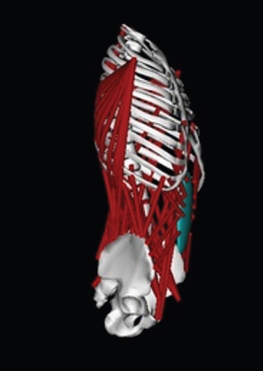
Musculoskeletal model of the lumbar spine. The model includes a rigid pelvis and sacrum, five lumbar vertebrae, and the torso, including the thoracic spine and rib cage.
Fig. 2.
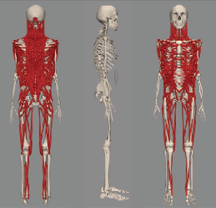
The thoracolumbar spine and rib cage model, including vertebrae from T1 to L5. It has three degrees of motion for rotational movement. This model also includes the head, neck, upper extremities, and most major muscles of the lumbar spine, abdomen, and intercostal muscles.
Fig. 3.
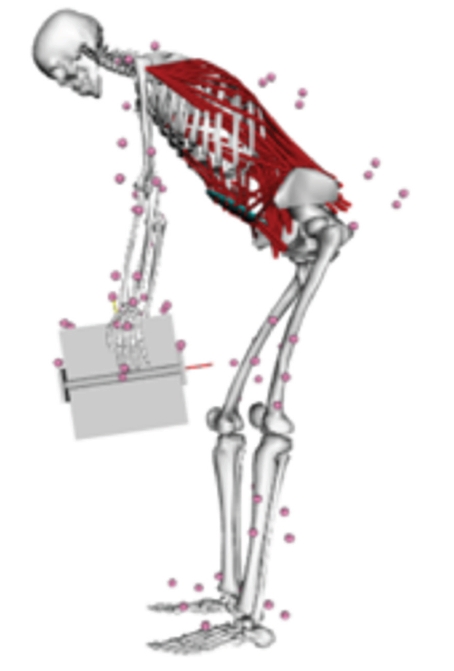
Spine model for estimating spinal loading during lifting and lowering tasks. Passive structures and kinematic constraints were incorporated to improve the accuracy of spinal load prediction during dynamic tasks.
Fig. 4.
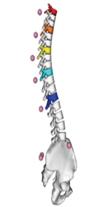
Articulated kinematic model of the thoracic and lumbar spine. The thoracic vertebrae were modelled separately to increase the degrees of freedom.
Fig. 5.
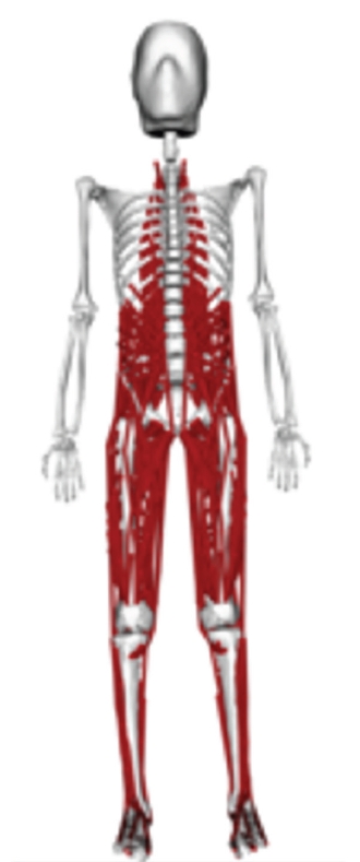
London lumbar spine model. This model includes the lumbar spine and lower limbs and was created to analyse muscle forces and joint contact forces during daily activities, particularly lifting.
Fig. 6.
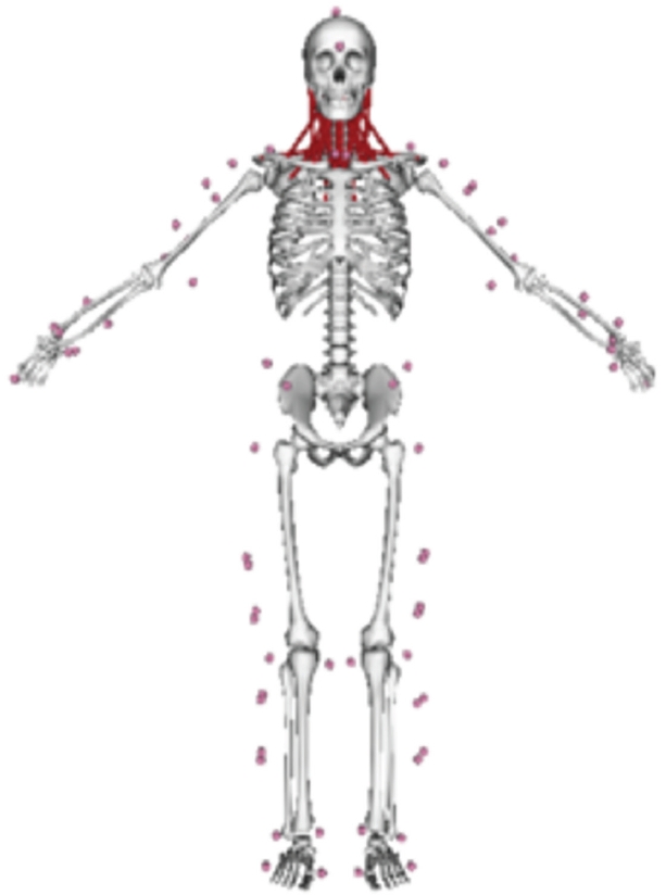
MASI model of the cervical spine for injury mechanism analysis and simulation in sport. The model includes the scapula and clavicle, allowing coupled scapular and clavicular motion with humeral elevation.
Fig. 7.
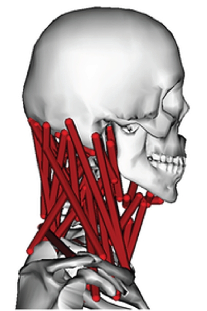
Hyoid model of the cervical spine. The accuracy of cervical moment estimates was improved by incorporating the hyoid muscles into the model.
Table 1.
Various models of spine developed for OpenSim software
- 1. Roggio F, Ravalli S, Maugeri G, et al. Technological advancements in the analysis of human motion and posture management through digital devices. World J Orthop 2021;12:467-84.ArticlePubMedPMC
- 2. Kim W, Kim M. Sports motion analysis system using wearable sensors and video cameras. In: Proceedings of the 2017 International Conference on Information and Communication Technology Convergence (ICTC); 2017 Oct; Jeju, Korea. ICTC; 2017. p. 1089-91.Article
- 3. Delp SL, Anderson FC, Arnold AS, et al. OpenSim: open-Source software to create and analyze dynamic simulations of movement. IEEE Trans Biomed Eng 2007;54:1940-50.ArticlePubMed
- 4. Favier CD, Finnegan ME, Quest RA, Honeyfield L, McGregor AH, Phillips ATM. An open-source musculoskeletal model of the lumbar spine and lower limbs: a validation for movements of the lumbar spine. Comput Methods Biomech Biomed Engin 2021;24:1310-25.ArticlePubMed
- 5. Silvestros P, Preatoni E, Gill HS, et al. Musculoskeletal modelling of the human cervical spine for the investigation of injury mechanisms during axial impacts. PLoS One 2019;14:e0216663. ArticlePubMedPMC
- 6. Vasavada AN, Li S, Delp SL. Influence of muscle morphometry and moment arms on the moment-generating capacity of human neck muscles. Spine (Phila Pa 1976) 1998;23:412-22.ArticlePubMed
- 7. Christophy M, Faruk Senan NA, Lotz JC, O'Reilly OM. A musculoskeletal model for the lumbar spine. Biomech Model Mechanobiol 2012;11:19-34.ArticlePubMed
- 8. Bruno AG, Bouxsein ML, Anderson DE. Development and validation of a musculoskeletal model of the fully articulated thoracolumbar spine and rib cage. J Biomech Eng 2015;137:081003.ArticlePubMedPMCPDF
- 9. Schmid S, Burkhart KA, Allaire BT, Grindle D, Anderson DE. Musculoskeletal full-body models including a detailed thoracolumbar spine for children and adolescents aged 6-18 years. J Biomech 2020;102:109305.ArticlePubMed
- 10. Akhavanfar M, Mir-Orefice A, Uchida TK, Graham RB. An enhanced spine model validated for simulating dynamic lifting tasks in OpenSim. Ann Biomed Eng 2024;52:259-69.ArticlePubMedPDF
- 11. Wang K, Wang L, Deng Z, Jiang C, Niu W, Zhang M. Influence of passive elements on prediction of intradiscal pressure and muscle activation in lumbar musculoskeletal models. Comput Methods Programs Biomed 2019;177:39-46.ArticlePubMed
- 12. Barrett JM, McKinnon CD, Dickerson CR, Callaghan JP. An electromyographically driven cervical spine model in OpenSim. J Appl Biomech 2021;37:481-93.ArticlePubMed
- 13. Bruno AG, Burkhart K, Allaire B, Anderson DE, Bouxsein ML. Spinal loading patterns from biomechanical modeling explain the high incidence of vertebral fractures in the thoracolumbar region. J Bone Miner Res 2017;32:1282-90.ArticlePubMedPMCPDF
- 14. Caruthers EJ, Thompson JA, Chaudhari AM, et al. Muscle forces and their contributions to vertical and horizontal acceleration of the center of mass during sit-to-stand transfer in young, healthy adults. J Appl Biomech 2016;32:487-503.ArticlePubMed
- 15. Cazzola D, Holsgrove TP, Preatoni E, Gill HS, Trewartha G. Cervical spine injuries: a whole-body musculoskeletal model for the analysis of spinal loading. PLoS One 2017;12:e0169329. ArticlePubMedPMC
- 16. Beaucage-Gauvreau E, Robertson WSP, Brandon SCE, et al. Validation of an OpenSim full-body model with detailed lumbar spine for estimating lower lumbar spine loads during symmetric and asymmetric lifting tasks. Comput Methods Biomech Biomed Engin 2019;22:451-64.ArticlePubMed
- 17. Mortensen JD, Vasavada AN, Merryweather AS. The inclusion of hyoid muscles improve moment generating capacity and dynamic simulations in musculoskeletal models of the head and neck. PLoS One 2018;13:e0199912. ArticlePubMedPMC
- 18. Kuo C, Fanton M, Wu L, Camarillo D. Spinal constraint modulates head instantaneous center of rotation and dictates head angular motion. J Biomech 2018;76:220-8.ArticlePubMed
- 19. Kuo C, Sheffels J, Fanton M, Yu IB, Hamalainen R, Camarillo D. Passive cervical spine ligaments provide stability during head impacts. J R Soc Interface 2019;16:20190086.ArticlePubMedPMCPDF
- 20. Lintern TO, Nash MP, Kelly P, Bloomfield FH, Taberner AJ, Nielsen PMF. Probabilistic description of infant head kinematics in abusive head trauma. Comput Methods Biomech Biomed Engin 2017;20:1633-42.ArticlePubMed
- 21. Hoy MG, Zajac FE, Gordon ME. A musculoskeletal model of the human lower extremity: the effect of muscle, tendon, and moment arm on the moment-angle relationship of musculotendon actuators at the hip, knee, and ankle. J Biomech 1990;23:157-69.ArticlePubMed
- 22. Hamner SR, Seth A, Delp SL. Muscle contribution to propulsion and support during running. J Biomech 2010;43:2709-16.PubMedPMC
- 23. Rajagopal A, Dembia CL, DeMers MS, Delp DD, Hicks JL, Delp SL. Full-body musculoskeletal model for muscle-driven simulation of human gait. IEEE Trans Biomed Eng 2016;63:2068-79.ArticlePubMedPMCPDF
- 24. Raabe ME, Chaudhari AMW. An investigation of jogging biomechanics using the full-body lumbar spine model: model development and validation. J Biomech 2016;49:1238-43.ArticlePubMedPMC
- 25. Damsgaard M, Rasmussen J, Christensen ST, Surma E, de Zee M. Analysis of musculoskeletal systems in the AnyBody Modeling System. Simul Model Pract Theory 2006;14:1100-11.Article
- 26. Seth A, Hicks JL, Uchida TK, et al. OpenSim: Simulating musculoskeletal dynamics and neuromuscular control to study human and animal movement. PLoS Comput Biol 2018;14:e1006223. ArticlePubMedPMC
References
Figure & Data
REFERENCES
Citations
Citations to this article as recorded by 
 ePub Link
 Cite
Cite XML Download
Spine OpenSim models: a narrative review of the literature
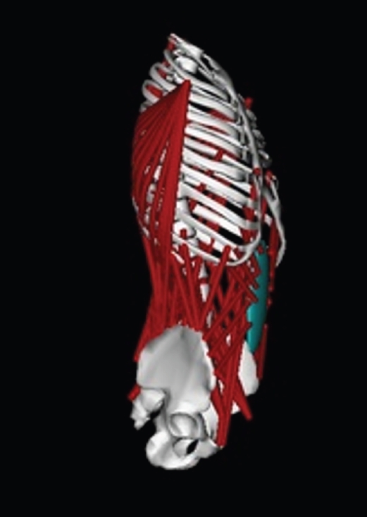
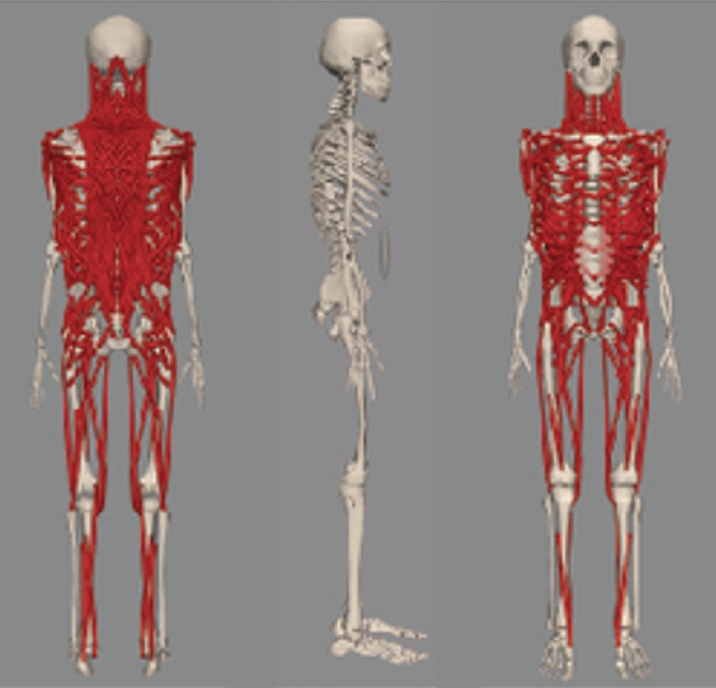
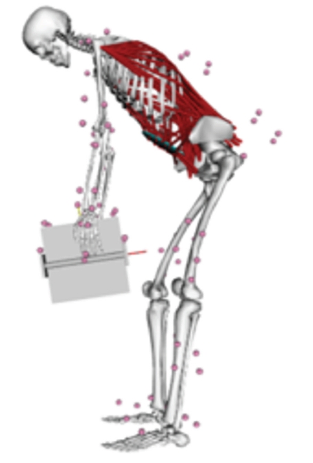
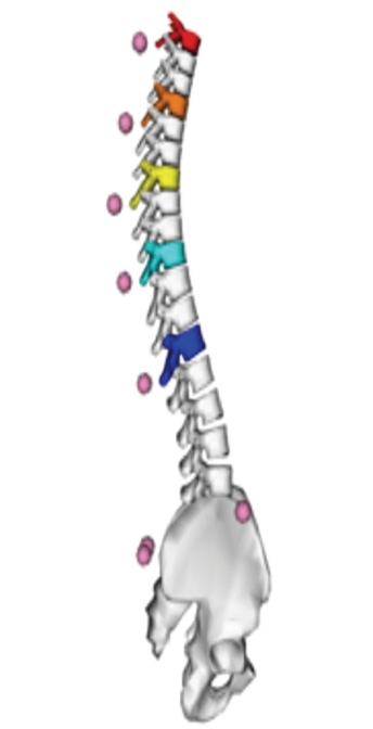
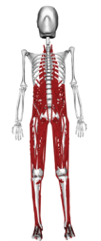
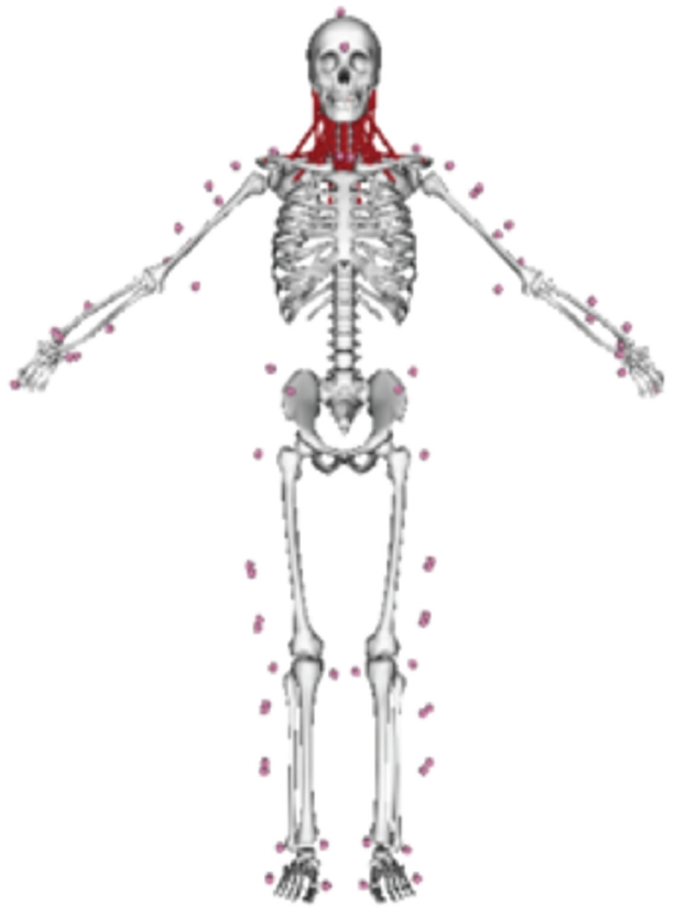
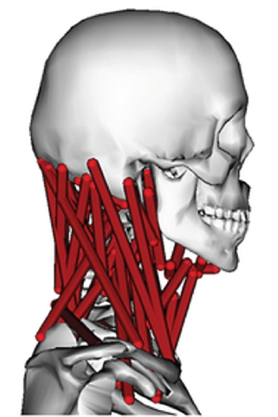
Fig. 1. Musculoskeletal model of the lumbar spine. The model includes a rigid pelvis and sacrum, five lumbar vertebrae, and the torso, including the thoracic spine and rib cage.
Fig. 2. The thoracolumbar spine and rib cage model, including vertebrae from T1 to L5. It has three degrees of motion for rotational movement. This model also includes the head, neck, upper extremities, and most major muscles of the lumbar spine, abdomen, and intercostal muscles.
Fig. 3. Spine model for estimating spinal loading during lifting and lowering tasks. Passive structures and kinematic constraints were incorporated to improve the accuracy of spinal load prediction during dynamic tasks.
Fig. 4. Articulated kinematic model of the thoracic and lumbar spine. The thoracic vertebrae were modelled separately to increase the degrees of freedom.
Fig. 5. London lumbar spine model. This model includes the lumbar spine and lower limbs and was created to analyse muscle forces and joint contact forces during daily activities, particularly lifting.
Fig. 6. MASI model of the cervical spine for injury mechanism analysis and simulation in sport. The model includes the scapula and clavicle, allowing coupled scapular and clavicular motion with humeral elevation.
Fig. 7. Hyoid model of the cervical spine. The accuracy of cervical moment estimates was improved by incorporating the hyoid muscles into the model.
Fig. 1.
Fig. 2.
Fig. 3.
Fig. 4.
Fig. 5.
Fig. 6.
Fig. 7.
Spine OpenSim models: a narrative review of the literature
| Model | Validity test | Output | Usage |
|---|---|---|---|
| Musculoskeletal model of the lumbar spine | No information | IK, ID | It is not suitable for complex motions of the spine that involve motion across two or more planes. |
| Thoracolumbar spine and rib cage model | The validity of the muscle forces and joint contact forces in this model was evaluated using data from the literature | IK, ID, SO | This model can be used for designing exoskeletons and determining spinal loading in various lifting conditions. |
| Full-body models with detailed thoracolumbar spine for children and adolescents | The validity of the outputs of this model was evaluated with literature. | IK, ID | This model can be used for evaluation of the motion of the spine in the subjects with age 6-18 years. |
| Model for estimating spinal loading during lifting and lowering tasks | The outputs of the model were validated using nine dynamic lifting tasks. | IK, ID, SO, FD | The model can effectively estimate spinal loads during lifting and lowering tasks. |
| Articulated kinematic model of the thoracic and lumbar spine | No information | IK, ID | This dynamic articulated model of the lumbar and thoracic spine was designed for kinematic analysis. |
| Lumbar spine with passive elements | No information | IK, ID | This model allows for the simulation of the rotational moment/force generated by the intervertebral disc, facet articulations, and ligaments |
| London lumbar spine model | The outputs of the models were validated with literature. | IK, ID | The outputs of the model regarding joint contact forces aligned well with existing literature. |
| University of Waterloo cervical spine model | The compressive force at C7‒T1 was calculated in this model and was compared to values from other models. | IK, ID | The results revealed discrepancies between the forces in this model and those reported in existing literature, ranging from 25.5 to 368.1 N. |
| Personalised scoliotic spine models | No information | IK, ID | This project aimed to create specific model of spine for scoliotic subjects. |
| Model to determine muscle forces in sit-to-stand transfer | No information | IK, ID | In this project a model was developed to analyse sit-to-stand transfer motion and also muscle forces. |
| MASI cervical spine model | The validity of MASI model was established based on dynamic verification, kinematic validation, and dynamic validation. | IK, ID, CMC, FD | By use of this model, joint contact force and muscle forces can be analysed in impact activities. |
| Rugby cervical spine model | The validity of the Rugby models was established based on dynamic verification, kinematic validation, and dynamic validation. | IK, ID, CMC, FD | By use of this model, joint contact force and muscle forces can be analysed in impact activities. |
| Full-body lifting model | The validity of the model was assessed through various procedures, including comparisons with EMG muscle data, evaluation of intradiscal pressure, and comparison of contact forces with vertebral body implant measurements | IK, ID, SO | It appears that this model has acceptable validity for evaluating changes in lumbar loading during lifting. |
| Model of neck to determine impact loading | No information | IK, ID | The kinematics of head movement was evaluated in this study in various loading conditions. |
| Head and neck musculoskeletal biomechanics | No information | IK, ID | This model has a high level of validity in determining the ROM. |
| Infant head kinematics during abusive head trauma model | No information | IK, ID | This model was developed to determine the kinematics of head movement in infants during shaking. |
| Hyoid model | No information | IK, ID, CMC, FD, RRA | The accuracy of cervical moments based on this model has been improved by incorporating hyoid muscles into the models. |
Table 1. Various models of spine developed for OpenSim software
All listed models are publicly available in OpenSim. IK, inverse kinematics; ID, inverse dynamics; SO, static optimization; FD, forward dynamics; CMC, computer muscle control; RRA, residual reduction algorithm; ROM, range of motion.


