 E-submission
 TOTA
TOTA TOTS
TOTS
Articles
- Page Path
- HOME > J Musculoskelet Trauma > Volume 33(3); 2020 > Article
- Review Article Current Concepts of Bone Healing
- Dong Hun Suh, Bong Mo Koo, Jong Woo Kang
-
Journal of Musculoskeletal Trauma 2020;33(3):171-177.
DOI: https://doi.org/10.12671/jkfs.2020.33.3.171
Published online: July 31, 2020
Department of Orthopedic Surgery, Korea University Ansan Hospital, Korea University College of Medicine, Ansan, Korea
- 2,517 Views
- 105 Download
- 0 Crossref
- 0 Scopus
Abstract
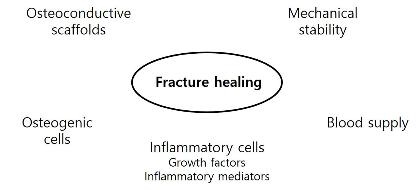
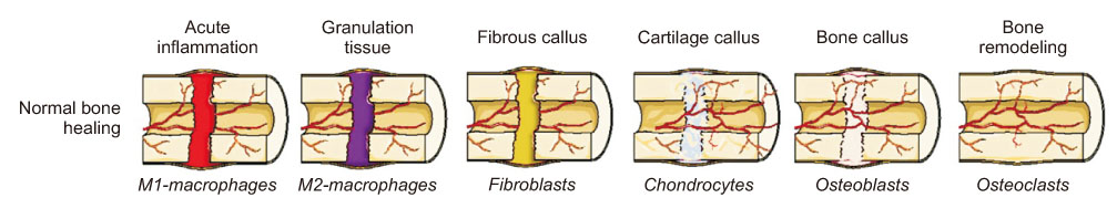
Bone injuries induce an inflammatory response that promotes bone healing. On the other hand, an aberrant process, where inflammation becomes chronic, can inhibit the healing of injured bone. At the first stage of the bone healing process, inflammatory cells, such as neutrophils and macrophages, are assembled and secrete various cytokines, chemokines, and growth factors. During callus formation, cells differentiated from mesenchymal stem cells, such as osteoblasts and chondrocytes, play leading roles in bone healing. Currently, various treatment modalities have been developed through the known mechanism of bone healing, and the clinical outcomes of bone defect and fracture nonunion have been good.
J Korean Fract Soc. 2020 Jul;33(3):171-177. Korean.
Published online Jul 24, 2020.
https://doi.org/10.12671/jkfs.2020.33.3.171
Published online Jul 24, 2020.
https://doi.org/10.12671/jkfs.2020.33.3.171
Copyright © 2020 The Korean Fracture Society. All rights reserved.
Review
Current Concepts of Bone Healing

Abstract
Bone injuries induce an inflammatory response that promotes bone healing. On the other hand, an aberrant process, where inflammation becomes chronic, can inhibit the healing of injured bone. At the first stage of the bone healing process, inflammatory cells, such as neutrophils and macrophages, are assembled and secrete various cytokines, chemokines, and growth factors. During callus formation, cells differentiated from mesenchymal stem cells, such as osteoblasts and chondrocytes, play leading roles in bone healing. Currently, various treatment modalities have been developed through the known mechanism of bone healing, and the clinical outcomes of bone defect and fracture nonunion have been good.
Keywords
Bone healing, Fractures, Inflammation, Tissue engineering
Figures
Tables
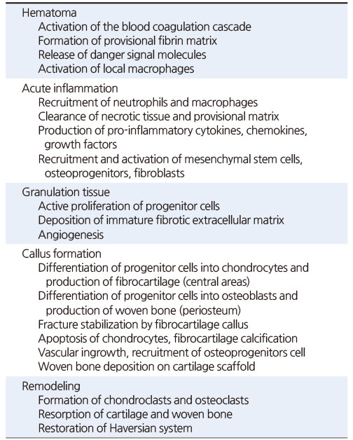
Table 1
Key Events during Secondary Fracture Healing
Notes
Financial support:None.
Conflict of interests:None.
References
-
Miranda MA, Moon MS. Treatment strategy for nonunions and malunions. In: Stannard JP, Schmidt AH, Kregor PJ, editors. Surgical treatment of orthopaedic trauma. New York: Thieme; 2007. pp. 77-100.
-
-
Hak DJ, Fitzpatrick D, Bishop JA, et al. Delayed union and nonunions: epidemiology, clinical issues, and financial aspects. Injury 2014;45 Suppl 2:S3–S7.
-
-
National Osteoporosis Foundation. What is osteoporosis and what causes it? [Internet]. Arlington: National Osteoporosis Foundation; [cited 2020 Apr 15].Available from: https://www.nof.org/patients/what-
is- osteoporosis/.
-
-
Mountziaris PM, Spicer PP, Kasper FK, Mikos AG. Harnessing and modulating inflammation in strategies for bone regeneration. Tissue Eng Part B Rev 2011;17:393–402.
-
-
Ortega-Gómez A, Perretti M, Soehnlein O. Resolution of inflammation: an integrated view. EMBO Mol Med 2013;5:661–674.
-
-
Kumagai K, Vasanji A, Drazba JA, Butler RS, Muschler GF. Circulating cells with osteogenic potential are physiologically mobilized into the fracture healing site in the parabiotic mice model. J Orthop Res 2008;26:165–175.
-
-
Fong EL, Chan CK, Goodman SB. Stem cell homing in musculoskeletal injury. Biomaterials 2011;32:395–409.
-
-
Osta B, Benedetti G, Miossec P. Classical and paradoxical effects of TNF-α on bone homeostasis. Front Immunol 2014;5:48
-
-
Giannoudis PV, Einhorn TA, Marsh D. Fracture healing: the diamond concept. Injury 2007;38 Suppl 4:S3–S6.
-
-
Jagodzinski M, Krettek C. Effect of mechanical stability on fracture healing--an update. Injury 2007;38 Suppl 1:S3–S10.
-
-
Claes L, Recknagel S, Ignatius A. Fracture healing under healthy and inflammatory conditions. Nat Rev Rheumatol 2012;8:133–143.
-
-
McKibbin B. The biology of fracture healing in long bones. J Bone Joint Surg Br 1978;60-B:150–162.
-
-
Perren SM. Physical and biological aspects of fracture healing with special reference to internal fixation. Clin Orthop Relat Res 1979;(138):175–196.
-
-
Loi F, Córdova LA, Pajarinen J, Lin TH, Yao Z, Goodman SB. Inflammation, fracture and bone repair. Bone 2016;86:119–130.
-
-
Harwood PJ, Newman JB, Michael ALR. (ii) An update on fracture healing and non-union. Orthop Trauma 2010;24:9–23.
-
-
Gerstenfeld LC, Cullinane DM, Barnes GL, Graves DT, Einhorn TA. Fracture healing as a post-natal developmental process: molecular, spatial, and temporal aspects of its regulation. J Cell Biochem 2003;88:873–884.
-
-
Uhthoff HK, Rahn BA. Healing patterns of metaphyseal fractures. Clin Orthop Relat Res 1981;(160):295–303.
-
-
Aspenberg P, Sandberg O. Distal radial fractures heal by direct woven bone formation. Acta Orthop 2013;84:297–300.
-
-
Chen WT, Han da C, Zhang PX, et al. A special healing pattern in stable metaphyseal fractures. Acta Orthop 2015;86:238–242.
-
-
Claes L, Reusch M, Göckelmann M, et al. Metaphyseal fracture healing follows similar biomechanical rules as diaphyseal healing. J Orthop Res 2011;29:425–432.
-
-
Kolar P, Schmidt-Bleek K, Schell H, et al. The early fracture hematoma and its potential role in fracture healing. Tissue Eng Part B Rev 2010;16:427–434.
-
-
Grundnes O, Reikerås O. The importance of the hematoma for fracture healing in rats. Acta Orthop Scand 1993;64:340–342.
-
-
Alexander KA, Chang MK, Maylin ER, et al. Osteal macrophages promote in vivo intramembranous bone healing in a mouse tibial injury model. J Bone Miner Res 2011;26:1517–1532.
-
-
Raggatt LJ, Wullschleger ME, Alexander KA, et al. Fracture healing via periosteal callus formation requires macrophages for both initiation and progression of early endochondral ossification. Am J Pathol 2014;184:3192–3204.
-
-
Pettit AR, Chang MK, Hume DA, Raggatt LJ. Osteal macrophages: a new twist on coupling during bone dynamics. Bone 2008;43:976–982.
-
-
Kon T, Cho TJ, Aizawa T, et al. Expression of osteoprotegerin, receptor activator of NF-kappaB ligand (osteoprotegerin ligand) and related proinflammatory cytokines during fracture healing. J Bone Miner Res 2001;16:1004–1014.
-
-
Bielby R, Jones E, McGonagle D. The role of mesenchymal stem cells in maintenance and repair of bone. Injury 2007;38 Suppl 1:S26–S32.
-
-
Wu AC, Raggatt LJ, Alexander KA, Pettit AR. Unraveling macrophage contributions to bone repair. Bonekey Rep 2013;2:373
-
-
Schindeler A, McDonald MM, Bokko P, Little DG. Bone remodeling during fracture repair: the cellular picture. Semin Cell Dev Biol 2008;19:459–466.
-
-
Malizos KN, Papatheodorou LK. The healing potential of the periosteum molecular aspects. Injury 2005;36 Suppl 3:S13–S19.
-
-
Keramaris NC, Calori GM, Nikolaou VS, Schemitsch EH, Giannoudis PV. Fracture vascularity and bone healing: a systematic review of the role of VEGF. Injury 2008;39 Suppl 2:S45–S57.
-
-
Gaston MS, Simpson AH. Inhibition of fracture healing. J Bone Joint Surg Br 2007;89:1553–1560.
-
-
Komatsu DE, Warden SJ. The control of fracture healing and its therapeutic targeting: improving upon nature. J Cell Biochem 2010;109:302–311.
-
-
Silverman SL, Siris E, Kendler DL, et al. Persistence at 12 months with denosumab in postmenopausal women with osteoporosis: interim results from a prospective observational study. Osteoporos Int 2015;26:361–372.
-
-
Pietschmann P, Mechtcheriakova D, Meshcheryakova A, Föger-Samwald U, Ellinger I. Immunology of osteoporosis: a minireview. Gerontology 2016;62:128–137.
-
-
Franceschi C, Bonafè M, Valensin S, et al. I nflamm-aging. Inflamm-aging. An evolutionary perspective on immunosenescence. Ann N Y Acad Sci 2000;908:244–254.
-
-
Lencel P, Magne D. Inflammaging: the driving force in osteoporosis? Med Hypotheses 2011;76:317–321.
-
-
Topini C, Topini D, Cerica G, Nardocci F, Topini G. Osteoporosis and risk of fracture, analysis on a population admitted in rehabilitation post-acute. Clin Cases Miner Bone Metab 2014;11:129–131.
-
-
Panteli M, Pountos I, Jones E, Giannoudis PV. Biological and molecular profile of fracture non-union tissue: current insights. J Cell Mol Med 2015;19:685–713.
-
-
Kloen P, Doty SB, Gordon E, Rubel IF, Goumans MJ, Helfet DL. Expression and activation of the BMP-signaling components in human fracture nonunions. J Bone Joint Surg Am 2002;84:1909–1918.
-
-
Fernandez-Bances I, Perez-Basterrechea M, Perez-Lopez S, et al. Repair of long-bone pseudoarthrosis with autologous bone marrow mononuclear cells combined with allogenic bone graft. Cytotherapy 2013;15:571–577.
-
-
Patterson TE, Kumagai K, Griffith L, Muschler GF. Cellular strategies for enhancement of fracture repair. J Bone Joint Surg Am 2008;90 Suppl 1:111–119.
-
-
Wang M, Zhang G, Wang Y, et al. Crosstalk of mesenchymal stem cells and macrophages promotes cardiac muscle repair. Int J Biochem Cell Biol 2015;58:53–61.
-
-
Spiller KL, Anfang RR, Spiller KJ, et al. The role of macrophage phenotype in vascularization of tissue engineering scaffolds. Biomaterials 2014;35:4477–4488.
-
-
Spiller KL, Vunjak-Novakovic G. Clinical translation of controlled protein delivery systems for tissue engineering. Drug Deliv Transl Res 2015;5:101–115.
-
-
Wernike E, Montjovent MO, Liu Y, et al. VEGF incorporated into calcium phosphate ceramics promotes vascularisation and bone formation in vivo. Eur Cell Mater 2010;19:30–40.
-
-
Aro HT, Govender S, Patel AD, et al. Recombinant human bone morphogenetic protein-2: a randomized trial in open tibial fractures treated with reamed nail fixation. J Bone Joint Surg Am 2011;93:801–808.
-
-
Heckman JD, Ryaby JP, McCabe J, Frey JJ, Kilcoyne RF. Acceleration of tibial fracture-healing by non-invasive, low-intensity pulsed ultrasound. J Bone Joint Surg Am 1994;76:26–34.
-
-
Kristiansen TK, Ryaby JP, McCabe J, Frey JJ, Roe LR. Accelerated healing of distal radial fractures with the use of specific, low-intensity ultrasound. A multicenter, prospective, randomized, double-blind, placebo-controlled study. J Bone Joint Surg Am 1997;79:961–973.
-
-
Leung KS, Lee WS, Tsui HF, Liu PP, Cheung WH. Complex tibial fracture outcomes following treatment with low-intensity pulsed ultrasound. Ultrasound Med Biol 2004;30:389–395.
-
-
Emami A, Petrén-Mallmin M, Larsson S. No effect of low-intensity ultrasound on healing time of intramedullary fixed tibial fractures. J Orthop Trauma 1999;13:252–257.
-
-
Mayr E, Rudzki MM, Rudzki M, Borchardt B, Häusser H, Rüter A. [Does low intensity, pulsed ultrasound speed healing of scaphoid fractures?]. Handchir Mikrochir Plast Chir 2000;32:115–122.German.
-
-
Lubbert PH, van der, Hoorntje LE, van der. Low-intensity pulsed ultrasound (LIPUS) in fresh clavicle fractures: a multi-centre double blind randomised controlled trial. Injury 2008;39:1444–1452.
-
-
Watanabe Y, Matsushita T, Bhandari M, Zdero R, Schemitsch EH. Ultrasound for fracture healing: current evidence. J Orthop Trauma 2010;24 Suppl 1:S56–S61.
-
-
Handolin L, Kiljunen V, Arnala I, Pajarinen J, Partio EK, Rokkanen P. The effect of low intensity ultrasound and bioabsorbable self-reinforced poly-L-lactide screw fixation on bone in lateral malleolar fractures. Arch Orthop Trauma Surg 2005;125:317–321.
-
-
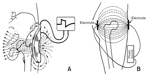
Aleem IS, Aleem I, Evaniew N, et al. Efficacy of electrical stimulators for bone healing: a meta-analysis of randomized shamcontrolled trials. Sci Rep 2016;6:31724
-
-
Goldstein C, Sprague S, Petrisor BA. Electrical stimulation for fracture healing: current evidence. J Orthop Trauma 2010;24 Suppl 1:S62–S65.
-
 Cite
Cite
