 E-submission
 TOTA
TOTA TOTS
TOTS
Articles
- Page Path
- HOME > J Musculoskelet Trauma > Volume 25(1); 2012 > Article
-
Case Report
- Treatment of a 3rd Lumbar Vertebra Translational Injury Combined with Incomplete Cauda Equina Syndrome in Ankylosing Spondylitis: A Case Report
- Jin-Wan Kim, M.D., Young-Chul Ko, M.D., Chul-Young Jung, M.D., Il-Soo Eun, M.D., Young-June Kim, M.D., Chang-Kyu Kim, M.D.
-
Journal of the Korean Fracture Society 2012;25(1):77-81.
DOI: https://doi.org/10.12671/jkfs.2012.25.1.77
Published online: January 31, 2012
Department of Orthopedic Surgery, Busan Medical Center, Busan, Korea.
- Address reprint requests to: Young-Chul Ko, M.D. Department of Orthopedic Surgery, Busan Medical Center, 1330, Geoje 2-dong, Yeonje-gu, Busan 611-072, Korea. Tel: 82-51-607-2862, Fax: 82-51-507-3001, drgo1973@nate.com
• Received: August 25, 2011 • Revised: November 23, 2011 • Accepted: December 9, 2011
Copyright © 2012 The Korean Fracture Society
- 709 Views
- 1 Download
Abstract
- Ankylosing spondylitis is a rheumatic disease in which mainly the spinal and sacroiliac joints are affected. Patients with ankylosing spondylitis are at significant risk for spinal fracture when exposed to even minor trauma. Most spinal fractures with ankylosing spondylitis occur in the cervical spine, whereas spinal fractures in thoracic or lumbar spine are rare, especially in the lower lumbar spine. Furthermore, neurologic symptoms in cases of lower lumbar spine fracture are rarer than in cases of cervical and thoracic spinal fracture. We have experienced a case of translation injury of the 3rd lumbar vertebra accompanied by incomplete cauda equine syndrome in ankylosing spondylitis and the authors gained good clinical results with surgical treatment. We have reported here on this case and have included a review of the relevant literature.
- 1. Chung YK, Yoo JH, Park YW, Baik SC. Fracture of the fifth lumbar vertebra in ankylosing spondylitis. J Korean Soc Spine Surg, 1996;3:280-284.
- 2. Fast A, Parikh S, Marin EL. Spine fractures in ankylosing spondylitis. Arch Phys Med Rehabil, 1986;67:595-597.
- 3. Ghozlani I, Ghazi M, Nouijai A, et al. Prevalence and risk factors of osteoporosis and vertebral fractures in patients with ankylosing spondylitis. Bone, 2009;44:772-776.Article
- 4. Murray GC, Persellin RH. Cervical fracture complicating ankylosing spondylitis: a report of eight cases and review of the literature. Am J Med, 1981;70:1033-1041.
- 5. Olerud C, Frost A, Bring J. Spinal fractures in patients with ankylosing spondylitis. Eur Spine J, 1996;5:51-55.ArticlePDF
- 6. Osgood CP, Abbasy M, Mathews T. Multiple spine fractures in ankylosing spondylitis. J Trauma, 1975;15:163-166.Article
- 7. Patel SN, Turtz A, Dixon A, Yocom S. Neurologically intact lumbar spine displaced fracture with ankylosing spondylitis. West J Emerg Med, 2011;12:142-143.
- 8. Chaudhary SB, Hullinger H, Vives MJ. Management of acute spinal fractures in ankylosing spondylitis. ISRN Rheumatol, 2011;2011:150484. ArticlePDF
- 9. Shen FH, Samartzis D. Successful nonoperative treatment of a three-column thoracic fracture in a patient with ankylosing spondylitis: existence and clinical significance of the fourth column of the spine. Spine (Phila Pa 1976), 2007;32:E423-E427.
- 10. Thorngren KG, Liedberg E, Aspelin P. Fractures of the thoracic and lumbar spine in ankylosing spondylitis. Arch Orthop Trauma Surg, 1981;98:101-107.ArticlePDF
REFERENCES
Fig. 1
(A, B) Preoperative anteroposterior view and lateral view show third lumbar vertebrae translational injury traversing across all three columns in a 55-year-old man with ankylosing spondylitis.

Fig. 2
(A, B) Preoperative T2 weighted sagittal and axial MRI show translational injury in the L3 vertebral body with canal encroachment and posterior longitudinal ligament tear in the posterior column.


Figure & Data
REFERENCES
Citations
Citations to this article as recorded by 
 Cite
CiteTreatment of a 3rd Lumbar Vertebra Translational Injury Combined with Incomplete Cauda Equina Syndrome in Ankylosing Spondylitis: A Case Report
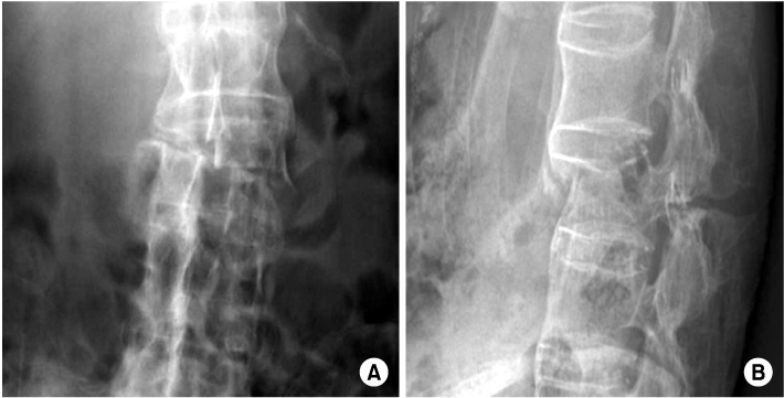
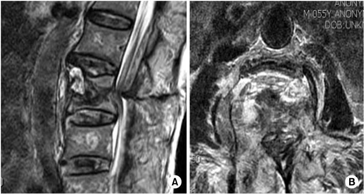
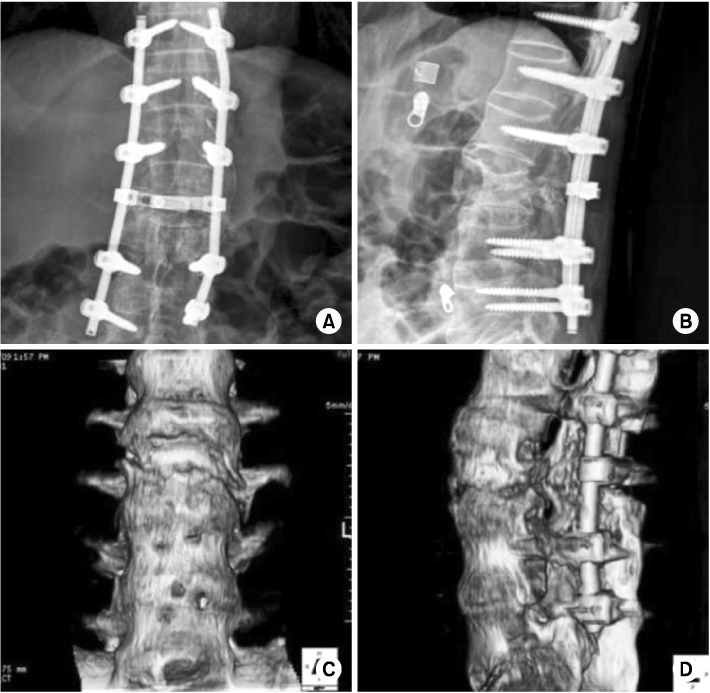

Fig. 1
(A, B) Preoperative anteroposterior view and lateral view show third lumbar vertebrae translational injury traversing across all three columns in a 55-year-old man with ankylosing spondylitis.
Fig. 2
(A, B) Preoperative T2 weighted sagittal and axial MRI show translational injury in the L3 vertebral body with canal encroachment and posterior longitudinal ligament tear in the posterior column.
Fig. 3
(A~D) At a postoperative 12 weeks, anteroposterior and lateral plain X-ray show correction of height loss and anteroposterior and lateral 3D reconstruction image shows maintenance of reduced fracture.
Fig. 4
(A, B) At a postoperative 12 months, anteroposterior and lateral plain X-ray show reduced kyphosis and fracture.
Fig. 1
Fig. 2
Fig. 3
Fig. 4
Treatment of a 3rd Lumbar Vertebra Translational Injury Combined with Incomplete Cauda Equina Syndrome in Ankylosing Spondylitis: A Case Report

