 E-submission
 TOTA
TOTA TOTS
TOTS
Search
- Page Path
- HOME > Search
Original Article
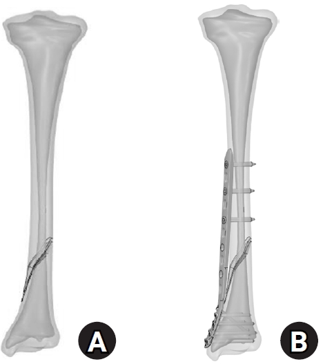
- Biomechanical analysis of medial distal tibial locking plate fixation for distal-third spiral tibial shaft fractures
- Yao-Jen Liu
- J Musculoskelet Trauma 2026;39(2):140-146. Published online April 10, 2026
- DOI: https://doi.org/10.12671/jmt.2026.00094
-
 Abstract
 PDF
PDF - Background
Distal spiral fractures of the tibial shaft present fixation challenges, particularly in patients who are not suitable candidates for intramedullary nailing. This study evaluated the biomechanical stability of medial minimally invasive percutaneous plating osteosynthesis (MIPO) under various physiological loading conditions.
Methods
A finite-element model of a distal AO/OTA 42-A1.1c spiral fracture of the tibia was created using computed tomography data. A precontoured titanium medial distal tibia locking compression plate with nine locking screws was simulated. Material properties were assigned to cortical and cancellous bone. The loading conditions included axial compression (750 N), varus/valgus bending (300 N at a 9° offset), and internal/external torsion (7.5 N·m). von Mises stress and fracture displacement were analyzed.
Results
Axial loading produced a peak plate stress of 508.06 MPa and a displacement of 2.17 mm. Valgus and varus loading generated stresses of 490.17 MPa and 324.08 MPa, respectively, with corresponding displacements of 3.86 mm and 2.01 mm. External and internal torsion resulted in stresses of 354.23 MPa and 358.9 MPa, respectively, with corresponding displacements of 2.64 mm and 2.22 mm.
Conclusions
Medial distal tibial plating demonstrated favorable biomechanical performance in this finite-element model; however, clinical extrapolation should be made cautiously. Level of evidence: V.
- 299 View
- 16 Download

Case Reports
- Concurrent Posterolateral Corner Injury Associated with a Schatzker Type 2 Tibial Plateau Fracture: A Case Report
- Jae Cheon Sim, Choong Won Jung, Tae Seok Nam
- J Korean Fract Soc 2020;33(1):27-31. Published online January 31, 2020
- DOI: https://doi.org/10.12671/jkfs.2020.33.1.27
-
Abstract
PDF
- Isolated posterolateral corner (PLC) injury associated with a Schatzker type 2 fracture is a very rare combination of injuries. A male who was driving a motor vehicle was injured after a collision accident. The plain radiographs and computed tomography scans of the knee showed a Schatzker type 2 fracture of the tibial plateau, mostly in the anterolateral portion of tibial plateau, and an avulsion fragment on the fibular tip. Magnetic resonance imaging showed no injury to cruciate ligaments, medial collateral ligament, or any meniscal injury. We performed an open reduction operation and internal fixation for treating the fracture. Six months later, he complained of instability. At 11 months later after initial operation, we performed the second operation for stabilizing the PLC. We present here a rare case of an isolated PLC injury associated with a Schatzker type 2 fracture. We discuss the mechanism of injury and review similar cases.
- 1,348 View
- 5 Download
- Calcified Anterior Tibial Artery Entrapment in Distal Third Tibial Fracture: A Case Report
- Kyu Hyun Yang, Yougun Won, Sang Bum Kim, Won Kuen Park, You Sun Jung
- J Korean Fract Soc 2016;29(1):68-72. Published online January 31, 2016
- DOI: https://doi.org/10.12671/jkfs.2016.29.1.68
-
Abstract
PDF
- In the distal third of the tibia, the anterior tibial artery runs close to the anterolateral surface of the tibial cortex. In a clinical situation, without vascular evaluation, injury or entrapment of the anterior tibial artery is difficult to detect. Because, an intact dorsalis pedis pulse is supplied with the collateral vessels of the posterior tibial artery. An entrapped anterior tibial artery can be injured during closed reduction in an emergency room or open reduction and internal fixation in the operating room. Care must be taken to prevent iatrogenic anterior tibial artery. In this case, an entrapped anterior tibial artery was observed in a simple radiograph and computed tomograph without contrast media for the vessel. We report on a rare case of calcified anterior tibial artery entrapment in a distal tibial fracture.
-
Citations
Citations to this article as recorded by
- Comparison of Time to Operation and Efficacies of Ultrasound-Guided Nerve Block and General Anesthesia in Emergency External Fixation of Lower Leg Fractures (AO 42, 43, 44)
Chan Kang, Sang-Bum Kim, Youn-Moo Heo, You-Gun Won, Byung-Hak Oh, June-Bum Jun, Gi-Soo Lee
The Journal of Foot and Ankle Surgery.2017; 56(5): 1019. CrossRef
- Comparison of Time to Operation and Efficacies of Ultrasound-Guided Nerve Block and General Anesthesia in Emergency External Fixation of Lower Leg Fractures (AO 42, 43, 44)
- 1,085 View
- 5 Download
- 1 Crossref
Original Article
- Intramedullary Nailing of Distal Tibial Fractures with Percutaneous Reduction by Pointed Reduction Forceps
- Jae Kwang Hwang, Chung Hwan Kim, Young Joon Choi, Gi Won Lee, Hyun Il Lee, Tae Kyung Kim
- J Korean Fract Soc 2014;27(2):144-150. Published online April 30, 2014
- DOI: https://doi.org/10.12671/jkfs.2014.27.2.144
-
Abstract
PDF
- PURPOSE
The purpose of this study is to analyze the radiographic and clinical results of intramedullary nailing after percutaneous reduction using pointed reduction forceps for spiral or oblique fractures of the distal tibia. The benefit of percutaneous reduction using pointed reduction forceps in anatomical reduction and maintenance was assessed.
MATERIALS AND METHODS
From January 2005 to December 2009, 47 cases of distal one-third tibial fracture were managed by intramedullary nailing using pointed reduction forceps. Thirty-eight cases were spiral fracture and nine cases were oblique fracture. In all cases, the percutaneous reduction was achieved using pointed reduction forceps under fluoroscopy control. While maintaining the reduction with the pointed reduction forceps, the intramedullary nail was inserted. The pointed reduction forceps were removed after insertion of proximal and distal inter-locking screws. Alignment was evaluated with anterior-posterior and lateral radiographs taken immediately post-operation and at the time of union.
RESULTS
At immediate post-operation, the mean displacement of valgus and anterior angulation was 0.57degrees and 0.24degrees, respectively. That of valgus and anterior angulation at bone union was 0.37degrees and 0.16degrees, respectively. The average duration of bone union was 16.1 weeks.
CONCLUSION
Intramedullary nailing with percutaneous reduction using pointed reduction forceps for distal tibial fractures was an easy and effective method for achievement of accurate alignment intra-operatively. Accurate alignment was successfully maintained until bone union.
- 804 View
- 2 Download
Case Report
- Minimally Invasive Percutaneous Plate Stabilization Using a Medial Locking Plate for Proximal Tibial Fractures: Technical Note
- Jae Ang Sim, Beom Koo Lee, Kwang Hui Kim, Yong Seuk Lee
- J Korean Fract Soc 2013;26(4):327-332. Published online October 31, 2013
- DOI: https://doi.org/10.12671/jkfs.2013.26.4.327
-
Abstract
PDF
- Minimally invasive plate osteosynthesis (MIPO) is beneficial for proximal tibial fractures since these injuries are mostly caused by high energy traumas. The advantages of MIPO are minimization of soft tissue dissection and preservation of periosteal vascularization. Lateral plating has mostly developed as MIPO for proximal tibial fractures. We introduce minimal invasive percutaneous plate stabilization using a medial locking plate as alternative treatment for proximal tibial fractures.
-
Citations
Citations to this article as recorded by- Effect of Korean Medicine Treatments in Patients with Proximal Tibia Fracture: A Retrospective Observational Study
Jung Min Lee, Eun-Jung Lee
Journal of Korean Medicine Rehabilitation.2020; 30(3): 141. CrossRef - Medial Minimally Invasive Percutaneous Plate Osteosynthesis in Proximal Tibial Comminuted Fractures
Jae-Ang Sim, Kwang-Hui Kim, Yong-Seuk Lee, Sang-Jin Lee, Beom-Koo Lee
Journal of the Korean Orthopaedic Association.2014; 49(4): 278. CrossRef
- Effect of Korean Medicine Treatments in Patients with Proximal Tibia Fracture: A Retrospective Observational Study
- 1,255 View
- 35 Download
- 2 Crossref
Original Articles
- Treatment for Bone Defect of Open Tibial Fractures by Using Intramedullary Nail Fixation with Autogenous Iliac Bone Graft
- Hyub Sakong, Ki Cheor Bae, Chul Hyun Cho, Kyung Jae Lee, Eun Seok Son, Du Han Kim
- J Korean Fract Soc 2012;25(4):288-294. Published online October 31, 2012
- DOI: https://doi.org/10.12671/jkfs.2012.25.4.288
-
Abstract
PDF
- PURPOSE
This study was conducted to evaluate the results of intramedullary nail fixation with autogenous iliac bone graft for defects of bone after tibial fractures.
MATERIALS AND METHODS
Ten patients with bone defects in tibial fractures who had been treated with intramedullary nail fixation with autogenous iliac bone graft between May 2005 and September 2008 with more than 12 month follow-up were subject to study. Of the 10 patients, 8 were male and 2 were female, and the mean age was 50.2 years (29~76 years). By cause of accident, motor vehicle accidents caused 9 cases, a crush caused 1 case, and the average follow-up period was 21.9 months (12~42 months). Radiologically, we analyzed the union of the bone defect on simple x-ray and clinical evaluation was performed using the estimate method of Mekhali.
RESULTS
This study reveals that there was radiological union in all 10 cases and the mean time to union was 8.4 months (5~18 months). By clinical evaluation according to Mekhali's estimate method, 9 patients had excellent outcomes and 1 patient had limitation of motion in the ankle joint rated as a fair clinical result. None of patients developed complications post-operatively.
CONCLUSION
Our study demonstrated that the intramedullary nail fixation with autogenous iliac bone graft can be a useful operative method because it can remove external fixators early and reduce complications, and autogenous bones have exceptional osteoconduction, osteoinduction, and bone-forming ability resulting in excellent union of bones.
- 811 View
- 7 Download
- Alteration of the Patella Tendon Length after Intramedullary Nail in Tibial Shaft Fractures
- Dong Eun Shin, Ki Shik Nam, Jin Young Bang, Ji Hoon Chang
- J Korean Fract Soc 2012;25(4):283-287. Published online October 31, 2012
- DOI: https://doi.org/10.12671/jkfs.2012.25.4.283
-
Abstract
PDF
- PURPOSE
To compare and analyze length change of patella tendon after intramedullary nailing of tibial shaft fracture using transtendinous approach.
MATERIALS AND METHODS
Thirty-two cases were analyzed from December, 1999 to December, 2005. Insall Salvati ratios were estimated. Severity of initial trauma, duration of nail retension, knee function and pain on change of length of patellar tendon was evaluated.
RESULTS
Mean duration of nail retention was twenty-two months. The shortening of patella tendon was observed in 25 cases (p<0.001). The effect of AO type and the duration of nail retension on the decrease of Insall Salvati ratio was not significant (p>0.05, p=0.778). Lysholom score decrease to 89.5. There was no significant difference between the shortening of patellar tendon length and knee pain (p=0.058).
CONCLUSION
After intramedullary nailing for closed tibia fracture, shortening of patellar tendon length is observed. That is irrelevant to the fracture type and the duration of nail retension. The shortening of patella tendon length may contribute to decreasing of knee function, but it was no significance of knee pain after intramedullary nailing.
- 792 View
- 0 Download
- Treatment of Tibial Fractures In Children With Pin and Plaster Technique
- Byoung Ho Suh, Gyu Min Kong, Sang Ho Moon, Dong Joon Kim, Jin Woo Kwon, Se Won Park
- J Korean Fract Soc 2005;18(3):325-329. Published online July 31, 2005
- DOI: https://doi.org/10.12671/jkfs.2005.18.3.325
-
Abstract
PDF
- PURPOSE
To evaluate the result of tibial shaft fractures in children treated with pin and plaster method.
MATERIALS AND METHODS
From March 1998 to February 2003, Tibial shaft fractures in thirty six pediatric patients which were treated with pin and plaster method were clinically and radiologicaly evaluated retrospectively.
RESULTS
Mean bony union duration was 9.8 weeks. All fractures healed within acceptable angulations. There was neither delayed union nor nonunion. There were complications related to the pins, including superficial and deep infection, skin sloughing. There were 7 cases of tibial overgrowth but they had no functional disability.
CONCLUSION
Pin and plaster method can substitute other operative methods in tibial fractures in children which is difficult to reduce or maintain reduction by conservative treatment.
- 594 View
- 2 Download
- Treatment of Open Tibial Shaft Fractures using Unreamed Nailing
- Jong Keon Oh, Chang Wug Oh, Kwon Jae Roh, Duk Moon Chung
- J Korean Fract Soc 2005;18(1):22-28. Published online January 31, 2005
- DOI: https://doi.org/10.12671/jkfs.2005.18.1.22
-
Abstract
PDF
- PURPOSE
To report the results of unreamed nailing using a nail with the largest possible diameter for the management of the open tibial shaft fractures.
MATERIALS AND METHODS
Nineteen patients with open tibial shaft fractures underwent unreamed nailing with the largest possible diameter according to the isthmic diameter measured on preoperative radiography. There were 1 Grade I, 6 Grade II, 9 Grade IIIa, 3 Grade IIIb open fractures. There were 4 type A, 12 type B, 3 type C fractures according to the OTA classification. Fractures were classified as The nail was introduced after gentle passage of a 7 to 8 millimeter-hand reamer.
RESULTS
Union was obtained in all cases. However 9 (47%) fractures required an additional procedures before union. In 6 cases, dynamization was done. Two of them were required exchange nailing for nonunion, 1 of two gained bony union through additional bone graft. Three of the others had gained union through exchange nailing, bone graft, bone transport respectively. There were one rotational malunion, one superfical and one deep infection. Interlocking screw breakage developed only in one patient.
CONCLUSION
Our data indicate that unreamed nailing in the management of open tibial fractures is safe and reliable method. Using a tight fitting nail with the largest possible diameter is a safe and effective way to avoid the problems of screw breakage. -
Citations
Citations to this article as recorded by- Treatment of Type IIIb Open Tibial Fractures
Seong Yeon Lim, Il Jae Lee, Jae Ho Joe, Hyung Keun Song
Journal of the Korean Fracture Society.2014; 27(4): 267. CrossRef - Management of Open Tibial Fractures: Role of Internal Fixation
Yerl-Bo Sung
Journal of the Korean Fracture Society.2007; 20(4): 349. CrossRef
- Treatment of Type IIIb Open Tibial Fractures
- 911 View
- 0 Download
- 2 Crossref
- Minimally Invasive Percutaneous Plate Stabilization of Proximal Tibial Fractures
- Chang Wug Oh, Jong Keon Oh, In Ho Jeon, Hee Soo Kyung, Il Hyung Park, Joo Chul Ihn, Yeon Ki Woo, Ho Sung Jung
- J Korean Fract Soc 2004;17(3):224-229. Published online July 31, 2004
- DOI: https://doi.org/10.12671/jkfs.2004.17.3.224
-
Abstract
PDF
- PURPOSE
Despite of various treatment methods, proximal tibial fractures are common injuries that may be associated with poor outcomes and high rates of complications. To improve this, percutaneous plating technique was performed in the proximal tibial fractures.
MATERIALS AND METHODS
Twenty-four proximal tibial fractures (AO 41A; 5, AO 41C; 12, AO 42; 7) were treated by percutaneous plating with either or both sides without bone graft. One was open fracture.
RESULTS
All fractures were healed. The average time for fracture healing was 16.5 weeks (range, 8~24 weeks). Complications included a 1cm shortened case and two mal-alignments; a 6 degree valgus case and 5 degree varus case. There was one case of superficial infection, which healed after plate removal. But, there was no deep infection. Results were evaluated by modified Rasmussen score system, all patients had excellent or good result.
CONCLUSION
Minimally invasive percutaneous plating technique can provide favorable results in the treatment of proximal tibial fractures. -
Citations
Citations to this article as recorded by- MINIMALLY INVASIVE OSTEOSYNTHESIS WITH PLATE OR NAIL FOR META-DIAPHYSEAL TIBIAL FRACTURES - WHAT IS BETTER?
B. Makelov
Trakia Journal of Sciences.2023; 21(4): 357. CrossRef - Medial Minimally Invasive Percutaneous Plate Osteosynthesis in Proximal Tibial Comminuted Fractures
Jae-Ang Sim, Kwang-Hui Kim, Yong-Seuk Lee, Sang-Jin Lee, Beom-Koo Lee
Journal of the Korean Orthopaedic Association.2014; 49(4): 278. CrossRef - Minimally Invasive Percutaneous Plate Stabilization Using a Medial Locking Plate for Proximal Tibial Fractures - Technical Note -
Jae Ang Sim, Beom Koo Lee, Kwang Hui Kim, Yong Seuk Lee
Journal of the Korean Fracture Society.2013; 26(4): 327. CrossRef - Clinical Outcomes of Locking Compression Plate Fixation through Minimally Invasive Percutaneous Plate Osteosynthesis in the Treatment of Distal Tibia Fracture
Jae-Sung Yoo, Hyun-Woo Park
Journal of the Korean Fracture Society.2012; 25(2): 117. CrossRef - Minimally Invasive Plate Osteosynthesis for Proximal Tibial Shaft Fracture
Young-Soo Byun, Ki-Chul Park, Hyun-Jong Bong, Chang-Hoon Lee
Journal of the Korean Fracture Society.2011; 24(1): 23. CrossRef - Minimally Invasive Plate Osteosynthesis for the Upper Extremity Fracture Using a Lumbar Spreader - Surgical Technique -
Gu-Hee Jung, Chyul-Hyun Cho, Jae-Do Kim
Journal of the Korean Fracture Society.2011; 24(1): 83. CrossRef - Management of Fractures of Distal Tibia by Minimally Invasive Plate Osteosynthesis through an Anterior Approach
Gu-Hee Jung, Jae-Do Kim, Jae-Ho Jang, Sung-Keun Heo, Dong-won Lee
Journal of the Korean Orthopaedic Association.2010; 45(6): 473. CrossRef - The Comparison of Minimally Invasive Plate Osteosynthesis and Intramedullary Nailing in the Treatment of the Proximal and Distal Tibia Fracture
Joon Soon Kang, Seung Rim Park, Sang Rim Kim, Yong Geun Park, Jae Ho Jung, Sung Wook Choi
Journal of the Korean Fracture Society.2010; 23(2): 172. CrossRef - Staged Minimally Invasive Plate Osteosynthesis of Proximal Tibial Fracture
Joon-Woo Kim, Chang-Wug Oh, Jong-Keon Oh, Hee-Soo Kyung, Woo-Kie Min, Byung-Chul Park, Kyung-Hoon Kim, Hee-Joon Kim
Journal of the Korean Fracture Society.2009; 22(1): 6. CrossRef - Proximal Tibia Fracture: Plating
Ki-Chul Park
Journal of the Korean Fracture Society.2009; 22(3): 206. CrossRef
- MINIMALLY INVASIVE OSTEOSYNTHESIS WITH PLATE OR NAIL FOR META-DIAPHYSEAL TIBIAL FRACTURES - WHAT IS BETTER?
- 823 View
- 2 Download
- 10 Crossref
- Biomechanical Analysis of Hybrid External Fixation for the Distal Tibial Fractures: A FEM Study
- Duk Young Jung, Boug Ju Kim, Seok Bae Ryu, Jong Keon Oh
- J Korean Fract Soc 2004;17(2):160-166. Published online April 30, 2004
- DOI: https://doi.org/10.12671/jkfs.2004.17.2.160
-
Abstract
PDF
- PURPOSE
To analyze the biomechanical effects of different frame configurations of the hybrid external fixators for distal tibial fractures on the frame stiffness and stress distribution with a finite element method (FEM).
MATERIALS AND METHODS
Five configurations were simulated: Group I: two wires with convergence angle of 60degrees, Group II: 3rd wire on a bisector axis of the group I. Group III: two wires with 30degrees. Group IV: 3rd wire on a bisector axis. Group V: two wires with 30degree and a half pin on the distal articular fragment. Each group was simulated under compression, torsion, anterior-posterior and lateral-medial bending load. Stiffness, stress and deformation values were calculated.
RESULTS
The overall stiffness was increased by 15~30% with the addition of a third wire, and by 150~400% with a anteromedial half pin on the articular fragment. The half pin decreased the stress level of the frame by about 43% and the deformation of the 5/8 ring by about 30%.
CONCLUSION
The addition of a half pin on the articular fragment is not only a method of increasing the stiffness but also a way of decreasing the stress concentration and the deformation of the frame.
- 608 View
- 1 Download
- Unreamed interlocking nailing in tibia fracture
- You Sung Suh, Young Il Cho, Ho Won Jung, Yeon Il Kim
- J Korean Soc Fract 2002;15(4):470-476. Published online October 31, 2002
- DOI: https://doi.org/10.12671/jksf.2002.15.4.470
-
Abstract
PDF
- PURPOSE
To evaluate of clinical results and malunion according to nail insertion site and early ambulation after unreamed interlocking intramedullary nailing for the treatment of tibial fractures, MATERIALS AND METHODS: We reviewed 46 tibial fractures that were treated with unreamed static intramedullary nailing prospectively from March 1997 to May 2001. Nail insertion site and angulation of fracture site were reviewed by radiograph. All of 46 cases, ambulation was started at postoperative 2 weeks, and then clinical outcomes were reveiwed RESULTS: In all 46 cases, union was achieved at average 18.2 weeks clinically and average 19.4 weeks radiographically. There is no significant difference in angulation according to nail insertion site, i,.e. after central/medial/lateral insertion, outcome was 2 . 4 5 degrees +/-2 . 1 7 / 2 . 2 2 degrees +/-1 . 8 4 / 1 . 7 3 degrees +/-1.33(p; 0.705) in last follow up anterioposterior view, and 1.81 degrees +/-1 . 1 3 / 2 . 6 7 degrees +/-1 . 6 2 / 2 . 0 0 degrees +/-1.64(p; 0.320) in last follow up lateral view. No breakage of intramedullary nails and no stiffness on adjacent joints.
CONCLUSION
We confirmed that unreamed interlocking nailing in tibial fractures is one of the effective method for low recurrence of malunion and early ambulation
- 500 View
- 0 Download
- The Angular Deformity of Interlocking Nailing in Tibial Fractures
- Hwa Jae Jeong, Kyung Chul Kim, Jae Yeul Choi, Bon Seop Koo, Jung Hee Oh
- J Korean Soc Fract 2000;13(4):905-911. Published online October 31, 2000
- DOI: https://doi.org/10.12671/jksf.2000.13.4.905
-
Abstract
PDF
- PURPOSE
We studied the relationship between angular deformity and possibly contributing factors in the treatment of tibial fractures with interlocking nailing.
MATERIALS AND METHODS
Intramedullary nailing of the tibia was performed on 49 cases and were followed for the minimum of 12 months. We analyzed relationship between angular deformity and postoperative tibial alignment, operative technique and other factors.
RESULTS
Of the 49 cases, 19(38%) were angulated. Angular deformity was seen in 60%, 51.8% and 11.8% in the proximal, distal and middle third of tibial fractures respectively. With AO classification, Group A,B,C were angulated in 32.4%, 55.6%, 66.7%. In group A, 43.8% of spiral fractures, 28.6% of oblique fractures and 14.3% of transverse fractures were angulated. The cases combined with fibular fracture showed higher incidence of angular deformity than the cases with intact fibula. The opening of fracture and the nail insertion site were not significant to angular deformity.
CONCLUSION
Angular deformity of interlocking nailing in tibial fractures were more common in proximal, comminuted and spiral fractures. Precise attentions to operative technique i. e. accurate anatomical reduction and centromedullary nail orientation are recommended to prevent angular deformity. In proximal third tibial shaft fractures where muscles and patellar tendon has deforming force on fracture fragment, authors believe that use of interlocking nailing must be limited with fracture pattern.
- 641 View
- 3 Download
- Brooker Intramedually Nailing for the Treatment of Distal 1/3 Tibial Fractures with Compromised Soft Tissue
- Chung Nam Kang, Jong Oh Kim, Yeo Hon Yun, Dong Wook Kim, Young Do Koh, Jae Doo Yoo, Jong Keon Oh, Ki Woong Lee
- J Korean Soc Fract 1999;12(4):924-931. Published online October 31, 1999
- DOI: https://doi.org/10.12671/jksf.1999.12.4.924
-
Abstract
PDF
- The treatment of distal tibial fractures with compromised soft tissue poses many problems that usually occurs from the high-energy trauma, and the results are often unsatisfactory following lots of complications like loss of reduction, malunion, and inlection. We studied to evaluate the treatment results of Brooker intramedually nailing for the distal 1/3 tibial fractures with compromised soft tissue. Twenty-three cases of distal tibial fractures with comprolnised soft tissue were reviewed and we analyzed the results of surgical treatment in the viewpoint of union time, loss of reduction, malunion, complication and its final outcome. The range of follow-up was 24 months to 38 months with mean 29 months follow-up. Most of patients were between twenty and sixty years, and average age was 43.2 years. Acording to Gustilo and Andersons classification, 3 were Type I, 2 were Type II of 5 open fractures. According to Tschernes classification, 13 were Grade I, 5 were Grade II of 18 closed fractures. The average to union was 15 weeks with range 11 to 20 weeks. The healing was slowest in Tschernes Type II and fastest in Tschernes Type I fracture. There were 3 cases of malunion, more than 5 degrees. All of the 3 cases were posterior angulation. Only 1 case was the loss of reduction. This case was 3 to 10 degrees of varus angulation. There were 3 cases of superficial infection. The infection was controlled with antibiotic therapy. Only 1 case was acceptable of the final outcome. This case waf limping gait because of pain and loss of ankle dorsiflexion to 15 degrees. But, the limitation of ordinary work was not seen. And 18 cases were excellent and 4 cases were good. We recommand that wherever possible, Brooker intramedually nailing can be used for distal tibial fractures with compromised soft tissue. And a high rate of union and a low rate of complication can be expected with thit treatment modality.
- 508 View
- 0 Download
 First
First Prev
Prev

