 E-submission
 TOTA
TOTA TOTS
TOTS
Search
- Page Path
- HOME > Search
Original Articles
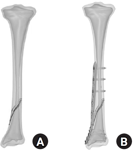
- Biomechanical analysis of medial distal tibial locking plate fixation for distal-third spiral tibial shaft fractures
- Yao-Jen Liu
- J Musculoskelet Trauma 2026;39(2):140-146. Published online April 10, 2026
- DOI: https://doi.org/10.12671/jmt.2026.00094
-
 Abstract
 PDF
PDF - Background
Distal spiral fractures of the tibial shaft present fixation challenges, particularly in patients who are not suitable candidates for intramedullary nailing. This study evaluated the biomechanical stability of medial minimally invasive percutaneous plating osteosynthesis (MIPO) under various physiological loading conditions.
Methods
A finite-element model of a distal AO/OTA 42-A1.1c spiral fracture of the tibia was created using computed tomography data. A precontoured titanium medial distal tibia locking compression plate with nine locking screws was simulated. Material properties were assigned to cortical and cancellous bone. The loading conditions included axial compression (750 N), varus/valgus bending (300 N at a 9° offset), and internal/external torsion (7.5 N·m). von Mises stress and fracture displacement were analyzed.
Results
Axial loading produced a peak plate stress of 508.06 MPa and a displacement of 2.17 mm. Valgus and varus loading generated stresses of 490.17 MPa and 324.08 MPa, respectively, with corresponding displacements of 3.86 mm and 2.01 mm. External and internal torsion resulted in stresses of 354.23 MPa and 358.9 MPa, respectively, with corresponding displacements of 2.64 mm and 2.22 mm.
Conclusions
Medial distal tibial plating demonstrated favorable biomechanical performance in this finite-element model; however, clinical extrapolation should be made cautiously. Level of evidence: V.
- 311 View
- 16 Download

- Triplane Fracture Management: Prediction of Periosteal Entrapment and the Need for Open Reduction by Measurements of the Physeal Fracture Gap in Preoperative Computed Tomography Scans
- Dae Hee Lee, Joo Han Kwon, Jae Uk Jung
- J Korean Fract Soc 2024;37(1):1-7. Published online January 31, 2024
- DOI: https://doi.org/10.12671/jkfs.2024.37.1.1
-
Abstract
PDF
- Purpose
This study measured the physeal fracture gap on preoperative ankle computed tomography (CT) to predict the periosteal entrapment that requires an open reduction in distal tibia triplane fractures.
Materials and Methods
This study retrospectively reviewed patients who had undergone internal fixation for a triplane fracture from April 2004 to September 2022. The demographic data, including age,body mass index, and past medical history, were analyzed. In the radiographic evaluations, ankle CT and ankle simple radiographs, including anteroposterior (AP), lateral, and mortise views, were taken preoperatively. Postoperatively, simple ankle radiographs were obtained periodically, including AP, mortise, and lateral views. The physeal fracture gap was measured on ankle CT, and the larger gap between the coronal and sagittal view of CT was selected. The residual physeal gap <2 mm was considered an adequate reduction.
Results
Of 17 cases, three demonstrated successful reduction using closed reduction techniques. Periosteal entrapment was observed in 14 cases open reduction cases. In all three closed reduction cases, the physeal gap estimated on preoperative ankle CT was under 3 mm with a mean gap of 2.4±0.2 mm (range, 2.1-2.5 mm). In the remaining 14 open reduction cases, the measured physeal gap was over 3 mm, averaging 5.0±2.7 mm (range, 3.1-12.2 mm). There was a significant difference in the preoperative physeal gap between the two groups (p<0.01). Overall, good reduction was achieved in all 17 cases; the postoperative physeal gap was under 2 mm with a mean of 1.0±0.5 mm (closed reduction group, 0.5±0.2 mm; open reduction group, 1.1±0.5 mm).
Conclusion
Open reduction is strongly recommended for triplane fractures with a physeal fracture gap of 3 mm or more in preoperative ankle CT, suggesting the possibility of an entrapped periosteum in the fracture gap. -
Citations
Citations to this article as recorded by
- Diagnostic values of radiographic indices for predicting periosteal entrapment in pediatric proximal phalangeal base physeal fractures of toes
Ho Young Park, Jeong-Seok Moon, Kiwook Kim
Skeletal Radiology.2026; 55(1): 97. CrossRef
- Diagnostic values of radiographic indices for predicting periosteal entrapment in pediatric proximal phalangeal base physeal fractures of toes
- 1,865 View
- 26 Download
- 1 Crossref
- Intra-Articular Alterations after Suprapatellar Nailing in Tibial Shaft Fractures: An Arthroscopic Evaluation
- GwangChul Lee, Sung Hun Yang, Sung Min Jo, Jeong Min Kook
- J Korean Fract Soc 2022;35(4):129-134. Published online October 31, 2022
- DOI: https://doi.org/10.12671/jkfs.2022.35.4.129
-
Abstract
PDF
- Purpose
This study attempted to study the intra-articular changes due to intramedullary nailing through the suprapatellar approach by evaluating the joint cartilage damage and presence of foreign bodies through a comparison of the pre- and post-operative status evaluated by knee arthroscopy.
Materials and Methods
This retrospective study analyzed fifteen patients who underwent intramedullary nailing through the suprapatellar approach for proximal tibial shaft fracture from January 2017 to March 2020. The condition of the joint cartilage and the presence of foreign substances in the patellofemoral joint were evaluated. The cartilage status of the patellofemoral joint was evaluated using the International Cartilage Repair Society (ICRS) grading system. Data from the ICRS grading and the visual analogue scale (VAS) scores of the femoral and patellar cartilage were compared to each independent variable surveyed.
Results
All the intra-articular structures before nailing were normal. In all cases after nailing, articular cartilage damage of the patellofemoral joint and intra-articular debris were observed. The average VAS score was 0.6 (0-1) before surgery and 2.27 (0-4) after surgery. There were no statistically significant differences except for the correlation in the diameter of the tibia nail and femoral ICRS grade (p=0.001) and the damage to the cartilage was greater in the femoral cartilage than that to the patella (p=0.001).
Conclusion
Intra-articular damage appears to be unavoidable in suprapatellar nailing. Further research is needed on the long-term effects of intra-articular damage and on methods to reduce this damage.
- 740 View
- 11 Download
Case Report
- Concurrent Posterolateral Corner Injury Associated with a Schatzker Type 2 Tibial Plateau Fracture: A Case Report
- Jae Cheon Sim, Choong Won Jung, Tae Seok Nam
- J Korean Fract Soc 2020;33(1):27-31. Published online January 31, 2020
- DOI: https://doi.org/10.12671/jkfs.2020.33.1.27
-
Abstract
PDF
- Isolated posterolateral corner (PLC) injury associated with a Schatzker type 2 fracture is a very rare combination of injuries. A male who was driving a motor vehicle was injured after a collision accident. The plain radiographs and computed tomography scans of the knee showed a Schatzker type 2 fracture of the tibial plateau, mostly in the anterolateral portion of tibial plateau, and an avulsion fragment on the fibular tip. Magnetic resonance imaging showed no injury to cruciate ligaments, medial collateral ligament, or any meniscal injury. We performed an open reduction operation and internal fixation for treating the fracture. Six months later, he complained of instability. At 11 months later after initial operation, we performed the second operation for stabilizing the PLC. We present here a rare case of an isolated PLC injury associated with a Schatzker type 2 fracture. We discuss the mechanism of injury and review similar cases.
- 1,371 View
- 5 Download
Original Article
- Comparative Analysis of Minimally Invasive Plate Osteosynthesis and Intramedullary Nailing in the Treatment of the Distal Tibia Fractures
- Ho Min Lee, Young Sung Kim, Jong Pil Kim, Phil Hyun Chung, Suk Kang, Kaung Suk Jo
- J Korean Fract Soc 2018;31(3):94-101. Published online July 31, 2018
- DOI: https://doi.org/10.12671/jkfs.2018.31.3.94
-
Abstract
PDF
- PURPOSE
This study compared the radiological and clinical results of minimally invasive plate osteosynthesis (MIPO) and intramedullary nailing (IMN) of distal tibial fractures, which were classified as the simple intra-articular group and extra-articular group.
MATERIALS AND METHODS
Fifty patients with distal tibial fractures, who could be followed-up more than 12 months, were evaluated. Group A consisted of 19 patients treated with MIPO and group B consisted of 31 patients treated with IMN. The results of each group were analyzed by radiological and clinical assessments.
RESULTS
The mean operation times in groups A and B were 72.4 minutes and 65.7 minutes, respectively. The mean bone union times in groups A and B were 16.4 weeks and 15.7 weeks, respectively. The bone union rate in groups A and B were 100% and 93%, respectively. The ranges of ankle motion were similar in the two groups at the last follow-up. The mean American Orthopaedic Foot and Ankle Society score was similar: 90.1 in group A and 90.5 in group B. The radiological and clinical results were similar in the intra and extra-articular groups. In groups A and B, two cases of posterior angulation and five cases of valgus deformity of more than 5° were encountered.
CONCLUSION
Both MIPO and IMN achieved satisfactory results in extra-articular AO type A and simple articular extension type C1 and C2 distal tibia fractures. -
Citations
Citations to this article as recorded by- Intramedullary Nailing versus Minimally Invasive Plate Osteosynthesis for Distal Tibia Shaft Fractures: Retrospective Comparison of Functional and Cosmetic Outcomes
Kahyun Kim, In Hee Kim, Geon Jung Kim, SungJoon Lim, Ji Young Yoon, Jong Won Kim, Yong Min Kim
Journal of Korean Foot and Ankle Society.2023; 27(3): 93. CrossRef
- Intramedullary Nailing versus Minimally Invasive Plate Osteosynthesis for Distal Tibia Shaft Fractures: Retrospective Comparison of Functional and Cosmetic Outcomes
- 774 View
- 2 Download
- 1 Crossref
Case Report
- Calcified Anterior Tibial Artery Entrapment in Distal Third Tibial Fracture: A Case Report
- Kyu Hyun Yang, Yougun Won, Sang Bum Kim, Won Kuen Park, You Sun Jung
- J Korean Fract Soc 2016;29(1):68-72. Published online January 31, 2016
- DOI: https://doi.org/10.12671/jkfs.2016.29.1.68
-
Abstract
PDF
- In the distal third of the tibia, the anterior tibial artery runs close to the anterolateral surface of the tibial cortex. In a clinical situation, without vascular evaluation, injury or entrapment of the anterior tibial artery is difficult to detect. Because, an intact dorsalis pedis pulse is supplied with the collateral vessels of the posterior tibial artery. An entrapped anterior tibial artery can be injured during closed reduction in an emergency room or open reduction and internal fixation in the operating room. Care must be taken to prevent iatrogenic anterior tibial artery. In this case, an entrapped anterior tibial artery was observed in a simple radiograph and computed tomograph without contrast media for the vessel. We report on a rare case of calcified anterior tibial artery entrapment in a distal tibial fracture.
-
Citations
Citations to this article as recorded by- Comparison of Time to Operation and Efficacies of Ultrasound-Guided Nerve Block and General Anesthesia in Emergency External Fixation of Lower Leg Fractures (AO 42, 43, 44)
Chan Kang, Sang-Bum Kim, Youn-Moo Heo, You-Gun Won, Byung-Hak Oh, June-Bum Jun, Gi-Soo Lee
The Journal of Foot and Ankle Surgery.2017; 56(5): 1019. CrossRef
- Comparison of Time to Operation and Efficacies of Ultrasound-Guided Nerve Block and General Anesthesia in Emergency External Fixation of Lower Leg Fractures (AO 42, 43, 44)
- 1,105 View
- 5 Download
- 1 Crossref
Original Article
- Intramedullary Nailing of Distal Tibial Fractures with Percutaneous Reduction by Pointed Reduction Forceps
- Jae Kwang Hwang, Chung Hwan Kim, Young Joon Choi, Gi Won Lee, Hyun Il Lee, Tae Kyung Kim
- J Korean Fract Soc 2014;27(2):144-150. Published online April 30, 2014
- DOI: https://doi.org/10.12671/jkfs.2014.27.2.144
-
Abstract
PDF
- PURPOSE
The purpose of this study is to analyze the radiographic and clinical results of intramedullary nailing after percutaneous reduction using pointed reduction forceps for spiral or oblique fractures of the distal tibia. The benefit of percutaneous reduction using pointed reduction forceps in anatomical reduction and maintenance was assessed.
MATERIALS AND METHODS
From January 2005 to December 2009, 47 cases of distal one-third tibial fracture were managed by intramedullary nailing using pointed reduction forceps. Thirty-eight cases were spiral fracture and nine cases were oblique fracture. In all cases, the percutaneous reduction was achieved using pointed reduction forceps under fluoroscopy control. While maintaining the reduction with the pointed reduction forceps, the intramedullary nail was inserted. The pointed reduction forceps were removed after insertion of proximal and distal inter-locking screws. Alignment was evaluated with anterior-posterior and lateral radiographs taken immediately post-operation and at the time of union.
RESULTS
At immediate post-operation, the mean displacement of valgus and anterior angulation was 0.57degrees and 0.24degrees, respectively. That of valgus and anterior angulation at bone union was 0.37degrees and 0.16degrees, respectively. The average duration of bone union was 16.1 weeks.
CONCLUSION
Intramedullary nailing with percutaneous reduction using pointed reduction forceps for distal tibial fractures was an easy and effective method for achievement of accurate alignment intra-operatively. Accurate alignment was successfully maintained until bone union.
- 812 View
- 2 Download
Case Report
- Minimally Invasive Percutaneous Plate Stabilization Using a Medial Locking Plate for Proximal Tibial Fractures: Technical Note
- Jae Ang Sim, Beom Koo Lee, Kwang Hui Kim, Yong Seuk Lee
- J Korean Fract Soc 2013;26(4):327-332. Published online October 31, 2013
- DOI: https://doi.org/10.12671/jkfs.2013.26.4.327
-
Abstract
PDF
- Minimally invasive plate osteosynthesis (MIPO) is beneficial for proximal tibial fractures since these injuries are mostly caused by high energy traumas. The advantages of MIPO are minimization of soft tissue dissection and preservation of periosteal vascularization. Lateral plating has mostly developed as MIPO for proximal tibial fractures. We introduce minimal invasive percutaneous plate stabilization using a medial locking plate as alternative treatment for proximal tibial fractures.
-
Citations
Citations to this article as recorded by- Effect of Korean Medicine Treatments in Patients with Proximal Tibia Fracture: A Retrospective Observational Study
Jung Min Lee, Eun-Jung Lee
Journal of Korean Medicine Rehabilitation.2020; 30(3): 141. CrossRef - Medial Minimally Invasive Percutaneous Plate Osteosynthesis in Proximal Tibial Comminuted Fractures
Jae-Ang Sim, Kwang-Hui Kim, Yong-Seuk Lee, Sang-Jin Lee, Beom-Koo Lee
Journal of the Korean Orthopaedic Association.2014; 49(4): 278. CrossRef
- Effect of Korean Medicine Treatments in Patients with Proximal Tibia Fracture: A Retrospective Observational Study
- 1,271 View
- 35 Download
- 2 Crossref
Original Articles
- Arthroscopic Assisted Intra-Articular Reduction and Internal Fixation of Tibia Plateau Fracture
- Dong Hwi Kim, Gwang Chul Lee, Kwi Youn Choi, Sung Won Cho, Sang Ho Ha
- J Korean Fract Soc 2013;26(3):191-198. Published online July 31, 2013
- DOI: https://doi.org/10.12671/jkfs.2013.26.3.191
-
Abstract
PDF
- PURPOSE
We evaluated the results of arthroscopic intra-articular reduction and internal fixation of tibial plateau fractures without cortical window along with any additional bone grafts.
MATERIALS AND METHODS
From March 2006 to March 2009, twelve patients with arthroscopic intra-articular reduction and internal fixation of tibial plateau fractures over 5 mm in depression and displacement on the articular surface in computed tomography (CT) were enrolled in this study. We reduced or removed the depressed fracture fragment using freer without making a cortical window. Then, we accomplished internal fixation by a cannulated screw. All cases have not received bone graft. Both the postoperative clinical and radiological results were evaluated by the Rasmussen system.
RESULTS
The fractures were healed completely in an average of 9 (range from 7 to 12) weeks. According to Rasmussen classification, we obtained satisfactory clinical results as excellent in 8 cases, good in 3 cases, and fair in 1 case; and radiological results were excellent in 7 cases and good in 5 cases.
CONCLUSION
We consider that arthroscopic intra-articular reduction and internal fixation of tibial plateau fractures without cortical window and any additional bone grafts is are a useful methods for attaining satisfactory results. -
Citations
Citations to this article as recorded by- Current Concepts in Management of Tibia Plateau Fracture
Sang Hak Lee, Kang-Il Kim
Journal of the Korean Fracture Society.2014; 27(3): 245. CrossRef
- Current Concepts in Management of Tibia Plateau Fracture
- 897 View
- 4 Download
- 1 Crossref
- Treatment for Bone Defect of Open Tibial Fractures by Using Intramedullary Nail Fixation with Autogenous Iliac Bone Graft
- Hyub Sakong, Ki Cheor Bae, Chul Hyun Cho, Kyung Jae Lee, Eun Seok Son, Du Han Kim
- J Korean Fract Soc 2012;25(4):288-294. Published online October 31, 2012
- DOI: https://doi.org/10.12671/jkfs.2012.25.4.288
-
Abstract
PDF
- PURPOSE
This study was conducted to evaluate the results of intramedullary nail fixation with autogenous iliac bone graft for defects of bone after tibial fractures.
MATERIALS AND METHODS
Ten patients with bone defects in tibial fractures who had been treated with intramedullary nail fixation with autogenous iliac bone graft between May 2005 and September 2008 with more than 12 month follow-up were subject to study. Of the 10 patients, 8 were male and 2 were female, and the mean age was 50.2 years (29~76 years). By cause of accident, motor vehicle accidents caused 9 cases, a crush caused 1 case, and the average follow-up period was 21.9 months (12~42 months). Radiologically, we analyzed the union of the bone defect on simple x-ray and clinical evaluation was performed using the estimate method of Mekhali.
RESULTS
This study reveals that there was radiological union in all 10 cases and the mean time to union was 8.4 months (5~18 months). By clinical evaluation according to Mekhali's estimate method, 9 patients had excellent outcomes and 1 patient had limitation of motion in the ankle joint rated as a fair clinical result. None of patients developed complications post-operatively.
CONCLUSION
Our study demonstrated that the intramedullary nail fixation with autogenous iliac bone graft can be a useful operative method because it can remove external fixators early and reduce complications, and autogenous bones have exceptional osteoconduction, osteoinduction, and bone-forming ability resulting in excellent union of bones.
- 820 View
- 7 Download
- Alteration of the Patella Tendon Length after Intramedullary Nail in Tibial Shaft Fractures
- Dong Eun Shin, Ki Shik Nam, Jin Young Bang, Ji Hoon Chang
- J Korean Fract Soc 2012;25(4):283-287. Published online October 31, 2012
- DOI: https://doi.org/10.12671/jkfs.2012.25.4.283
-
Abstract
PDF
- PURPOSE
To compare and analyze length change of patella tendon after intramedullary nailing of tibial shaft fracture using transtendinous approach.
MATERIALS AND METHODS
Thirty-two cases were analyzed from December, 1999 to December, 2005. Insall Salvati ratios were estimated. Severity of initial trauma, duration of nail retension, knee function and pain on change of length of patellar tendon was evaluated.
RESULTS
Mean duration of nail retention was twenty-two months. The shortening of patella tendon was observed in 25 cases (p<0.001). The effect of AO type and the duration of nail retension on the decrease of Insall Salvati ratio was not significant (p>0.05, p=0.778). Lysholom score decrease to 89.5. There was no significant difference between the shortening of patellar tendon length and knee pain (p=0.058).
CONCLUSION
After intramedullary nailing for closed tibia fracture, shortening of patellar tendon length is observed. That is irrelevant to the fracture type and the duration of nail retension. The shortening of patella tendon length may contribute to decreasing of knee function, but it was no significance of knee pain after intramedullary nailing.
- 804 View
- 1 Download
- Staged Minimally Invasive Plate Osteosynthesis of Distal Tibial Fractures
- Sung Ki Park, Chang Wug Oh, Jong Keon Oh, Kyung Hoon Kim, Woo Kie Min, Byung Chul Park, Won Ju Jeong, Joo Chul Ihn
- J Korean Fract Soc 2010;23(3):289-295. Published online July 31, 2010
- DOI: https://doi.org/10.12671/jkfs.2010.23.3.289
-
Abstract
PDF
- PURPOSE
To assess the result of staged minimally invasive plate osteosynthesis (MIPO) for distal tibial fracture with an open wound or injured soft tissue.
MATERIALS AND METHODS
In 20 patients (mean age, 47.8 year-old) with distal tibial fractures, there were 4 type A fractures and 16 type C fractures based on the AO classification system. Eight of the 20 patients had open fractures. MIPO was performed on average 23.9 days after bridging external fixation. At the final follow-up, we assessed the radiological results of bone union and alignment. Functional results were also evaluated by measuring the degrees of ankle motion and the American Orthopedic Foot & Ankle Society (AOFAS) scores.
RESULTS
Seventeen of 20 cases (85%) achieved primary union at an average of 21.3 weeks. There were 3 cases of nonunion requiring a bone graft. The mean AOFAS score was 88.5 (range, 67~92) and the average range of ankle motion was 49.2degrees (plantarflexion: 37.4degrees, dorsiflexion: 11.8degrees). Complication included 2 cases of minor mal-alignment, 1 case of claw toe and 1 case of peroneal neuropathy. Patients over the age of 60 had lower functional results. Additional factors did not affect the final results.
CONCLUSION
Staged MIPO may achieve satisfactory results in distal tibial fractures with soft tissue compromise, decreasing deep infections and soft tissue complications. -
Citations
Citations to this article as recorded by- Anterolateral Minimally Invasive Plate Osteosynthesis of Distal Tibial Fractures Using an Anterolateral Locking Plate
Dongwhan Suh, Hwan Hee Lee, Young Hoon Han, Jae Jung Jeong
Journal of Korean Foot and Ankle Society.2020; 24(1): 19. CrossRef - Minimally Invasive Osteosynthesis with Locking Compression Plate for Distal Tibia Fractures
Sung-Kyu Kim, Keun-Bae Lee, Keun-Young Lim, Eun-Sun Moon
Journal of the Korean Fracture Society.2011; 24(1): 33. CrossRef
- Anterolateral Minimally Invasive Plate Osteosynthesis of Distal Tibial Fractures Using an Anterolateral Locking Plate
- 1,293 View
- 6 Download
- 2 Crossref
- Minimally Invasive Percutaneous Plate Osteosynthesis Using a Lateral Plate in Distal Tibial Fracture
- Oog Jin Shon, Dae Sung Kim
- J Korean Fract Soc 2010;23(1):42-49. Published online January 31, 2010
- DOI: https://doi.org/10.12671/jkfs.2010.23.1.42
-
Abstract
PDF
- PURPOSE
To evaluate the efficacy of minimally invasive percutaneous plate osteosynthesis (MIPPO) using a lateral plate (Zimmer, Periarticular Lateral Distal Tibial Plates, USA) in distal tibial fracture within 3 cm to plafond, associated with medial soft tissue damage.
MATERIALS AND METHODS
From January 2005 to December 2007, 15 patients with distal tibial fracture treated by MIPPO technique using a lateral plate were analyzed. The duration of follow-up was more than 1 year. We evaluated union time by simple X-ray, clinical results by IOWA ankle rating system, and complication.
RESULTS
The bone union was achieved in all cases at average 16.7 weeks. Evaluation of the ankle function test showed an average of 90.3 points, resulting in satisfactory. At the last follow-up, there was no non-union, angular deformity more than 5 degrees or infection.
CONCLUSION
We concluded that MIPPO technique using a lateral plate is a efficient method for high functional recovery with good bone healing and low complication in distal tibial fracture within 3 cm to plafond, associated with medial soft tissue damage. -
Citations
Citations to this article as recorded by- Anatomically Percutaneous Wiring Reduction in Minimally Invasive Plate Osteosynthesis for Distal Tibial Fractures
Young-Mo Kim, Chan Kang, Deuk-Soo Hwang, Yong-Bum Joo, Woo-Yong Lee, Jung-Mo Hwang
Journal of the Korean Fracture Society.2011; 24(3): 230. CrossRef - Minimally Invasive Osteosynthesis with Locking Compression Plate for Distal Tibia Fractures
Sung-Kyu Kim, Keun-Bae Lee, Keun-Young Lim, Eun-Sun Moon
Journal of the Korean Fracture Society.2011; 24(1): 33. CrossRef
- Anatomically Percutaneous Wiring Reduction in Minimally Invasive Plate Osteosynthesis for Distal Tibial Fractures
- 1,061 View
- 3 Download
- 2 Crossref
Case Report
- Ipsilateral Femoral Segmental and Tibial Fractures : A Case Report
- Oog Jin Sohn, Chul Hyun Park, Sang Keun Bae
- J Korean Fract Soc 2009;22(3):193-196. Published online July 31, 2009
- DOI: https://doi.org/10.12671/jkfs.2009.22.3.193
-
Abstract
PDF
- The ipsilateral femoral segmental and tibial fractures seldom occur such as traffic accidents needed high energy mechanisms. For these fractures, surgical stabilization and early mobilization of joint produce can be the best clinical outcomes. We have experienced a case of ipsilateral femoral segmental and tibial fracture and gained good clinical results with surgical treatment. We have reported here on this case and included a review of the relevant literature.
-
Citations
Citations to this article as recorded by- Clinical Outcome after Treatment of Tibia Segmental Fracture with Intramedullary Nailing and Minimal Invasive Plate Osteosynthesis
Jun Young Lee, Hyung Seok Park, Dong Hyuk Cha
Journal of the Korean Fracture Society.2020; 33(3): 142. CrossRef
- Clinical Outcome after Treatment of Tibia Segmental Fracture with Intramedullary Nailing and Minimal Invasive Plate Osteosynthesis
- 1,188 View
- 11 Download
- 1 Crossref
Original Articles
- Staged Minimally Invasive Plate Osteosynthesis of Proximal Tibial Fracture
- Joon Woo Kim, Chang Wug Oh, Jong Keon Oh, Hee Soo Kyung, Woo Kie Min, Byung Chul Park, Kyung Hoon Kim, Hee Joon Kim
- J Korean Fract Soc 2009;22(1):6-12. Published online January 31, 2009
- DOI: https://doi.org/10.12671/jkfs.2009.22.1.6
-
Abstract
PDF
- PURPOSE
To assess the results of staged MIPO (Minimally Invasive Plate Osteosynthesis) for proximal tibial fractures with compromised soft tissue.
MATERIALS AND METHODS
Eighteen proximal tibial fractures (AO 41:9 cases, AO 42:9 cases) included this study. Ten were open fractures. After temporary external fixation until soft tissue healed (mean 27.3 days), MIPO was performed secondarily without bone graft. We assessed the bony union and knee function, and affecting factors of the results were investigated.
RESULTS
All fractures united at 20 weeks (range, 11~32) except 1 case. Mean range of knee flexion was 134.4degrees and mean IOWA knee score was 89.1. There were 2 superficial and 2 delayed deep infections from open fractures (grade II:1 case, grade III:3 cases), although they healed after implant removal. Open fractures seem to influence the infection rate. Otherwise, there was no related factor affecting the results.
CONCLUSION
MIPO after temporary external fixation can provide favorable results in proximal tibial fractures with soft tissue injuries, but attention of delayed infection should be paid in open fractures. -
Citations
Citations to this article as recorded by- MINIMALLY INVASIVE OSTEOSYNTHESIS WITH PLATE OR NAIL FOR META-DIAPHYSEAL TIBIAL FRACTURES - WHAT IS BETTER?
B. Makelov
Trakia Journal of Sciences.2023; 21(4): 357. CrossRef - Effect of Korean Medicine Treatments in Patients with Proximal Tibia Fracture: A Retrospective Observational Study
Jung Min Lee, Eun-Jung Lee
Journal of Korean Medicine Rehabilitation.2020; 30(3): 141. CrossRef - Comparison of Time to Operation and Efficacies of Ultrasound-Guided Nerve Block and General Anesthesia in Emergency External Fixation of Lower Leg Fractures (AO 42, 43, 44)
Chan Kang, Sang-Bum Kim, Youn-Moo Heo, You-Gun Won, Byung-Hak Oh, June-Bum Jun, Gi-Soo Lee
The Journal of Foot and Ankle Surgery.2017; 56(5): 1019. CrossRef - Minimally Invasive Plate Osteosynthesis for Proximal Tibial Shaft Fracture
Young-Soo Byun, Ki-Chul Park, Hyun-Jong Bong, Chang-Hoon Lee
Journal of the Korean Fracture Society.2011; 24(1): 23. CrossRef - The Use of Fresh Frozen Allogenic Bone Graft in the Impacted Tibial Plateau Fractures
Yeung Jin Kim, Soo Uk Chae, Jung Hwan Yang, Ji Wan Lee, Dae Han Wi, Duk Hwa Choi
Journal of the Korean Fracture Society.2010; 23(1): 26. CrossRef - Management of Open Fracture
Gu-Hee Jung
Journal of the Korean Fracture Society.2010; 23(2): 236. CrossRef - Staged Minimally Invasive Plate Osteosynthesis of Distal Tibial Fractures
Sung-Ki Park, Chang-Wug Oh, Jong-Keon Oh, Kyung-Hoon Kim, Woo-Kie Min, Byung-Chul Park, Won-Ju Jeong, Joo-Chul Ihn
Journal of the Korean Fracture Society.2010; 23(3): 289. CrossRef - Intramedullary Nailing of Proximal Tibial Fractures
Young-Soo Byun, Dong-Ju Shin
Journal of the Korean Fracture Society.2009; 22(3): 197. CrossRef - Proximal Tibia Fracture: Plating
Ki-Chul Park
Journal of the Korean Fracture Society.2009; 22(3): 206. CrossRef
- MINIMALLY INVASIVE OSTEOSYNTHESIS WITH PLATE OR NAIL FOR META-DIAPHYSEAL TIBIAL FRACTURES - WHAT IS BETTER?
- 1,010 View
- 0 Download
- 9 Crossref
- Mid-term Results of Distal Tibial Fractures Treated with Ilizarov External Fixator
- Suk Kyu Choo, Kyung Wook Nha, Hyoung Keun Oh, Dong Bong Lee
- J Korean Fract Soc 2007;20(4):323-329. Published online October 31, 2007
- DOI: https://doi.org/10.12671/jkfs.2007.20.4.323
-
Abstract
PDF
- PURPOSE
We analyed the mid-term results of distal tibial fractures treated with ilizarov external fixator and functional results according to delayed metaphyseal healing and fracture pattern.
MATERIALS AND METHODS
We reviewed 23 distal tibial fractures treated with ilizarov external fixator followed for minimum two year (mean 53 months). There were 10 A fractures, 2 B fractures, and 11 C fractures according to the AO classification. Radiographically, we analyzed bony union time according to translation of diaphyseal-metaphyseal fracture line and assessed arthritic score. Functional results was assessed with AOFAS score and analyzed according to delayed healing and fracture pattern.
RESULTS
Average union time was 21 weeks. Delayed healing of metaphyseal fracture line was associated translational displacement >3 mm (p=0.01). AOFAS scrore was averaged to 68 and there was no stastical significance between delayed metaphyseal healing and functional results (p=0.31). But, low AOFAS score and arthritis score was related to fracture type (p=0.02). In 11 C fractures, radiographic arthritic change were developed in 6 cases (55%).
CONCLUSION
The main prognosis of distal tibial fractures depends on articular involvement and to shorten the external fixation time, metaphyseal fracture should be reduced within 3mm.
- 1,035 View
- 3 Download
- Internal Fixation with Two Lowprofile Plates in Fractures of the Distal Tibia
- Dong Eun Shin, Duck Yun Cho, Hyung Ku Yoon, Tae Hyung Kim
- J Korean Fract Soc 2006;19(2):170-175. Published online April 30, 2006
- DOI: https://doi.org/10.12671/jkfs.2006.19.2.170
-
Abstract
- PURPOSE
To evaluate the functional results after internal fixation with two low profile plates in fractures of the distal tibia.
MATERIALS AND METHODS
From March 1998 to October 2002, twelve patients with fractures of the distal tibia were treated with internal fixation using two low profile plates and followed for at least one year. Fractures according to AO/OTA classification were one Type A1, four Type A2, two Type C1, two Type C2 and three Type C3. We analyzed the functional results by the Olerud and Molander ankle scoring system and the postoperative complications.
RESULTS
The average functional score was 81.2 points and the results were three excellent, six good, one fair and two poor. Bony union was achieved in all cases. There was 1 case of superficial wound infection as a complication.
CONCLUSION
Internal fixation with two low profile plates in fractures of the distal tibia may minimize the incidence of soft tissue complications and provide good bony union and functional results. Therefore, we consider internal fixation with two low profile plates as a good alternative treatment of the distal tibial fracture.
- 589 View
- 0 Download
- A Comparison according to Insertion Method for Intramedullary Nailing in Proximal Tibial Fractures
- Sang Ho Moon, Byoung Ho Suh, Chung Soo Hwang, Tae Hyun Yoon
- J Korean Fract Soc 2006;19(1):17-23. Published online January 31, 2006
- DOI: https://doi.org/10.12671/jkfs.2006.19.1.17
-
Abstract
- PURPOSE
To compare clinical and radiological results between standard insertion method and semiextended method which was designed to improve proximal fixation and alignment in proximal tibia fracture.
MATERIALS AND METHODS
A retrospective review from May 2000 to February 2004, identified 24 extraarticular fractures in proximal tibia, initially treated with locked intramedullary nails at least 1 year follow up. There were 12 open injuries, 4 segmental, 3 butterfly fragments and 17 comminuted. Semiextended method was used in 10 fratures and standard insertion method which is cephalad to tibial tubercle in 14. Follow up clinical assessment consisted of review of associated injuries and complications and these two methods were compared by postoperative angulation and displacement in anteroposterior and lateral radiographs. Data were analysed by t-tests.
RESULTS
In semiextended group, average angulation was 2.3 degrees in coronal and 2.8 degrees in sagittal plane and average displacement was 4.5 mm in coronal and 5.3 mm in sagittal. In ordinary group, average angulation was 5.1 degrees in coronal and 7.4 degrees in sagittal plane and average displacement was 6.1 mm in coronal and 5.3 mm in sagittal. In semiextended group, there were significant reduction in coronal angulation (p=0.006) and sagittal angulation (p=0.001), but there was no significant difference in coronal (p=0.344) and sagittal (p=0.99) displacement. Both groups showed anterior, valgus angulation and posterolateral displacement in most cases. There were 14 associated injuries and one patient developed nonunion and was treated by nail exchange with autogenous bone graft.
CONCLUSION
Our retrospective analysis demonstrated that semiextended method is effective for reducing coronal and sagittal angulation, but is not helpful for reducing displacement in both planes.
- 520 View
- 0 Download
- Treatment of Tibial Fractures In Children With Pin and Plaster Technique
- Byoung Ho Suh, Gyu Min Kong, Sang Ho Moon, Dong Joon Kim, Jin Woo Kwon, Se Won Park
- J Korean Fract Soc 2005;18(3):325-329. Published online July 31, 2005
- DOI: https://doi.org/10.12671/jkfs.2005.18.3.325
-
Abstract
PDF
- PURPOSE
To evaluate the result of tibial shaft fractures in children treated with pin and plaster method.
MATERIALS AND METHODS
From March 1998 to February 2003, Tibial shaft fractures in thirty six pediatric patients which were treated with pin and plaster method were clinically and radiologicaly evaluated retrospectively.
RESULTS
Mean bony union duration was 9.8 weeks. All fractures healed within acceptable angulations. There was neither delayed union nor nonunion. There were complications related to the pins, including superficial and deep infection, skin sloughing. There were 7 cases of tibial overgrowth but they had no functional disability.
CONCLUSION
Pin and plaster method can substitute other operative methods in tibial fractures in children which is difficult to reduce or maintain reduction by conservative treatment.
- 601 View
- 2 Download
- Treatment of Open Tibial Shaft Fractures using Unreamed Nailing
- Jong Keon Oh, Chang Wug Oh, Kwon Jae Roh, Duk Moon Chung
- J Korean Fract Soc 2005;18(1):22-28. Published online January 31, 2005
- DOI: https://doi.org/10.12671/jkfs.2005.18.1.22
-
Abstract
PDF
- PURPOSE
To report the results of unreamed nailing using a nail with the largest possible diameter for the management of the open tibial shaft fractures.
MATERIALS AND METHODS
Nineteen patients with open tibial shaft fractures underwent unreamed nailing with the largest possible diameter according to the isthmic diameter measured on preoperative radiography. There were 1 Grade I, 6 Grade II, 9 Grade IIIa, 3 Grade IIIb open fractures. There were 4 type A, 12 type B, 3 type C fractures according to the OTA classification. Fractures were classified as The nail was introduced after gentle passage of a 7 to 8 millimeter-hand reamer.
RESULTS
Union was obtained in all cases. However 9 (47%) fractures required an additional procedures before union. In 6 cases, dynamization was done. Two of them were required exchange nailing for nonunion, 1 of two gained bony union through additional bone graft. Three of the others had gained union through exchange nailing, bone graft, bone transport respectively. There were one rotational malunion, one superfical and one deep infection. Interlocking screw breakage developed only in one patient.
CONCLUSION
Our data indicate that unreamed nailing in the management of open tibial fractures is safe and reliable method. Using a tight fitting nail with the largest possible diameter is a safe and effective way to avoid the problems of screw breakage. -
Citations
Citations to this article as recorded by- Treatment of Type IIIb Open Tibial Fractures
Seong Yeon Lim, Il Jae Lee, Jae Ho Joe, Hyung Keun Song
Journal of the Korean Fracture Society.2014; 27(4): 267. CrossRef - Management of Open Tibial Fractures: Role of Internal Fixation
Yerl-Bo Sung
Journal of the Korean Fracture Society.2007; 20(4): 349. CrossRef
- Treatment of Type IIIb Open Tibial Fractures
- 920 View
- 0 Download
- 2 Crossref
- Treatment of Tibial Shaft Fractures in Children Using K-wires Fixation
- Phil Hyun Chung, Chung Soo Hwang, Suk Kang, Jong Pil Kim, Ho Jun Cheon
- J Korean Fract Soc 2004;17(4):384-388. Published online October 31, 2004
- DOI: https://doi.org/10.12671/jkfs.2004.17.4.384
-
Abstract
PDF
- PURPOSE
To report the effectiveness of Kirschner wire fixation for the treatment of unstable tibial shaft fractures in children.
MATERIALS AND METHODS
We analyzed 15 cases of pediatric tibial shaft fractures treated at our hospital with fixation using K-wire and followed up for more than 1 year from July 1998 to January 2002. The subjects included 11 boys and 4 girls. The ages ranged from 3 to 10 years at the time of injury, with the average age being 7.9 years. We examined the presence of angulation, leg length discrepancy, joint motion limitation, and complications.
RESULTS
Bony fusion was obtained in all patients by an average of postoperative 9.5 weeks. At the time of last follow-up (by an average of postoperative 1 year and 4 months), anterior and posterior radiographs showed an average of 4.2degree angulation, and lateral radiographs showed an average of 4.4degree angulation. The affected leg was extended by an average of 3.7 mm compared to the opposite leg according to Bell-Thompson's radiographs. As for complications, infection was developed around the pin in 3 cases but treated with the administration of oral antibiotics and sterilization around the site without progressing to deep infection. We could not observe joint motion limitation, pain and difficulties related with discrepancy in leg length.
CONCLUSION
We concluded that fixation using K-wire for children with tibial shaft fractures was a safe and effective method of surgery that could be performed easily, did not require secondary surgery to remove the wire, and showed sufficient stability after fixation.
- 728 View
- 6 Download
- Anterolateral Approach for the Distal Metaphyseal Fracture of the Tibia
- Taek Soo Jeon, Jae Woo Lim, Whan Yong Chung, Woo Suk Lee, Woo Sik Kim, Cheol Mog Hwang, Yong Chan Kim, Nam Hyun Kim, Yong Sang Kim, Sung Kwan Jo
- J Korean Fract Soc 2004;17(3):243-248. Published online July 31, 2004
- DOI: https://doi.org/10.12671/jkfs.2004.17.3.243
-
Abstract
PDF
- PURPOSE
The purpose of this study is to evaluate the effectiveness of anterolateral approach of the ankle for the distal tibial fracture in aspect of preventing complication and acquiring union.
MATERIALS AND METHODS
Authors reviewed 21 patients of distal metaphyseal fracture of the tibia treated by anterolateral approach and lateral plating method from February, 2000 to May, 2002. Mean follow-up period was 17 months (12~29 months). There were twelve type A, two type B, and four type C patients according to AO/OTA classification. We have analyzed the bone union rate and Ovadia`s functional scale. We also reviewed the complication rate, such as soft tissue problem and postoperative infection.
RESULTS
In all cases union was achieved and mean time to union were 16 weeks. The functional result by Ovadia's scale were 17 excellent cases and 4 good cases in objective evaluation, and 19 excellent cases and 2 good cases in subjective evaluation. Wound infection occurred in one case, but the infection was controlled after plate removal and the union was acquired through cast immobilization. There was no other complication, such as soft tissue necrosis.
CONCLUSION
The anterolateral approach is a safe and worthwhile method for distal tibia fracture while avoiding some of the complication associated with standard anteromedial approach and plating method.
- 770 View
- 24 Download
- Minimally Invasive Percutaneous Plate Stabilization of Proximal Tibial Fractures
- Chang Wug Oh, Jong Keon Oh, In Ho Jeon, Hee Soo Kyung, Il Hyung Park, Joo Chul Ihn, Yeon Ki Woo, Ho Sung Jung
- J Korean Fract Soc 2004;17(3):224-229. Published online July 31, 2004
- DOI: https://doi.org/10.12671/jkfs.2004.17.3.224
-
Abstract
PDF
- PURPOSE
Despite of various treatment methods, proximal tibial fractures are common injuries that may be associated with poor outcomes and high rates of complications. To improve this, percutaneous plating technique was performed in the proximal tibial fractures.
MATERIALS AND METHODS
Twenty-four proximal tibial fractures (AO 41A; 5, AO 41C; 12, AO 42; 7) were treated by percutaneous plating with either or both sides without bone graft. One was open fracture.
RESULTS
All fractures were healed. The average time for fracture healing was 16.5 weeks (range, 8~24 weeks). Complications included a 1cm shortened case and two mal-alignments; a 6 degree valgus case and 5 degree varus case. There was one case of superficial infection, which healed after plate removal. But, there was no deep infection. Results were evaluated by modified Rasmussen score system, all patients had excellent or good result.
CONCLUSION
Minimally invasive percutaneous plating technique can provide favorable results in the treatment of proximal tibial fractures. -
Citations
Citations to this article as recorded by- MINIMALLY INVASIVE OSTEOSYNTHESIS WITH PLATE OR NAIL FOR META-DIAPHYSEAL TIBIAL FRACTURES - WHAT IS BETTER?
B. Makelov
Trakia Journal of Sciences.2023; 21(4): 357. CrossRef - Medial Minimally Invasive Percutaneous Plate Osteosynthesis in Proximal Tibial Comminuted Fractures
Jae-Ang Sim, Kwang-Hui Kim, Yong-Seuk Lee, Sang-Jin Lee, Beom-Koo Lee
Journal of the Korean Orthopaedic Association.2014; 49(4): 278. CrossRef - Minimally Invasive Percutaneous Plate Stabilization Using a Medial Locking Plate for Proximal Tibial Fractures - Technical Note -
Jae Ang Sim, Beom Koo Lee, Kwang Hui Kim, Yong Seuk Lee
Journal of the Korean Fracture Society.2013; 26(4): 327. CrossRef - Clinical Outcomes of Locking Compression Plate Fixation through Minimally Invasive Percutaneous Plate Osteosynthesis in the Treatment of Distal Tibia Fracture
Jae-Sung Yoo, Hyun-Woo Park
Journal of the Korean Fracture Society.2012; 25(2): 117. CrossRef - Minimally Invasive Plate Osteosynthesis for Proximal Tibial Shaft Fracture
Young-Soo Byun, Ki-Chul Park, Hyun-Jong Bong, Chang-Hoon Lee
Journal of the Korean Fracture Society.2011; 24(1): 23. CrossRef - Minimally Invasive Plate Osteosynthesis for the Upper Extremity Fracture Using a Lumbar Spreader - Surgical Technique -
Gu-Hee Jung, Chyul-Hyun Cho, Jae-Do Kim
Journal of the Korean Fracture Society.2011; 24(1): 83. CrossRef - Management of Fractures of Distal Tibia by Minimally Invasive Plate Osteosynthesis through an Anterior Approach
Gu-Hee Jung, Jae-Do Kim, Jae-Ho Jang, Sung-Keun Heo, Dong-won Lee
Journal of the Korean Orthopaedic Association.2010; 45(6): 473. CrossRef - The Comparison of Minimally Invasive Plate Osteosynthesis and Intramedullary Nailing in the Treatment of the Proximal and Distal Tibia Fracture
Joon Soon Kang, Seung Rim Park, Sang Rim Kim, Yong Geun Park, Jae Ho Jung, Sung Wook Choi
Journal of the Korean Fracture Society.2010; 23(2): 172. CrossRef - Staged Minimally Invasive Plate Osteosynthesis of Proximal Tibial Fracture
Joon-Woo Kim, Chang-Wug Oh, Jong-Keon Oh, Hee-Soo Kyung, Woo-Kie Min, Byung-Chul Park, Kyung-Hoon Kim, Hee-Joon Kim
Journal of the Korean Fracture Society.2009; 22(1): 6. CrossRef - Proximal Tibia Fracture: Plating
Ki-Chul Park
Journal of the Korean Fracture Society.2009; 22(3): 206. CrossRef
- MINIMALLY INVASIVE OSTEOSYNTHESIS WITH PLATE OR NAIL FOR META-DIAPHYSEAL TIBIAL FRACTURES - WHAT IS BETTER?
- 837 View
- 2 Download
- 10 Crossref
- Biomechanical Analysis of Hybrid External Fixation for the Distal Tibial Fractures: A FEM Study
- Duk Young Jung, Boug Ju Kim, Seok Bae Ryu, Jong Keon Oh
- J Korean Fract Soc 2004;17(2):160-166. Published online April 30, 2004
- DOI: https://doi.org/10.12671/jkfs.2004.17.2.160
-
Abstract
PDF
- PURPOSE
To analyze the biomechanical effects of different frame configurations of the hybrid external fixators for distal tibial fractures on the frame stiffness and stress distribution with a finite element method (FEM).
MATERIALS AND METHODS
Five configurations were simulated: Group I: two wires with convergence angle of 60degrees, Group II: 3rd wire on a bisector axis of the group I. Group III: two wires with 30degrees. Group IV: 3rd wire on a bisector axis. Group V: two wires with 30degree and a half pin on the distal articular fragment. Each group was simulated under compression, torsion, anterior-posterior and lateral-medial bending load. Stiffness, stress and deformation values were calculated.
RESULTS
The overall stiffness was increased by 15~30% with the addition of a third wire, and by 150~400% with a anteromedial half pin on the articular fragment. The half pin decreased the stress level of the frame by about 43% and the deformation of the 5/8 ring by about 30%.
CONCLUSION
The addition of a half pin on the articular fragment is not only a method of increasing the stiffness but also a way of decreasing the stress concentration and the deformation of the frame.
- 610 View
- 1 Download
- Unreamed interlocking nailing in tibia fracture
- You Sung Suh, Young Il Cho, Ho Won Jung, Yeon Il Kim
- J Korean Soc Fract 2002;15(4):470-476. Published online October 31, 2002
- DOI: https://doi.org/10.12671/jksf.2002.15.4.470
-
Abstract
PDF
- PURPOSE
To evaluate of clinical results and malunion according to nail insertion site and early ambulation after unreamed interlocking intramedullary nailing for the treatment of tibial fractures, MATERIALS AND METHODS: We reviewed 46 tibial fractures that were treated with unreamed static intramedullary nailing prospectively from March 1997 to May 2001. Nail insertion site and angulation of fracture site were reviewed by radiograph. All of 46 cases, ambulation was started at postoperative 2 weeks, and then clinical outcomes were reveiwed RESULTS: In all 46 cases, union was achieved at average 18.2 weeks clinically and average 19.4 weeks radiographically. There is no significant difference in angulation according to nail insertion site, i,.e. after central/medial/lateral insertion, outcome was 2 . 4 5 degrees +/-2 . 1 7 / 2 . 2 2 degrees +/-1 . 8 4 / 1 . 7 3 degrees +/-1.33(p; 0.705) in last follow up anterioposterior view, and 1.81 degrees +/-1 . 1 3 / 2 . 6 7 degrees +/-1 . 6 2 / 2 . 0 0 degrees +/-1.64(p; 0.320) in last follow up lateral view. No breakage of intramedullary nails and no stiffness on adjacent joints.
CONCLUSION
We confirmed that unreamed interlocking nailing in tibial fractures is one of the effective method for low recurrence of malunion and early ambulation
- 505 View
- 0 Download
- Comparative Analysis of Interlocking IM Nailing and LC-DCP fixation in the Treatment of Distal Tibial Fracture
- Hwa Yeop Na, Young Jun Park, Sang Hoon Ko, Wahn Sub Choe, Young Sang Lee, Kyung Dong Yoon
- J Korean Soc Fract 2002;15(2):152-158. Published online April 30, 2002
- DOI: https://doi.org/10.12671/jksf.2002.15.2.152
-
Abstract
PDF
- PURPOSE
To compare the clinical results between interlocking IM nailing and LC-DCP fixation in the treatment of distal tibial shaft fracture.
MATERIALS AND METHODS
From August 1998 to August 2001, 23 patients were treated by interlocking IM nail and 15 patients were treated by LC-DCP for distal tibial shaft fracture.
RESULTS
Accoding to Robinson classification, there were 12 type 1 fractures (52.1%) and 11 type 2a fractures (47.8%) in the interlocking IM nailing group, and 4 type 1 fractures (26.7%), 8 type 2a fractures (53.4%) and 3 type 2c fractures (20.07%) in the LC-DCP fixation group. The average time to bony union was 16 weeks in the patients treated with interlocking IM nail and 12 weeks in the patients treated with LC-DCP. In the functional outcome (according to Klemm and Borner), 18 patients treated (78.2%) with interlocking IM nail showed satisfactory results and 13 patients (86.6%) treated with LC-DCP had satisfactory results.
CONCLUSION
We concluded that more satisfactory results could be obtained with LC-DCP fixation compared with interlocking IM nailing in the treatment of the distal tibial fracture. -
Citations
Citations to this article as recorded by- A Comparison of the Results between Intramedullary Nailing and Minimally Invasive Plate Osteosynthesis in Distal Tibia Fractures
Chul-Hyun Park, Chi-Bum Choi, Bum-Jin Shim, Dong-Chul Lee, Oog-Jin Shon
Journal of the Korean Orthopaedic Association.2014; 49(4): 285. CrossRef - Comparative Study Using of Treatment with Minimally Invasive Plate Osteosynthesis Using Periarticular Plate and Interlocking Intramedullary Nailing in Distal Tibia Fractures
Chang Hwa Hong, Jong Seok Park, Sang Seon Lee, Soo Ik Awe, Woo Jong Kim, Ki Jin Jung
Journal of the Korean Fracture Society.2010; 23(3): 296. CrossRef - A Comparison between Minimally Invasive Plate Osteosynthesis & Interlocking Intramedullary Nailing in Distal Tibia Fractures
Kee-Byung Lee, Si-Young Song, Duek-Joo Kwon, Yong-Beom Lee, Nam-Kyou Rhee, Jun-Ha Choi
Journal of the Korean Fracture Society.2008; 21(4): 286. CrossRef - Comparative Study of Intramedullary Nailing and Plate for Metaphyseal Fractures of the Distal Tibia
Hoon Jeong, Jae-Doo Yoo, Young-Do Koh, Hoon-Sang Sohn
Journal of the Korean Fracture Society.2007; 20(2): 154. CrossRef
- A Comparison of the Results between Intramedullary Nailing and Minimally Invasive Plate Osteosynthesis in Distal Tibia Fractures
- 842 View
- 0 Download
- 4 Crossref
- Comparison of External Fixation and Interlocking IM Nail in Open Tibial Fractures
- Hyung Jin Chung, Duck Kyu Kim, Yerl Bo Sung, Jong Guk Ahn
- J Korean Soc Fract 2001;14(4):632-642. Published online October 31, 2001
- DOI: https://doi.org/10.12671/jksf.2001.14.4.632
-
Abstract
PDF
- PURPOSE
To compare and analyze the results of the treatment based on the method of treatment between interlocking IM nail and external fixation of type II, III A, and III B open fractures of the tibia] shaft.
MATERIALS AND METHODS
A clinical analysis was performed on 57 cases of type II, III A, and III B open fractures of tibial shaft from January 1994 to October 1999 those studies are followed at least 1 year or more. The results was analyzed according to complications and functional results based on operative methods of types of open fractures.
RESULTS
In aspect of delayed union and nonunion, interlocking IM nail indicate a great results(p = 0.036) in type II. In angulation, interlocking IM nail marks an outstanding results in case of type II. There is no difference between interlocking IM nail and external fixation in infection. But, the delay of operation after injury increase the possibilities of infection.
CONCLUSION
At present, especially in type m, external fixation was preferred. But, interlocking IM nail has a good results in aspect of complications. Therefore unreamed intramedullary nailing for open tibia shaft fractures is a good treatment method to be recommended.
- 580 View
- 1 Download
- The Angular Deformity of Interlocking Nailing in Tibial Fractures
- Hwa Jae Jeong, Kyung Chul Kim, Jae Yeul Choi, Bon Seop Koo, Jung Hee Oh
- J Korean Soc Fract 2000;13(4):905-911. Published online October 31, 2000
- DOI: https://doi.org/10.12671/jksf.2000.13.4.905
-
Abstract
PDF
- PURPOSE
We studied the relationship between angular deformity and possibly contributing factors in the treatment of tibial fractures with interlocking nailing.
MATERIALS AND METHODS
Intramedullary nailing of the tibia was performed on 49 cases and were followed for the minimum of 12 months. We analyzed relationship between angular deformity and postoperative tibial alignment, operative technique and other factors.
RESULTS
Of the 49 cases, 19(38%) were angulated. Angular deformity was seen in 60%, 51.8% and 11.8% in the proximal, distal and middle third of tibial fractures respectively. With AO classification, Group A,B,C were angulated in 32.4%, 55.6%, 66.7%. In group A, 43.8% of spiral fractures, 28.6% of oblique fractures and 14.3% of transverse fractures were angulated. The cases combined with fibular fracture showed higher incidence of angular deformity than the cases with intact fibula. The opening of fracture and the nail insertion site were not significant to angular deformity.
CONCLUSION
Angular deformity of interlocking nailing in tibial fractures were more common in proximal, comminuted and spiral fractures. Precise attentions to operative technique i. e. accurate anatomical reduction and centromedullary nail orientation are recommended to prevent angular deformity. In proximal third tibial shaft fractures where muscles and patellar tendon has deforming force on fracture fragment, authors believe that use of interlocking nailing must be limited with fracture pattern.
- 645 View
- 3 Download
- Medullary Canal Widening Effect on Insertion of Tibial Intramedullary Nail bent anteriorly at distal portionJong Min Sohn
- Hyung Kwan Kim, Juhae Jahng, Dae Hyun Baek, Nan Kyoung Ha, Hyuk Je Lee
- J Korean Soc Fract 2000;13(2):267-271. Published online April 30, 2000
- DOI: https://doi.org/10.12671/jksf.2000.13.2.267
-
Abstract
PDF
- PURPOSE
: We have studied medullary canal widening effect on anteroposterior plane by intramedullary nails with distal anterior bend in the surgical treatment of tibial fracture and signified the clinical importance of the effect.
MATERIALS AND METHODS
: Ineramedullary tibial nails with distal anterior bend, Russell-Taylor(R)and AIMTMtibial nails were compared and the amount of medullary canal widening was calculated mathematically using the length and angle of distal anterior bend of the nails. the length and angle of distal anterior bend of Russell-Taylor(R)and AIMTMtibial nails were 64mm, 3 and 47.5mm, 5 , respectively.
RESULTS
: The amount of medullary canal widening on anteroposterior plane by Russell-Taylor(R)and AIMTM tibial nails were 2.81mm and 3.26 mm more than the nail diameter, respectively.
CONCLUSION
: On insertion of tibial nails with distal anterior bend, the medullary canal widening effect on anteroposterior plane by the nails should be carefully considered. We think it will be better to insert these nails with unreamed technique or to insery smaller diameter nails after minor reaming and forceful blow should be avoided especially when the nail passes down through the isthmic portion of the tibial diaphysis to prevent the fracture of isthmic portion.
- 527 View
- 0 Download
- Brooker Intramedually Nailing for the Treatment of Distal 1/3 Tibial Fractures with Compromised Soft Tissue
- Chung Nam Kang, Jong Oh Kim, Yeo Hon Yun, Dong Wook Kim, Young Do Koh, Jae Doo Yoo, Jong Keon Oh, Ki Woong Lee
- J Korean Soc Fract 1999;12(4):924-931. Published online October 31, 1999
- DOI: https://doi.org/10.12671/jksf.1999.12.4.924
-
Abstract
PDF
- The treatment of distal tibial fractures with compromised soft tissue poses many problems that usually occurs from the high-energy trauma, and the results are often unsatisfactory following lots of complications like loss of reduction, malunion, and inlection. We studied to evaluate the treatment results of Brooker intramedually nailing for the distal 1/3 tibial fractures with compromised soft tissue. Twenty-three cases of distal tibial fractures with comprolnised soft tissue were reviewed and we analyzed the results of surgical treatment in the viewpoint of union time, loss of reduction, malunion, complication and its final outcome. The range of follow-up was 24 months to 38 months with mean 29 months follow-up. Most of patients were between twenty and sixty years, and average age was 43.2 years. Acording to Gustilo and Andersons classification, 3 were Type I, 2 were Type II of 5 open fractures. According to Tschernes classification, 13 were Grade I, 5 were Grade II of 18 closed fractures. The average to union was 15 weeks with range 11 to 20 weeks. The healing was slowest in Tschernes Type II and fastest in Tschernes Type I fracture. There were 3 cases of malunion, more than 5 degrees. All of the 3 cases were posterior angulation. Only 1 case was the loss of reduction. This case was 3 to 10 degrees of varus angulation. There were 3 cases of superficial infection. The infection was controlled with antibiotic therapy. Only 1 case was acceptable of the final outcome. This case waf limping gait because of pain and loss of ankle dorsiflexion to 15 degrees. But, the limitation of ordinary work was not seen. And 18 cases were excellent and 4 cases were good. We recommand that wherever possible, Brooker intramedually nailing can be used for distal tibial fractures with compromised soft tissue. And a high rate of union and a low rate of complication can be expected with thit treatment modality.
- 510 View
- 0 Download
- Treatment of comminuted Tibial Fractures using Ilizarov Method
- Eui Hwan Ahn, Sung Tae Lee, Hyeon Seok Kang
- J Korean Soc Fract 1999;12(4):916-923. Published online October 31, 1999
- DOI: https://doi.org/10.12671/jksf.1999.12.4.916
-
Abstract
PDF
- From March 1996 to March 1999, thirty two cases of comminuted tibial fractures were treated with Ilizarov external fixator. 13 cases were closed fractures and 19 cases open fractures. Among 19 open fractures, there were 3 cases of Gustilo type I, 10 cases of type II and 6 cases of type III fractures. All the cases could not be initially treated by open reduction and internal fixation because of open wound or severe comminution. Among 32 cases, 4 were tibial condyles, 22 were tibial shafts, 6 were tibial plafonds. All the cases were followed up from a minimum 12 months up to 35 months with an dverage of 22 months. We obtained satisfactory bony union in ail cases with the average duration of 18.1 weeks. Bone graft was done initially in two cases. Numerous complications were encountered, most commonly, joint stiffness and pin tract infection were developed but they were treated well. To avoid such complications, careful management was needed. According to Tuckers clafsification, the result was graded as excellent in 8, good in 18, fair in 4 and poor in 2 cases. We conclude that Ilizarov external fixatior is a very useful method for initial treatment in getting reduction, maintenance of reduction, early ambulation and fracture healing in cases of communited tibial fractures whether open or closed.
- 666 View
- 0 Download
- Intramedullary Nailing in Distal Tibial Fracture
- Kyu Hyun Yang, Dae Yong Han, Seong Jin Park, Hwan Wook Yoo, Ki Won Yang
- J Korean Soc Fract 1999;12(4):901-908. Published online October 31, 1999
- DOI: https://doi.org/10.12671/jksf.1999.12.4.901
-
Abstract
PDF
- PURPOSE
: We evaluated the efficacy of intramedullary nailing in distal tibial fractures. Material and Method : Twenty-six patients with distal tibial fracture were treated with intramedullary nailing between Jan. 1996 and May 1998. Operation was done on the fracture table under skeletal traction. We evaluated the causes of trauma, type of fracture, location of fracture, time to union, malunion, nonunion, range of motion of knee and ankle, and degree of pain.
RESULTS
: There were 4 cases of open fracture and 4 cases of closed soft tissue injury at fracture site. The time to fracture union was 19 weeks on average. One case(3.8%) did not heal by 10 months and was classified as nonunion. The union rate was 96.2 % and the complication rate was 7.7%(one case of nonunion and one case of malunion). There was no infection and soft tissue disruption. The range of motion of knee was reduced in 1 case(3.8%) and 2 patients(7.7%) complained of mild pain at the knee joint. The range of motion of the ankle joint was reduced in 4 cases(15.5%), averaging 15.5 degrees in dorsiflexion and 9 cases(34.6%), averaging 21 degress in plantarflexion. Two patients complained of mild pain at the ankle joint.
CONCLUSION
: We had relatively good clinical and radiological results and concluded that closed intramedullary nailing is a safe and effective method of managing distal tibial fracture.
- 498 View
- 0 Download
- Proximal Tibiosbular Fracture associated with Popliteal Artery Injury
- Sang Soo Kim, Hong Jun Han, Dong Churl Kim, Dae Ho Ha, Hee Jun Yoo, Suk Kyun Park
- J Korean Soc Fract 1999;12(4):885-893. Published online October 31, 1999
- DOI: https://doi.org/10.12671/jksf.1999.12.4.885
-
Abstract
PDF
- Injury to the popliteal artery results in amputation more frequently than any other arterial injury. The major factor in the amputated limbs was a delay in diagnosis and therapy of the arterial injury associated with blunt trauma. The proximal tibial fractures produced the highest percentage of vascular complications and indicated immediate application of therapeutic measures. The purpose of this study is to investigate the long-term results and factors that influences the results of surgical treatment in patients with combined proximal tibial fracture and popliteal artery injury. Authors reviewed the records of 24 cases treated for this injury between January 1984 and May 1997. The age of the patients ranged from 17 to 70 years(average 45 years). Nine patients presented with life threatening injuries and classical signs of acute limb ischemia. Prolonged ischemic time ranged from 3 to 6 hours 30 minutes(average 4 hours 50 minutes). The most common cause of thoses injury was traffic accident in 16 cases. Five cases had neurologic deficit ; significant soft tissue injury was present in 14 extremities. Vascular procedures included saphenous vein interposition, end-to-end anastomosis, etc. Bony procedures were accomplished by external means in 14 cases and the others treated by immediate internal fixation in 5 cases. Intraoperative fasciotomy was performed in 5 patients with lower limb ischemia. The results suggested that limb salvage was possible in 63 percent of patients with combined proximal tibial fracture and popliteal artery injuries, but a history of life-threatening condition and severe associated injury with vascular compromise was an unfavorable prognostic factor. So a well organized multidisciplinary approach is necessary to ensure life and functional limb salvage.
- 548 View
- 3 Download
- Comparative Analysis of Interlocking Nail and Anatomical Plate in the Treatment of Distal Tibial Fracture
- Gun Il Im, Do Young Kim, Joo Ho Shin, Kang Seob Youn, Won Ho Cho
- J Korean Soc Fract 1999;12(3):632-637. Published online July 31, 1999
- DOI: https://doi.org/10.12671/jksf.1999.12.3.632
-
Abstract
PDF
- We studied 39 patients with distal tibial shaft fracture. Seventeen fractures(10 closed fractures and 7 open fractures: 5 type-I and 2 type II fractures, according to the classification of Gustilo et al.) were treated with interlocking nail, and 22 fractures(19 closed and 3 open fractures: 1 type I and 2 type II fractures) were treated with anatomical plate. The clinical results were analyzed according to treatment modality. All of the patients were followed up for more than 1 year. The average time to union was 18.1 weeks in the patients treated with interlocking nail and 23.7 weeks in the patients treated with anatomical plate. In the functional outcome(according to Klemm and Borner), twelve patients(70.6%) treated with interlocking nail showed excellent results and 10 patients(45.5%) treated with anatomical plate had excellent results. We concluded that more satisfactory results could be obtained with interlocking nail compared with anatomical plate in the treatment of the distal tibial fracture.
-
Citations
Citations to this article as recorded by- Comparative Analysis of Minimally Invasive Plate Osteosynthesis and Intramedullary Nailing in the Treatment of the Distal Tibia Fractures
Ho-Min Lee, Young-Sung Kim, Jong-Pil Kim, Phil-Hyun Chung, Suk Kang, Kaung Suk Jo
Journal of the Korean Fracture Society.2018; 31(3): 94. CrossRef - Comparative Analysis of Minimally Invasive Plate Osteosynthesis Using Periarticular Plate and Intramedullary Nailing in Distal Tibial Metaphyseal Fractures
Gwang Chul Lee, Jun Young Lee, Sang Ho Ha, Hong Moon Sohn, Yi Kyu Park
Journal of the Korean Fracture Society.2012; 25(1): 20. CrossRef - Comparative Study Using of Treatment with Minimally Invasive Plate Osteosynthesis Using Periarticular Plate and Interlocking Intramedullary Nailing in Distal Tibia Fractures
Chang Hwa Hong, Jong Seok Park, Sang Seon Lee, Soo Ik Awe, Woo Jong Kim, Ki Jin Jung
Journal of the Korean Fracture Society.2010; 23(3): 296. CrossRef - A Comparison between Minimally Invasive Plate Osteosynthesis & Interlocking Intramedullary Nailing in Distal Tibia Fractures
Kee-Byung Lee, Si-Young Song, Duek-Joo Kwon, Yong-Beom Lee, Nam-Kyou Rhee, Jun-Ha Choi
Journal of the Korean Fracture Society.2008; 21(4): 286. CrossRef
- Comparative Analysis of Minimally Invasive Plate Osteosynthesis and Intramedullary Nailing in the Treatment of the Distal Tibia Fractures
- 799 View
- 0 Download
- 4 Crossref
- Treatment of conuninuted Segmental Fracture of the Proximal Tibial Shaft with Retrograde Ender Nails
- Jung Geun Choi, Geun Sun Choi, Soo Jae Yim, Syung Ryul Yoon
- J Korean Soc Fract 1999;12(3):543-548. Published online July 31, 1999
- DOI: https://doi.org/10.12671/jksf.1999.12.3.543
-
Abstract
PDF
- PURPOSE
To present the result of retrograde Ender pinning technique for treatment of comminuted segmental fracture of the proximal tibial shaft.
MATERIALS and METHODS
From 1994 to 1998, we treated twenty-one cases of the comminuted segmental proximal tibial shaft fracture with the retrograde Ender pinning technique. We have followed up the clinical results.
RESULTS
The clinical bone union time was average eleven weeks, and the radiologic bone union time was average nineteen weeks.
CONCLUSIONS
In the treatment of comminuted segmental proximal tibial shaft fracture, the retrograde Ender pinning technique is useful. The method improves fixation of the proximal fragment, is simple and effective, provides good fixation of the tibia in patients in whom stabilization of several fractures is required, and can be used for fixation of tibial shaft fracture with soft tissue damage.
- 427 View
- 3 Download
- The Results and Complications After Treatment of Open Tibia Fractures in Children
- Chung Nam Kang, Jong Ho Kim, Dong Wook Kim, Young Do Gho, Jae Doo You, Jin Chang
- J Korean Soc Fract 1998;11(2):464-470. Published online April 30, 1998
- DOI: https://doi.org/10.12671/jksf.1998.11.2.464
-
Abstract
PDF
- We describe the results of treatment and complication of open tibial fractures in 44 children. There were 30 males and 14 females with an average age of the 6.7 years(range 3~2 years). The mean follow up period was 15 months(range 1.4~28month). According to the classification of Gustilo et al, Type I were 17 cases, Type II were 13 cases, Type IIIA were 9 cases and Type IIIB were 5 cases. All patient received tetanus prophylaxis, and systemic thirty-four with minimal soft tissue injury were closed primarily. The other 10 were initially left open; of these, 7 wounds were allowed to heal secondarily and 3 larger wounds required split skin grafts. The average time to healing of the fracture was 12.9weeks(range 6.9~22.4weeks). The complication included superficial infection(7%), malunion(5%), delayed union(2%), synostosis(2%), and leg length discrepancy(5%): these incidences are similar to those reported in adult. The osteomyelitis, compartment syndrome, and vascular injury didn't developed at any case. The K-wire fixation of open tibia fractures of the childrens is very useful to prevent the displacement and to care for the openwound without the risk of deep infection.
-
Citations
Citations to this article as recorded by- Analysis of Treatment Outcomes for Open Fractures of the Tibia in Children
Jong-Hyuk Park, Jung Ryul Kim, Dong Hun Ham, Hyung Suk Lee, Sung Jin Shin
Journal of the Korean Orthopaedic Association.2010; 45(6): 440. CrossRef
- Analysis of Treatment Outcomes for Open Fractures of the Tibia in Children
- 995 View
- 2 Download
- 1 Crossref
- A Technique for Intramedullary Nailing of Proximal Third Tibia Fractures
- Jung Jae Kim, Kyung Min Noh, Woo Shin Cho, Yung Tae Kim, Key Yong Kim
- J Korean Soc Fract 1998;11(2):456-463. Published online April 30, 1998
- DOI: https://doi.org/10.12671/jksf.1998.11.2.456
-
Abstract
PDF
- INTRODUCTION : Recently, intramedullary nailing is the treatment of choice in the management of fractures in the tibial diaphysis. But fractures of the proximal third of the tibial shaft including segmental fractures do not appear to respond as favorably to intramedullaryu nailing as do fractures in the distal 2/3 of the tibia. One of the msot frequent complications of intramedullary nailing of proximal third tibia is fracture malalignment leading to anterior angulation, anterior displacement or valgus angulation. There are several factors combine to make these fractures difficult to reduce when nailing. 1) The pull of the patellar tendon and muscles around the proximal tibia on the shory proximal fracture fragment. 2) The traditional medial entrance point of nailing in the proximal tibia. The author`s objective is to introduce a new technique to neutralize these factors so that intramedullary nailing can be consistently used to treat poximal third ribia fractures.
MATERIALS AND METHODS
: Five of proximal third tibial shaft including one segmental fracture were treated with a new technique for intramedullary nailing of these fractures. The clinical summary was s belows. 1) Inserted two 0.125inch Steinmann pins at the proximal tibial fragment, which authors called as 'blocking pin'. These pins were crossed with 10-15degree angle at midpoint of the proximal tibia anteroposteriorly and mediolaterally. 3) Made the entrance hole using awl which was introduced just anterior to the cross point of two blocking pins. 4) The AO unreamed tibial nail was inserted anterior to two blocking pins, After then, continued the nail insertion with closed technique. 5) Performed proximal(3 screws) and distal(2 or 3 screws) locking. 6) Removed two blocking pins finally. Outcomes were evaluated immediately postoperatively and at follow-up visit by measurement of alignment in both the anteroposterior and lateral planes. All patient were evaluated clinically also.
RESULTS
: The average anterior angulation and anterior displacement immediately postoperatively were 3.0degree (range 1degree to 5degree) and 2.8mm (range 1mm to 4mm) respectively. The average coronal plane alignement was 3.2degree valgus(range 2degree to4degree valgus). There was no complication from the use of this technique either intraoperatively or postoperatively. Fracture alignment at the time of last follow-up was unchanged from immediate postoperative measurements. All 5 cases healed clinically and radiologically. In 3 casesm bone graft was performed at postoperative 6 weeks due to delayed union. The average time to radiologic healing was 21weeks(17-26 weeks).
CONCLUSIONS
: Intramedullary nailing of proximal third tibial fractures including segmental fractures is technically demending and has a problem of the high rate of malalignments. However, through a new technique above mentioned which neutralize deforming factors, reliable alignments and healing were achieved successfully in proximal third tibial fractures. The authors introduce and recommend a new technique in intramedullary nailing to treat the authors introduce and recommend a new technique in intramedullary nailing to treat the fractures of the proximal third of the tibial shaft(esp. segmental fractures). -
Citations
Citations to this article as recorded by- Fractures of the Middle Third of the Tibia Treated with a Functional Brace
Augusto Sarmiento, Loren L. Latta
Clinical Orthopaedics & Related Research.2008; 466(12): 3108. CrossRef
- Fractures of the Middle Third of the Tibia Treated with a Functional Brace
- 1,012 View
- 22 Download
- 1 Crossref
- A Prospective Study of Fractures of the Tibial Shaft Treated with Intramedullary Interlocking Nail : Comparing One versus Two Distal Screws
- Eun Woo Lee, Ki Ser Kang, Soo Yong Kang, Eui Chan Jang, Jin Woo Lee
- J Korean Soc Fract 1997;10(2):303-308. Published online April 30, 1997
- DOI: https://doi.org/10.12671/jksf.1997.10.2.303
-
Abstract
PDF
- In non-randomized prospective study, 67 tibial fractures were treated with intramedullary inter-locking nail. Patients were divided into 2 groups based on the number of distal locking screw. Group I was consisted of 33 fractures treated with one distal locking screw Group II was consisted of 34 fractures treated with two distal locking screws. The patients were followed up for an average of 12 months. There was no statistically significant difference between group I and II with regard to total operation time, fracture union time. However fluoroscopic time was significantly longer at group II than group I. Serial radiographs of patients in both group were analyzed for change of hardware and fracture healing postoperatively. No significant difference was found between two groups in fracture union time, hardware failure and complications in proximal and middle tibial fracture. But the angulation and locking screw breakage were significant in group I in distal tibia fracture. We concluded that fracture of the proximal and middle third of the tibia that require interlocking nail can be successfully treated with a single distal locking screw. However, in fractures of the distal one third, two distal locking screws should be required to prevent of angular deformity in sagittal plane and for stablefixaton. The use of a single distal locking screw reduces operation time, radiation exposure, local soft tissue discomfort and cost without compromizing fracture union.
- 621 View
- 0 Download
- Treatment of Acute Compartment Syndrome with Tibial fracture
- Young Bae Pyoi, Dong Min Shin, Pan Ok Kim
- J Korean Soc Fract 1996;9(3):614-621. Published online July 31, 1996
- DOI: https://doi.org/10.12671/jksf.1996.9.3.614
-
Abstract
PDF
- It has been known that early recognition and prompt decompression is critical in acute compartment syndrome with tibial fracture because inappropriate treatment lead to signincant functional disabilities. We treated 567 cases of tibial fracture and experienced 21 cases of acute compartment syndrome between September 1988 and June 1994. The purpose of this study is to analysis the initial degree of intracompartmental pressure and duration between the diagnosis and decompression, to evaluate the functional results and to discuss the complications. The result obtained were as follows 1. Anterior intracompartmental pressure was ranged from 25mnHg to 81mmHg (average 43.4mmHg), and deep posterior intracompartmental pressure ranged from 19mmHg to 61mmHg (average 32.7mmHg). 2. Among the 21 cases, common peroneal neuropathy were developed in 19 cases. We experienced complete recovery in 5 cases, incomplete recovery in 13 cases and 1 case of no change. We found posterior tibial neuropathy in 8 cases, and experienced complete recovery in 2 cases, incomplete recovery in 5 cases and 1 case of no change. 3. As an complications, clawing of toe developed in 3 cases, equinovarus deformity of ankle in 2 cases, superficial wound infection, osteomyelitis and nonunion in 1 case. 4. We obtained good or excellent results in 16 cases(76.1%). 5. We thought that the most important factor to decide the prognosis seems to be duration of high level of tissue pressure and also it is neccessary early diagnosis and early treatments to obtain good results.
-
Citations
Citations to this article as recorded by- Is CT Angiography a Reliable Tool for Diagnosis of Traumatic Vessel Injury in the Lower Extremities?
Jong-Hyuk Park, Kwang-Bok Lee, Hyuk Park, Jun-Mo Lee
Journal of the Korean Fracture Society.2012; 25(1): 26. CrossRef
- Is CT Angiography a Reliable Tool for Diagnosis of Traumatic Vessel Injury in the Lower Extremities?
- 891 View
- 0 Download
- 1 Crossref
- A Clinical Analysis on Treatment of Tibial Fractures Using Ex-Fi-Re External Fixator
- Soo Kyun Rah, Dae Ik Kim, Chang Uk Choi
- J Korean Soc Fract 1995;8(2):354-360. Published online April 30, 1995
- DOI: https://doi.org/10.12671/jksf.1995.8.2.354
-
Abstract
PDF
- Since Malgaigne described a external fixators in 1853, it were widely used for open and communited fractures. Modern external fixators such as modified Hoffman, Denham and of hofix external fixators are commonly applied because it could obtain rigid fixation and compression of fracture site. Most of external fixators used only as a fixator after reduction, however new extemal fixator(Ex-F-Re) by Osteo is consists of two units, which are the correction and fixation units. This device is possible to manipulate fracture fragments under the complete control of stability and to obtain more rigid fixation and compression of fracture site. Among the 16 cases of tibial fractures treated with Ex-Fi-Re external fixator device from June 1992 to June 1993, 14 casrs had minimum 1 year follow up and the following results were obtained. 1. Among 14 cases,7 were open fractures,4 were closed periarticular communited fractures and 3 were infected nonunion of tibia. 2. Among 11 cases of fracture and 3 cases of infected nonunion, nonunion was happened in each one case. 3. Average bony union period was 24.4 weeks in fracture group and 44 weeks in infected nonunion group. 4. Ex-Fi-Re external fixator was very rigid fixator, easy to use, and possible to manipulate fracture fragments under the complete control of stability.
- 542 View
- 0 Download
- Treatment of tibial fractures by new tibial brooker interlocking nail system
- Churl Hong Chun, Deug Man Cho, Young Ok Min, Seung Ki You
- J Korean Soc Fract 1991;4(2):326-331. Published online November 30, 1991
- DOI: https://doi.org/10.12671/jksf.1991.4.2.326
- 528 View
- 0 Download
- Treatment of the defect nonunion of the tibia by Ilizarov internal transfer technique report of one case
- Kyu Hyun Yang, Jun Seop Jahng, Hui Wan Park, Jin Woo Lee
- J Korean Soc Fract 1991;4(1):106-111. Published online May 31, 1991
- DOI: https://doi.org/10.12671/jksf.1991.4.1.106
- 558 View
- 2 Download
- The Treatment of Type III Open Tibia Fractures with Ender Nails: Preliminary Report
- Sang Ho Ha, Sang Hong Lee, Dong Min Shin, Jin Seok Park
- J Korean Soc Fract 1989;2(2):219-228. Published online November 30, 1989
- DOI: https://doi.org/10.12671/jksf.1989.2.2.219
-
Abstract
PDF
- Type III open tibial fracture is difficult to treat beacause of frequent complications, and recently external fixation have been used successfully. Intramedullary nailing with flexible nails is now routinely performed in Type I and II open tibial fractures. Thirteen cases of Type III open tibial fractures teated with Ender nails from January 1 983 to December 1988 were identified and retrospectively reviewed. The results were as follows; 1. The patients were predominantly more common in male and the average age was 41 years, and most common cause was traffic accident. 2. According to the Gustilo and Andersons classification, Type III-A was 7 cases, TypeIII-B was 5 cases, and Type III-C was 1 case. All 13 cases were combined fibular fracture. 3. All 13 cases were used two Ender nails, nailing methods were two antegrade insertioni (7 cases), two retrograde insertion (3 cases), and one antegrade & retrograde insertion (3 cases). 8 cases were operated within one day of accident, 7 cases were used other combined metals, and 5 cases were treated with Rush pinning of fibular fracture. 4. Among the secondary additional operation required after Ender nailing, bone operation were 7 cases, soft tissue operation were 5 cases, and delayed amputation were 2 cases. 5. Among II cases except amputation, controversial complications were nonunion (6 cases), chronic osteomyelitis (1 case), delayed union (1 case). 6. A retrospective evaluation of 13 cases treated Ender nailing revealed that Ender nailing was simple, effective method in some selected cases of Type III open tibia fractures.
- 543 View
- 0 Download
- Analysis of Results of External Fixation in the Tibial Shaft Fracture
- Sang Soo Kim, Dae Moo Shim, Min Jong Lee, Yong Suk Shim
- J Korean Soc Fract 1989;2(1):60-70. Published online June 30, 1989
- DOI: https://doi.org/10.12671/jksf.1989.2.1.60
-
Abstract
PDF
- The fracture of the tibia shaft is difficult to treat because of high tendency of open fracture and complications such as osteomyelitis, soft tissue defect, delayed union and non-union. To evaluate that external fixation alone can be a final solution on treatment of the tibial fractures of the change of treament mode after external fixation, We studied 50 cases of fractures of the tibia shaft which were treated with external fixation between January 198 4 and June 1988. The results were as follows. 1. Among causes of injury, the traffic accidents were the most common. 2. Among the type of fractures, comminuted fractures were 26 cases(41%) and open fractures 47 cases(92%). By classification of Gustilo and Anderson most cases were open type III(60%). 3. The 4 cases(8%) were treated with external fixation alone and the time to union ave-raged 5.2 months. 4. For the 15 cases(30%), external fixators were removed, then applied cast and the time to union averaged 5.3 months. The 12 cases(24%) were treated with bone grafting and cast after removal of external fixator and the time to union averaged 6.8 months. 5. Among the secondary additional operatioin required after external fixation, bone operations were 34 cases, soft tissue operations 13 cases and comosite operations 3 cases. The bone 5 cases(39%) out of 13 cases of soft tissue operation. 6. The duration of external fixation was 22 days to 248 days, mean 101.6 days. 7. A retrospective evaluation of 50 cases treated external fixation revealed that external fixation was simple, safe and dffective method in the initial stage of fracture treatment but could not be a final Solution.
- 568 View
- 0 Download
 First
First Prev
Prev

