 E-submission
 TOTA
TOTA TOTS
TOTS
Search
- Page Path
- HOME > Search
Review Articles
- Current concepts and applications of bone graft substitutes in orthopedic surgery
- Jae Ho Cho, Hyung Keun Song
- J Musculoskelet Trauma 2025;38(4):169-177. Published online October 24, 2025
- DOI: https://doi.org/10.12671/jmt.2025.00248
-
 Abstract
 PDF
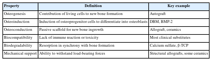
PDF - Bone defects, which often arise from high-energy injuries, infections, tumor resections, or nonunions, represent a persistent challenge in orthopedic trauma surgery. Autologous bone grafting remains the gold standard due to its unique combination of osteogenic, osteoinductive, and osteoconductive properties. However, issues such as donor site morbidity, limited graft volume, and increased surgical time have driven the development of bone graft substitutes. These substitutes vary widely in origin, composition, biological activity, and mechanical characteristics, encompassing allografts, xenografts, synthetic materials, and biologically enhanced constructs. This review outlines the fundamental biological principles underlying bone regeneration—including osteogenesis, osteoinduction, and osteoconduction—and addresses additional key factors such as biocompatibility, biodegradability, and mechanical strength. Current bone graft materials are classified by biological origin and functional characteristics, with an emphasis on their use in trauma surgery. Particular attention is given to the clinical applications, indications, and limitations of allograft-based solutions (such as structural allografts and demineralized bone matrix), synthetic ceramics (including calcium phosphate and bioactive glass), and biologically enhanced options, such as recombinant growth factors and stem cell therapies. In trauma settings, graft selection must be tailored to the characteristics of the defect, mechanical demands, the biological environment, and patient-specific factors. Integration with surgical technique and fixation is crucial for optimizing outcomes. Although modern substitutes show promise, none fully replicate the complex biology of autografts. Looking ahead, emerging technologies such as 3D printing, nanotechnology, and smart biomaterials offer exciting possibilities but face translational challenges. This review aims to provide practicing orthopedic surgeons with a concise, evidence-based overview of bone substitute options and their roles in trauma care. By applying core biological principles and clinical judgment, surgeons can better navigate the expanding array of graft materials to improve outcomes for patients with complex skeletal defects.
-
Citations
Citations to this article as recorded by
- Safety and Efficacy of rhBMP-2 for Treating Acute Traumatic Fractures of the Upper and Lower Extremities: A Multicenter Prospective Study
Seungyeob Sakong, Seokjun Hong, Wonseok Choi, Seonghyun Kang, Jae-Woo Cho, Whee Sung Son, Jeong-Seok Choi, Chang-Jin Yon, Won-Tae Cho, Jong-Keon Oh
Journal of Clinical Medicine.2026; 15(3): 1176. CrossRef
- Safety and Efficacy of rhBMP-2 for Treating Acute Traumatic Fractures of the Upper and Lower Extremities: A Multicenter Prospective Study
- 4,319 View
- 100 Download
- 1 Crossref

- Current Concepts of Vitamin D and Calcium in the Healing of Fractures
- Jihyo Hwang
- J Korean Fract Soc 2021;34(3):117-121. Published online July 31, 2021
- DOI: https://doi.org/10.12671/jkfs.2021.34.3.117
-
Abstract
PDF
- Fragile fractures, also known as osteoporosis fractures, insufficiency fractures, and senile fractures are a significant problem encountered by orthopedic surgeons. Calcium and vitamin D are essential for maintaining bone health and deficiencies in calcium and vitamin D are major risk factors for the development of osteoporosis. Sufficient amounts of calcium are also required for fracture-callus mineralization. Hence, compromised bone repair that is frequently observed in osteoporotic patients might be attributed to calcium and vitamin D deficiencies. Consequently, calcium and vitamin D supplementation represents a potential strategy for treating compromised fracture healing in osteoporotic patients. There is some clinical evidence of the positive effect of supplementation in fracture healing and posttraumatic bone turnover, but research in this area is ongoing. Calcium and vitamin D should be the primary treatment of choice in osteopenic patients with an insufficiency of calcium and vitamin D and for the prevention of secondary osteoporotic fractures. Calcium and vitamin D can also be used as addition to other primary osteoporotic medications such as antiresorptive or bone-forming agents. The role of calcium and vitamin D in fracture healing and the therapeutic potential of calcium and vitamin D supplementation is summarized in this context.
- 6,903 View
- 81 Download
Original Articles
- Intermittent Parathyroid Hormone Treatment for Stimulation of Callus Formation in Elderly Patients
- Hyung Keun Song, Sung Jun Kim, Jae Hoo Lee, Kyu Hyun Yang
- J Korean Fract Soc 2012;25(4):295-299. Published online October 31, 2012
- DOI: https://doi.org/10.12671/jkfs.2012.25.4.295
- Correction in: J Musculoskelet Trauma 2013;26(2):170
-
Abstract
PDF
- PURPOSE
The purpose of this study was to evaluate the effect of parathyroid hormone (PTH) on fracture healing in elderly patients.
MATERIALS AND METHODS
We analyzed the radiologic results in 14 patients. Group I (n=7) was administrated intermittent PTH after surgical treatment and group II (n=7) was treated only with surgery. We checked the time of initial callus formation, bridging callus formation, and bone union through periodic follow-up radiographs by a radiologist who did not know the patient's information.
RESULTS
The mean time to initial callus formation was 6 weeks for group I, compared with 6.7 weeks for group II. The mean time to bridging callus formation was 15.9 weeks for group I, compared with 23.0 weeks for group II. The mean time to bone union was 28.7 weeks for group I, compared with 41.9 weeks for group II. The difference in the cumulative detection rate (CDR) of the initial callus formation of group I and II was not statistically significant (p=0.793). However, the CDR of the bridging callus formation and bone union for group I were higher than those of group II (p=0.008, p=0.001, respectively).
CONCLUSION
The intermittent PTH administration after surgical treatment and maximum possible preservation of the periosteum in elderly patients accelerates fracture healing. -
Citations
Citations to this article as recorded by- Advances in Parathyroid Hormone-based medicines
Anne-Laure Bonnet, Lizaveta Aboishava, Michael Mannstadt
Journal of Bone and Mineral Research.2025; 40(11): 1195. CrossRef - Effects of Extracts from Cnidium officinale and Angelica sinensis on Bone Fusion in Mice with Femoral Fracture
Sang Woo Kim, Min-Seok Oh
Journal of Korean Medicine Rehabilitation.2024; 34(2): 1. CrossRef - Timing of osteoporosis therapies following fracture: the current status
Rajan Palui, Harsh Durgia, Jayaprakash Sahoo, Dukhabandhu Naik, Sadishkumar Kamalanathan
Therapeutic Advances in Endocrinology and Metabolism.2022;[Epub] CrossRef - Effect of Postoperative Parathyroid Hormone Administration on Osteoporotic Intertrochanteric Fractures of Females
Hyun Cheol Oh, Ju Hyung Yoo, Joong Won Ha, Yung Park, Sang Hoon Park, Han Kook Yoon
Journal of the Korean Orthopaedic Association.2020; 55(3): 237. CrossRef - The role of teriparatide in tuberosity healing after reverse shoulder arthroplasty in complex proximal humeral fragility fracture
Bancha Chernchujit, Renaldi Prasetia
Journal of Orthopaedic Surgery.2018;[Epub] CrossRef - Bone Substitutes and the Advancement for Enhancing Bone Healing
Dong-Hyun Lee, Ji Wan Kim
Journal of the Korean Fracture Society.2017; 30(2): 102. CrossRef - Current Role and Application of Teriparatide in Fracture Healing of Osteoporotic Patients: A Systematic Review
Sang-Min Kim, Kyung-Chung Kang, Ji Wan Kim, Seung-Jae Lim, Myung Hoon Hahn
Journal of Bone Metabolism.2017; 24(1): 65. CrossRef - The Effect of Teriparatide on Fracture Healing of Osteoporotic Patients: A Meta-Analysis of Randomized Controlled Trials
Shenghan Lou, Houchen Lv, Guoqi Wang, Licheng Zhang, Ming Li, Zhirui Li, Lihai Zhang, Peifu Tang
BioMed Research International.2016; 2016: 1. CrossRef - A systematic review on the use of daily subcutaneous administration of teriparatide for treatment of patients with osteoporosis at high risk for fracture in Asia
J.F. Chen, K. H. Yang, Z.L. Zhang, H.C. Chang, Y. Chen, H. Sowa, S. Gürbüz
Osteoporosis International.2015; 26(1): 11. CrossRef
- Advances in Parathyroid Hormone-based medicines
- 737 View
- 8 Download
- 9 Crossref
- Treatment of Open Tibial Shaft Fractures using Unreamed Nailing
- Jong Keon Oh, Chang Wug Oh, Kwon Jae Roh, Duk Moon Chung
- J Korean Fract Soc 2005;18(1):22-28. Published online January 31, 2005
- DOI: https://doi.org/10.12671/jkfs.2005.18.1.22
-
Abstract
PDF
- PURPOSE
To report the results of unreamed nailing using a nail with the largest possible diameter for the management of the open tibial shaft fractures.
MATERIALS AND METHODS
Nineteen patients with open tibial shaft fractures underwent unreamed nailing with the largest possible diameter according to the isthmic diameter measured on preoperative radiography. There were 1 Grade I, 6 Grade II, 9 Grade IIIa, 3 Grade IIIb open fractures. There were 4 type A, 12 type B, 3 type C fractures according to the OTA classification. Fractures were classified as The nail was introduced after gentle passage of a 7 to 8 millimeter-hand reamer.
RESULTS
Union was obtained in all cases. However 9 (47%) fractures required an additional procedures before union. In 6 cases, dynamization was done. Two of them were required exchange nailing for nonunion, 1 of two gained bony union through additional bone graft. Three of the others had gained union through exchange nailing, bone graft, bone transport respectively. There were one rotational malunion, one superfical and one deep infection. Interlocking screw breakage developed only in one patient.
CONCLUSION
Our data indicate that unreamed nailing in the management of open tibial fractures is safe and reliable method. Using a tight fitting nail with the largest possible diameter is a safe and effective way to avoid the problems of screw breakage. -
Citations
Citations to this article as recorded by- Treatment of Type IIIb Open Tibial Fractures
Seong Yeon Lim, Il Jae Lee, Jae Ho Joe, Hyung Keun Song
Journal of the Korean Fracture Society.2014; 27(4): 267. CrossRef - Management of Open Tibial Fractures: Role of Internal Fixation
Yerl-Bo Sung
Journal of the Korean Fracture Society.2007; 20(4): 349. CrossRef
- Treatment of Type IIIb Open Tibial Fractures
- 621 View
- 0 Download
- 2 Crossref
- The Effect of Bone Connecting Powder on Stimulation of Bone Healing (Biomechenical study using double-blind method)
- Kyu Hyun Yang, Sung Hoon Jung
- J Korean Soc Fract 2002;15(2):264-270. Published online April 30, 2002
- DOI: https://doi.org/10.12671/jksf.2002.15.2.264
-
Abstract
PDF
- PURPOSE
To investigate the effect of bone connecting powder on stimulation of bone healing, we performed a biomechenical study using the rats in double blinded method.
MATERIALS AND METHODS
One hundred ten-week-old korean rats were used. We performed closed intramedullary nailing with #2 Kirschner wire on the right femur and then transverse fracture was created on the right femoral shaft. The rats were divided into two groups in double blind method, one group was bone connecting powder feeding group and the other was placebo group. The rats were euthenized four weeks after fracture. We measured the ultimate load, stiffness, ultimate stress by 3-point bending test using electromechanical testing machine. The code used for double blinded method was disclosed after biomechanical test.
RESULT
Biomechenical test was performed at four weeks after fracture, in which there were 38 rats alive in the study group and 36 rats alive in the placebo group. There were 5 nonunion in study group, 7 in placebo group. The ultimate load was 40.77 +/- 28.09N for study group, 32.39 +/-25.10N for placebo group and stiffness was 49.98 +/- 45.32N/mm, 40.52 +/-36.61N/mm respectively. We calculated the ultimate stress to correct the difference from each bone's shape and thickness and it was 11.017 +/- 10.170N/mm 2 , 6.659 +/-6.670N/mm 2 for each other(p=0.041).
CONCLUSION
On the basis of this biomechenical study, it may be concluded that fracture healing is stimulated by bone connecting powder.
- 400 View
- 0 Download
- The Effects of aging process on fracture healing in rat callus
- Sang Ho Song, Young Hee Choi, Shim Chang Gu, Sang Ho Yoo, Young Euy Park
- J Korean Soc Fract 2001;14(2):135-144. Published online April 30, 2001
- DOI: https://doi.org/10.12671/jksf.2001.14.2.135
-
Abstract
PDF
- PURPOSE
Patient age significantly influences the rate of fracture healing. The rate of healing declines with increasing age. The authors compared the aging effect on fracture healing in the callus of rat femur by the light microscopy.
MATERIALS AND METHODS
In this study the unilateral, closed fractures were created in the femur of 18 Sprague-Dawley rats. The rats were killed in three age group(8 weeks:7, 32weeks:6, 70weeks:5) at 2 weeks after fracture. The composition of fracture callus(new bone, cartilage, mesenchymal layer) was measured by image analyzer with H-E stain. Immunohistochemical stain (PCNA, TUNEL, TRAP) positive cells were counted for the comparing of cellular activity according to the aging.
RESULTS
The percent of intramembranous new bone in the younger rat(8 week:22.32%) was higher than the older ones(30 week:7.09%, 70 week:5.37%). The percent of PCNA positive osteoblast in the newbone decreased according to the aging(8 week:64.25%, 30 week:57.40%, 70 week:29.54%). The number of osteoclast in the osteochondral junction at the 8 week(43) was more than that of 30 week(25.57) and 72 week(29.87). The number of TRAP positive osteoclast was not different as aging, but the number of osteoclast in the osteochondral junction(5.89) was more than that in the metapyseal area(2.08).
CONCLUSIONS
More new bone was found in younger rat. There was a strong correlation (p<0.05) between age and PCNA activity. More number of active osteoblast and osteoclast was found in younger rat femoral fracture callus, which indicated rapid fracture healing in younger age.
- 425 View
- 1 Download
- Thirty-five Degree Internal Oblique Radiographs in Assessment of Tibial Fracture Healing
- Eun Woo Lee, Ki Ser Kang, Soo Yong Kang, Jin Woo Lee
- J Korean Soc Fract 1996;9(2):475-479. Published online April 30, 1996
- DOI: https://doi.org/10.12671/jksf.1996.9.2.475
-
Abstract
PDF
- For the assessment of fracture healing, tomogram, computerized sonometry, resonant frequency analysis etc. were introduced recently, but most of orthopedic surgeons depend on plain X-ray and clinical experience. The progress of tibial fracture healing may be difficult to assess through routine radiological examination(AP and lateral). So, we intended to assess the healing of tibial fracture with 35° internal oblique view as well as AP and lateral. Five orthopedic surgeons assested the tibial fracture heating with only AP and lateral (group 1), and AP. lateral and 35° internal oblique view(group 2) in 45 tibial fractures. In the percent agreement of their assessment, Group 1 was 60% and group 2 was 76%. Group 2 was higher than group 1, especially in IM nailing and bone graft groups.The change of judgement between the two group was 18.7%, and it was higher in the distal tibial fracture, posterolateral bone graft and external device groups. In 11 Cases, the fibular fractures were overlapped with tibiai fracture in laterai view, in which cases 35° intelnal oblique view was useful for assessing the tibial fracture healing. We recommand 35° internal oblique view for assessment of tibial fracture healing before using more tophisticated and expensive procedure, especialiy in patients with posterolateral bone graft, distal libial fracture and combined fibular fracture, and probably in IM nailing and external device.
- 369 View
- 1 Download
 First
First Prev
Prev

