 E-submission
 TOTA
TOTA TOTS
TOTS
Search
- Page Path
- HOME > Search
Original Articles
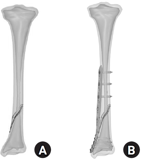
- Biomechanical analysis of medial distal tibial locking plate fixation for distal-third spiral tibial shaft fractures
- Yao-Jen Liu
- J Musculoskelet Trauma 2026;39(2):140-146. Published online April 10, 2026
- DOI: https://doi.org/10.12671/jmt.2026.00094
-
 Abstract
 PDF
PDF - Background
Distal spiral fractures of the tibial shaft present fixation challenges, particularly in patients who are not suitable candidates for intramedullary nailing. This study evaluated the biomechanical stability of medial minimally invasive percutaneous plating osteosynthesis (MIPO) under various physiological loading conditions.
Methods
A finite-element model of a distal AO/OTA 42-A1.1c spiral fracture of the tibia was created using computed tomography data. A precontoured titanium medial distal tibia locking compression plate with nine locking screws was simulated. Material properties were assigned to cortical and cancellous bone. The loading conditions included axial compression (750 N), varus/valgus bending (300 N at a 9° offset), and internal/external torsion (7.5 N·m). von Mises stress and fracture displacement were analyzed.
Results
Axial loading produced a peak plate stress of 508.06 MPa and a displacement of 2.17 mm. Valgus and varus loading generated stresses of 490.17 MPa and 324.08 MPa, respectively, with corresponding displacements of 3.86 mm and 2.01 mm. External and internal torsion resulted in stresses of 354.23 MPa and 358.9 MPa, respectively, with corresponding displacements of 2.64 mm and 2.22 mm.
Conclusions
Medial distal tibial plating demonstrated favorable biomechanical performance in this finite-element model; however, clinical extrapolation should be made cautiously. Level of evidence: V.
- 302 View
- 16 Download

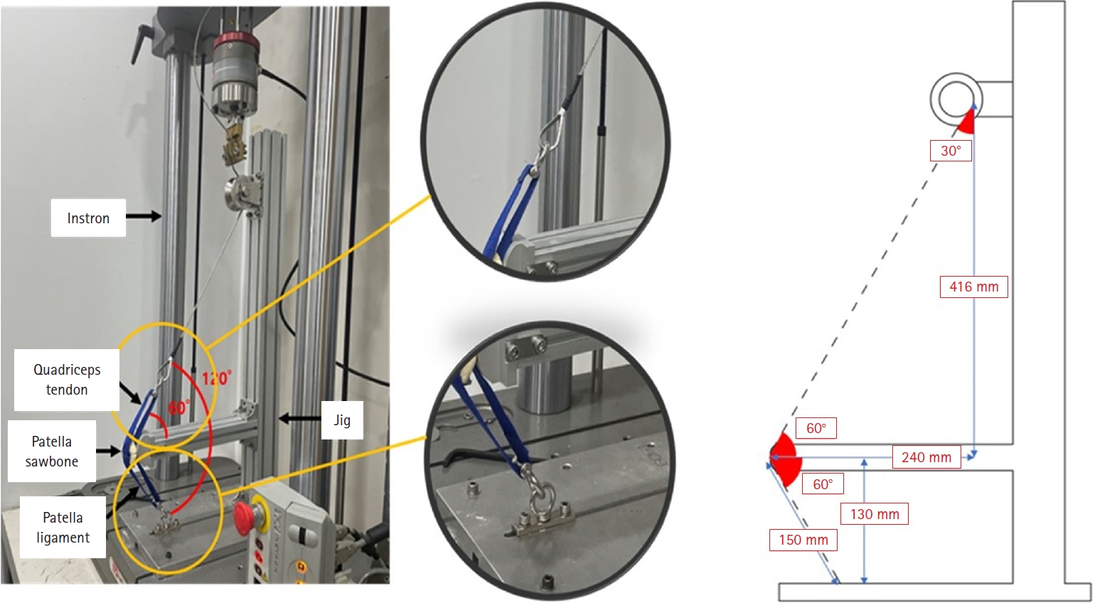
- Biomechanical comparison of anatomically precontoured patellar plate, anterior tension wiring through cannulated screws, and double-sided plating in patellar fractures using a synthetic bone model
- Abdullah M. Aljeaid, Wonseok Choi, Jeong-Seok Choi, Youngsig Choi, Jiyeon Bae, Jong-Keon Oh, Jae-Woo Cho
- J Musculoskelet Trauma 2026;39(2):130-139. Published online April 7, 2026
- DOI: https://doi.org/10.12671/jmt.2025.00353
-
Abstract
PDF
- Background
Patellar fractures are common injuries that require stable fixation to achieve optimal healing and restoration of knee function. This study aimed to analyze the mechanical properties of an anatomically precontoured patellar plate and to compare its maximum tensile load-bearing capacity with that of anterior tension wiring through cannulated screws and double-sided plating for the fixation of patellar fractures.
Methods
Artificial Sawbones with a standardized transverse fracture line were used to simulate patellar fractures. Each sawbone was attached to polyester bands, and this fracture model was applied consistently across all test samples. To evaluate mechanical properties of the anatomically precontoured patellar plate (model code 25-ANPA-209) made of ASTM F67 titanium, static tensile strength testing and dynamic tensile strength testing were performed, with seven samples prepared for each test. For comparison of maximum tensile load capacity among the anatomically precontoured patellar plate, anterior tension wiring through cannulated screws, and double-sided plating, five samples were prepared for each fixation group. All specimens were tested using a tension/compression testing machine.
Results
In the static tensile strength test, all seven samples exhibited a maximum tensile load capacity above 844 N without any fractures or failure points. The dynamic tensile strength test showed that all seven samples completed 10,000 cycles without deformation or damage to the anatomically precontoured patellar plate. When comparing maximum tensile load capacity, the anatomically precontoured patellar plate exhibited a significantly higher maximum tensile load-bearing capacity than anterior tension wiring through cannulated screws and double-sided plating.
Conclusions
The anatomically precontoured patellar plate demonstrated satisfactory mechanical performance, successfully meeting the criteria of both static and dynamic tensile strength testing, and showed superior maximum tensile load-bearing capacity compared with the other fixation methods evaluated. These findings suggest that the anatomically precontoured patellar plate may represent a reliable fixation option for the management of patellar fractures. Level of evidence: V.
- 581 View
- 22 Download
- Association between decreased bone mineral density and Pauwels angle in femoral neck fractures: a cross-sectional study
- Soo-Hwan Jung, Yong-Uk Kwon, Ji-Hun Park
- J Musculoskelet Trauma 2026;39(1):20-29. Published online January 25, 2026
- DOI: https://doi.org/10.12671/jmt.2025.00269

-
Abstract
PDF
 Supplementary Material
 - Background
Progressive osteoporosis reduces the trabecular structures of the proximal femur, whereas the primary compression trabeculae (PCTs) are relatively preserved. We hypothesize that the loss of the vertically oriented PCTs in osteoporosis, which act as a mechanical barrier, affects fracture line propagation and influences the Pauwels angle. This study investigated the association between bone mineral density (BMD) and Pauwels angles in low-energy femoral neck fractures (FNFs).
Methods
This cross-sectional study included 150 patients (mean age, 75.3 years; range, 50–94 years) diagnosed with intracapsular FNFs between May 2019 and May 2023. BMD was measured within 1 month of the injury date using dual-energy X-ray absorptiometry, and modified Pauwels angles were assessed using a computed tomography-based multiplanar reconstruction program. Multiple linear regression analysis was performed to evaluate the factors influencing the Pauwels angles. The dependent variable was the Pauwels angle, while the independent variables included sex, age, height, body weight, body mass index, American Society of Anesthesiologists score, Charlson comorbidity index score, smoking status, alcohol use, preinjury walking ability, and femoral neck BMD T-scores.
Results
Higher femoral neck BMD T-scores were significantly associated with increased Pauwels angles (β=3.449, P<0.001). Greater body weight was independently associated with increased Pauwels angles (β=0.213, P=0.007).
Conclusions
The Pauwels angle demonstrated a significant association with BMD, with lower BMD associated with less steep Pauwels angles. In the absence of BMD measurement, the Pauwels angle may indicate osteoporosis severity in patients with low-energy FNFs. Level of evidence: III.
- 875 View
- 23 Download
- The Effects of Extramedullary Reduction in Unstable Intertrochanteric Fracture: A Biomechanical Study Using Cadaver Bone
- Young Chang Park, Soon Phil Yoon, Kyu Hyun Yang
- J Korean Fract Soc 2018;31(3):79-86. Published online July 31, 2018
- DOI: https://doi.org/10.12671/jkfs.2018.31.3.79
-
Abstract
PDF
- PURPOSE
To prevent excessive sliding and subsequent fixation failures in unstable intertrochanteric fractures with posteromedial comminution, extramedullary reduction through overlapping of the anteromedial cortices of both proximal and distal fragments as a buttress has been introduced. The purpose of this study was to compare the biomechanical properties between two reduction methods-intramedullary reduction and extramedullary reduction-in treating unstable intertrochanteric fractures with posteromedial comminution (AO/OTA classification 31-A2.2).
MATERIALS AND METHODS
Eight pairs of frozen human cadaveric femora were used. The femora of each pair were randomly assigned to one of two groups: the intramedullary reduction group or the extramedullary reduction group. A single axial load-destruction test was conducted after cephalomedullary nailing. Axial stiffness, maximum load to failure, and energy absorbed to failure were compared between the two groups. Moreover, the pattern of mechanical failure was identified.
RESULTS
The mean axial stiffness in the extramedullary reduction group was 27.3% higher than that in the intramedullary reduction group (422.7 N/mm vs. 332.0 N/mm, p=0.017). Additionally, compared with the intramedullary reduction group, the mean maximum load to failure and mean energy absorbed to failure in the extramedullary group were 44.9% and 89.6% higher, respectively (2,848.7 N vs. 1,966.5 N, p=0.012 and 27,969.9 N·mm vs. 14,751.0 N·mm, p=0.012, respectively). In the intramedullary reduction group, the mechanical failure patterns were all sliding and varus deformities. In the extramedullary reduction group, sliding and varus deformities after external rotation were noted in 3 specimens, sliding and varus deformities after internal rotation were noted in 3 specimens, and medial slippage was noted in 2 specimens.
CONCLUSION
In unstable intertrochanteric fractures with posteromedial comminution, the biomechanical properties of extramedullary reduction are superior to those of intramedullary reduction. Anteromedial cortex could be the proper buttress, despite a comminuted posteromedial cortex. It could help enhance the stability of the bone-nail construct. -
Citations
Citations to this article as recorded by
- The effect of additional reduction screw fixation for basicervical femoral neck fracture: a finite element analysis
Kyoung-Joo Lee, Jihoon Ahn, Chul-Ho Kim
HIP International.2026; 36(2): 324. CrossRef - Comparison of the Dynamic Cut‐Out Failure Modes of Common Proximal Femoral Fixation Devices Using a Mesh‐Free Computational Method
Erica Ueda Boles, Sloan Kulper, Katie Whiffin, Marilyn Janice Oentaryo, Kerstin Schneider, Frankie Leung, Christian X. Fang
Journal of Orthopaedic Research.2026;[Epub] CrossRef - Cortical Support in Unstable Intertrochanteric Fracture without Fixation of Posteromedial Fragment
Ki-Tae Park, Dong-Hoon Lee, Kyung-Hoi Koo, Jung-Wee Park, Young-Kyun Lee
Clinics in Orthopedic Surgery.2026;[Epub] CrossRef - Which side should be taken care of when positioning a lag screw in intertrochanteric femoral fracture: right or left?
Min Uk Do, Kyeong Baek Kim, Sang-Min Lee, Hyun Tae Koo, Won Chul Shin
European Journal of Trauma and Emergency Surgery.2025;[Epub] CrossRef - Distal locking mechanism influences surgical and radiological outcomes in proximal femoral nailing using distal wedge versus distal screw designs
Aytek Hüseyin Çeliksöz, Büşra Tokmak, Ali Okan Tarlacık, Servet Igrek
Scientific Reports.2025;[Epub] CrossRef - The effect of anterior support screw (AS2) in unstable femoral trochanteric fractures: A multicenter randomized controlled trial
Takashi Maehara, Takashi Hayakawa, Shunsuke Mukoyama, Yoshihisa Anraku, Takahiro Hamada, Hiroyuki Suzuki, Takeshi Doi, Tomohiko Shimizu, Masanori Yorimitsu, Hidefumi Teramoto, Takao Mae, Yasunori Okamoto, Jun Hara, Kazushi Mihara, Koichi Kanekasu
Injury.2024; 55(10): 111725. CrossRef - Anteromedial cortical support reduction of intertrochanteric fractures–A review
Wenjun Xie, Liu Shi, Cheng Zhang, Xueliang Cui, Xiangxu Chen, Tian Xie, Sheng Zhang, Hui Chen, Yunfeng Rui
Injury.2024; 55(12): 111926. CrossRef - Anteromedial Cortical Support in Reduction of Trochanteric Hip Fractures
Wei Mao, Chen-Dong Liu, Shi-Min Chang, Ao-Lei Yang, Choon Chiet Hong
Journal of Bone and Joint Surgery.2024; 106(11): 1008. CrossRef - Five states of reduction in OTA/AO A1.3 intertrochanteric fractures of the femur a biomechanical study
Shu Li, Yong-Gang Bao, Rong-Hua Tian, Chun-Yang Meng, Hai-Bin Wang, Bin Wu, Xian-Min Bu
BMC Musculoskeletal Disorders.2024;[Epub] CrossRef - Intramedullary Impaction of the Basicervical Component Is Determinant of Fixation Failure in a Simple Two-Part Pertrochanteric Fracture
Seok Ha Hong, Kang Hun Yu, Seung Beom Han
Journal of Orthopaedic Trauma.2024; 38(4): 220. CrossRef - Flexible reamer use to overcome entry point errors in proximal femoral nail application in severe obese intertrochanteric fracture patients
Levent Horoz, Ali Ihsan Kilic, Cihan Kircil, Mehmet Fevzi Cakmak
BMC Musculoskeletal Disorders.2024;[Epub] CrossRef - Risk Factors Associated with Fixation Failure in Intertrochanteric Fracture Treated with Cephalomedullary Nail
Hyung-Gon Ryu, Dae Won Shin, Beom Su Han, Sang-Min Kim
Hip & Pelvis.2023; 35(3): 193. CrossRef - Positive or negative anteromedial cortical support of unstable pertrochanteric femoral fractures: A finite element analysis study
Qin Shao, Yue Zhang, Gui-Xin Sun, Chen-Song Yang, Na Liu, Da-Wei Chen, Biao Cheng
Biomedicine & Pharmacotherapy.2021; 138: 111473. CrossRef - Clinical and Radiologic Outcome of Intertrochanteric Fracture Treatment Using TFNA (Trochanteric Fixation Nail-Advanced)
Hyeon Joon Lee, Hyun Bai Choi, Ba Rom Kim, Seung Hwan Jo, Sang Hong Lee
Journal of the Korean Fracture Society.2021; 34(3): 105. CrossRef - Factors Associated with Mechanical Complications in Intertrochanteric Fracture Treated with Proximal Femoral Nail Antirotation
Oog-Jin Shon, Chang Hyun Choi, Chan Ho Park
Hip & Pelvis.2021; 33(3): 154. CrossRef - Additional Reduction Screw Fixation Technique for Pertrochanteric Hip Fractures: A Novel Method to Prevent Excessive Sliding in Cephalomedullary Nail Surgery
Chul-Ho Kim, Han Soul Kim, Dou Hyun Moon
Hip & Pelvis.2021; 33(3): 162. CrossRef - Comparison of sliding distance of lag screw and nonunion rate according to anteromedial cortical support in intertrochanteric fracture fixation: A systematic review and meta-analysis
Eic Ju Lim, Seungyeob Sakong, Whee Sung Son, Jae-Woo Cho, Jong-Keon Oh, Chul-Ho Kim
Injury.2021; 52(10): 2787. CrossRef - A new fluoroscopic view for evaluation of anteromedial cortex reduction quality during cephalomedullary nailing for intertrochanteric femur fractures: the 30° oblique tangential projection
Shi-Yi Chen, Shi-Min Chang, Rujan Tuladhar, Zhen Wei, Wen-Feng Xiong, Sun-Jun Hu, Shou-Chao Du
BMC Musculoskeletal Disorders.2020;[Epub] CrossRef - New Approach in the Treatment of Intertrochanteric Fracture Using a Cephalomedullary Nail
Junyoung Kim, Kihong Choi, Kyu Hyun Yang
Journal of the Korean Orthopaedic Association.2020; 55(3): 193. CrossRef - Effect of a synthetic osteoconductive bone graft substitute with zeta potential control (geneX®ds) in the treatment of intertrochanteric fracture: A single center experience of 115 consecutive proximal femoral nail antirotations
Won Chul Shin, Jae Hoon Jang, Jae Yoon Jeong, Kuen Tak Suh, Nam Hoon Moon
Journal of Orthopaedic Science.2019; 24(5): 842. CrossRef
- The effect of additional reduction screw fixation for basicervical femoral neck fracture: a finite element analysis
- 912 View
- 6 Download
- 20 Crossref
- Effect of Fracture Gap on Biomechanical Stability of Compression Bone-Plate Fixation System after Bone Fracture Augmentation
- Duk Young Jung, Sung Jae Lee, Seon Chil Kim, Jong Keon Oh
- J Korean Fract Soc 2010;23(2):220-226. Published online April 30, 2010
- DOI: https://doi.org/10.12671/jkfs.2010.23.2.220
-
Abstract
PDF
- PURPOSE
The goal of this study using the biomechanical test was to evaluate the mechanical stability of the bone-plate fixation system according to changes of the fracture gap sizes and widths.
MATERIALS AND METHODS
For mechanical test, four types with different fracture models simulating the clinical situations were constructed depending on the gap size (FGS, mm) and the gap width (FGW, %) at the fracture site: 0 mm/0%, 1 mm/100%, 4 mm/100%, 4 mm/50%. For analyzing the effects of fracture gap on the biomechanical stability of the bone-plate fixation system, 4-point bending test was performed under all same conditions.
RESULTS
It was found that the fracture gap sizes of 1 and 4 mm decreased mechanical stiffness by about 50~60% or more. Furthermore, even without fracture gap size, 50% or more fracture gap width considerably decreased mechanical stiffness and suggested the possibility of plate damage through strain results.
CONCLUSION
Our findings suggested that at least 50% contact of the fracture faces in a fracture surgery would be maintained to increase the mechanical stability of the bone-plate fixation system.
- 1,111 View
- 5 Download
- A Finite Element Analysis of Biomechanical Stability of Compression Plate Fixation System in according to Existing of Fracture Gap after Bone Fracture Augmentation
- Duk Young Jung, Bong Ju Kim, Jong Keon Oh
- J Korean Fract Soc 2010;23(1):83-89. Published online January 31, 2010
- DOI: https://doi.org/10.12671/jkfs.2010.23.1.83
-
Abstract
PDF
- PURPOSE
This study using the finite element analysis (FEA) focused on evaluating the biomechanical stability of the LC-DCP in accordance with existing of the fracture gap at the facture site after bone fracture augmentation.
MATERIALS AND METHODS
For FEM analysis, total eleven types with different fracture models considering clinical fracture cases were constructed according to the fracture gap sizes (0, 1, 4 mm)/widths (0, 25, 50, 75, 100%). Limited contact dynamic compression plate (LC-DCP) fixation system was used in this FEM analysis, and three types of load were applied to the bone-plate fixation system: compressive, torsional, bending load.
RESULTS
The results in FEM analysis showed that the 1, 4 mm fracture gap sizes and 75% or more fracture gap widths increased considerably the peak von Mises stress (PVMS) both the plate and the screw under all loading conditions. PVMS were concentrated on the center of the LC-DCP bone-plate, and around the necks of screws.
CONCLUSION
Based on the our findings, we recommend at least 50% contact of the fracture faces in a fracture surgery using the compression bone-plate system. Moreover, if x-ray observation after surgery finds 100% fracture gap or 50% or more fracture gap width, supplementary measures to improve biomechanical stability must be taken, such as restriction of walking of the patient or plastering. -
Citations
Citations to this article as recorded by- Application of Patient-Specific 3D-Printed Orthopedic Splint for Bone Fracture in Small Breed Dogs
Kwangsik Jang, Eun Joo Jang, Yo Han Min, Kyung Mi Shim, Chunsik Bae, Seong Soo Kang, Se Eun Kim
Journal of Veterinary Clinics.2023; 40(4): 268. CrossRef
- Application of Patient-Specific 3D-Printed Orthopedic Splint for Bone Fracture in Small Breed Dogs
- 973 View
- 1 Download
- 1 Crossref
- Evaluation of Osseointegration in Titanium Alloy Cortical Screws with the Passage of Time
- Jae Hyup Lee, Bong Soon Chang, Choon Ki Lee
- J Korean Fract Soc 2004;17(4):401-407. Published online October 31, 2004
- DOI: https://doi.org/10.12671/jkfs.2004.17.4.401
-
Abstract
PDF
- PURPOSE
To evaluate the osseointegration of titanium alloy cortical screws with the passage of time.
MATERIALS AND METHODS
Fifty four titanium alloy cortical screws (24 mm in length, 3.5 mm in diameter) were implanted bilaterally in the tibial diaphysis of adult mongrel male dogs of similar size and weight (30 +/-5 kg). The insertion torques, radiographs, undecalcified histology, histomorphometric analysis and extraction torques were evaluated at 2, 4 and 8 weeks after surgery.
RESULTS
The extraction torque at 2 weeks (1.14+/-0.470 cN. m) was significantly lower than the insertion torque (1.76+/-0.609 cN. m) (p=0.0071), the extraction torque at 4 weeks (2.57+/-1.36 cN. m) was slightly improved and the extraction torque at 8 weeks (3.18+/-0.499 cN. m) was significantly higher than insertion torque (p=0.0005). Direct bony contact in the early phase was poor and intervening fibrous tissue was observed at the bone-screw interface. However, the fixation between the bone and the screws improved with time. The percentage of bone-screw contact at 8 weeks (33.1+/-18.5%) was higher than that of 2 weeks (22.4+/-12.9%), but not statistically significant.
CONCLUSION
Because of thermal injury or pressure necrosis, the fixation strength of titanium alloy cortical screws at 2 weeks after implantation is significantly lower than that at the insertion time. So, we should keep in mind the initial phase weakness of screw fixation when we allow the patients the range of motion exercise or weight bearing and the improvement of the initial phase fixation is very important in clinical results.
- 572 View
- 4 Download
- Mechanical Properties of External Fixator according to Its Arrangement and Structure
- Hong Jun Han, Byung Chang Lee, Yeung Jin Kim, Byung Soo Jin, Gun Hyee Lee
- J Korean Soc Fract 2000;13(1):38-45. Published online January 31, 2000
- DOI: https://doi.org/10.12671/jksf.2000.13.1.38
-
Abstract
PDF
- PURPOSE
To obtain the accurate knowledge of the fundamental mechanical properties of the external fixator affected by variations in arrangements and structures. We used newly developed external fixator, Anyfix, universal testing machine and plastic padding bone model which had similar structural properties to human tibia. The measured performance for seven different configurations of external fixators was its ability to control the motion of the bone fragment at the fracture site. Based on a unit of applied load, the corresponding displacement measured at the fracture site was used to described the stiffness of the fixation device for each load. Three stiffness moduli can be determined as axial stiffness, anterior posterior bending stiffness and lateral bending stiffness.
RESULTS
In basic configuration, all three stiffnesses for unilateral two plane external fixator showed marked increase than those for unilateral one plane model. Axial compression stiffness and bending stiffness were increased when ring component were located far from the fracture site. In modified configuration, all three stiffnesses were increased when the number of pin was increased and small sized ring was used.
CONCLUSION
The stiffness of the external fixator can be substantially increased by using unilateral two plane, locating the ring at far portion from the fracture site, using a small sized ring and increasing the number of pins.
- 496 View
- 0 Download
- Biomechanical Analysis of Korean Radiolucent Carbon/Graphite Ring Fixator
- In Ho Choi, Jun kyung Kim, Kui won Choi, Chin Youb Chung, Tae Joon Cho, Ki Seok Lee
- J Korean Soc Fract 2000;13(1):1-12. Published online January 31, 2000
- DOI: https://doi.org/10.12671/jksf.2000.13.1.1
-
Abstract
PDF
- PURPOSE
The mechanical stiffness of Korean radiolucent carbon/graphite ring fixator(KRCRF) was analyzed and compared with those of conventional stainless steel Ilizarov system and the Smith- Nephew carbon fiber circular external fixator.
MATERIALS AND METHODS
The transfixing olive pins of the circular fixator on the acryl pylon were assembled in 90degrees- 90degrees and 135degrees- 45degrees configuration, respectively. And the fixator-pylon model was loaded with Instron model No. 8500 in three testing modes: axial compression, anteroposterior(AP) bending and lateral bending.
RESULTS
As compared with stainless steel Ilizarov fixator, the KRCRF was significantly more stiff on the axial compression test regardless of the ring size(140 mm and 200 mm diameters) and transfixation configuration. But, it was less stiff on the anteroposterior(AP) and lateral bending tests. When compared with the Smith-Nephew carbon fiber circular external fixator, the KRCRF was generally more stiff on the axial compression, AP and lateral bending tests regardless of the ring size(140 mm and 180 mm diameters) and configuration, except the AP bending stiffness in 90degrees- 90degrees configuration and lateral bending stffness in 135degrees- 45degrees configuration on the 180 mm diameter frame.
CONCLUSION
Considering the radiolucency, weight and biomechanical stffness, we think that the KRCRF is an excellent substitute for the imported circular fixators made of stainless steel or carbon/graphite. -
Citations
Citations to this article as recorded by- A Study on the Development of the Off-Line Software for Regulating the 6 D.O.F. Circular Fixator
Bum-Seok PARK, In-Ho CHOI, Jin-Woo KIM, Seung-Yeol LEE, Chang-Soo HAN
JSME International Journal Series C.2006; 49(4): 1123. CrossRef
- A Study on the Development of the Off-Line Software for Regulating the 6 D.O.F. Circular Fixator
- 886 View
- 0 Download
- 1 Crossref
- Biomechanical Analysis of Translucent Hexagonal External Fixator
- Duk Yong Lee, In Ho Choi, Chin Youb Chung, Tae Joon Cho, Yoon Keun Park
- J Korean Soc Fract 1997;10(2):379-387. Published online April 30, 1997
- DOI: https://doi.org/10.12671/jksf.1997.10.2.379
-
Abstract
PDF
- The mechanical stiffness of 4 configurations of the Translucent Hexagonal External Fixator(THEF) was analyzed and compared with conventional Ilizarov system in vitro. The advantage of the THEF was that it is less expensive, radio-translucent because it was made of carbon fiberepoxy. Stiffness in axial compression, torsion, A-P bending and lateral bending were measured in both fixators. The fixators were assembled into 90-90 and 45-135 configurations, respectively. In each configurations, two types of pin, smooth pins and olive pins, were used for transfixion. As compared with the Ilizarov fixator, the THEF was less stiff in axial compression when the two smooth pins were used for transfixion regardless of configuration, but was also less stiff in A-P and lateral bending except A-P bending when the smooth wires were assembled in 90-90 configuration, and lateral bending when the olive wires were assembled in 45-135 configuration. However, the THEF was more stiff in torsion regardless of configuration and type of wires used. When the olive wires were used, the THEF was more stiff than the Ilizarov fixator regardless of wire configuration in all loadiilg mode except AP bending. Changing the pin configuration from 90-90 to 45-135 decreased all stiffness of the Ilizarov fixator. However, lateral bending and axial compression stiffness with smooth wire and A-P bending stiffness regardless of types of wires were decreased in the THEF. Changing the smooth wires to olive wires increased the A-P and lateral bending stiffness in the Ilizarov fixator, while it increased all stiffnesses in the THEF. We believe that the results originated from the weakness of the material used. THEF may be an effective alternative for osteosynthesis, deformity correction in complex construct because of its radiolucency in spite of less favorable biomechanical properties in some loading mode.
- 502 View
- 0 Download
- Biomechanical study of rigidity in the domestic external fixator preliminary report about the mechanical charateristics of the domestic product comparing with the imported fixators
- Joo Chul Ihn, Young Il Youm, Il Key Lee, Myeun Whan Ahn, Jong Chul Ahn, Se Dong Kim, Do Sik Choo
- J Korean Soc Fract 1991;4(1):147-153. Published online May 31, 1991
- DOI: https://doi.org/10.12671/jksf.1991.4.1.147
- 623 View
- 0 Download
 First
First Prev
Prev

