Shoulder Quadruple Dislocation Fracture: Fracture of Glenoid Rim, Coracoid Process, Greater Tuberosity, Surgical Neck of Humerus Associated with Anterior Shoulder Dislocation: A Case Report

Article information

J Musculoskelet Trauma. 2019;32(1):47-51
Publication date (electronic) : 2019 January 25
doi : https://doi.org/10.12671/jkfs.2019.32.1.47
Department of Orthopaedic Surgery, Dankook University College of Medicine, Cheonan, Korea.
Correspondence to: Joong-Bae Seo, M.D., Ph.D. Department of Orthopaedic Surgery, Dankook University Hospital, 201 Manghyang-ro, Dongnam-gu, Cheonan 31116, Korea. Tel: +82-41-550-3060, Fax: +82-41-556-3238, ssjb1990@dku.edu
Received 2018 October 10; Revised 2018 November 21; Accepted 2018 November 21.

Abstract

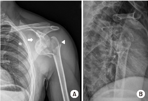
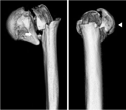
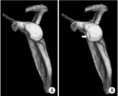
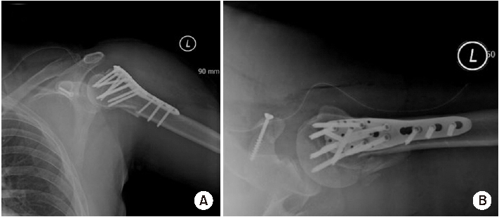
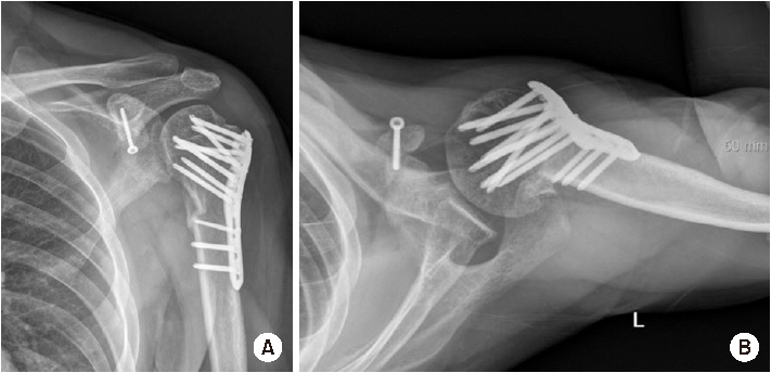
Shoulder joint dislocation has the most common incidence rate compare compared to other joints. It is reported that shoulder Shoulder dislocation couldmay be associated with glenoid rim, greater tuberosity of humerus and coracoid process fracture. There were have only been 2 cases of anterior shoulder dislocation simultaneously combined with simultaneous glenoid rim, coracoid process, and humerus greater tuberosity fracture worldwide and no report reports in Korea. We present a case of quadruple fracture (glenoid rim, coracoid process, greater tuberosity, surgical neck of humerus) associated with anterior shoulder dislocation and treated successfully by open reduction. In addition, with we provide the injury mechanism, diagnosis, treatment procedure and discussion.

Notes

Financial support

None.

Conflict of interests

None.

References

1. Liavaag S, Svenningsen S, Reikerås O, et al. The epidemiology of shoulder dislocations in Oslo. Scand J Med Sci Sports 2011;21:e334–e340.
2. Antonio GE, Griffith JF, Yu AB, Yung PS, Chan KM, Ahuja AT. First-time shoulder dislocation: high prevalence of labral injury and age-related differences revealed by MR arthrography. J Magn Reson Imaging 2007;26:983–991.
3. Green A, Izzi J Jr. Isolated fractures of the greater tuberosity of the proximal humerus. J Shoulder Elbow Surg 2003;12:641–649.
4. te Slaa RL, Verburg H, Marti RK. Fracture of the coracoid process, the greater tuberosity, and the glenoid rim after acute first-time anterior shoulder dislocation: a case report. J Shoulder Elbow Surg 2001;10:489–492.
5. Plachel F, Schanda JE, Ortmaier R, Auffarth A, Resch H, Bogner R. The “triple dislocation fracture”: anterior shoulder dislocation with concomitant fracture of the glenoid rim, greater tuberosity and coracoid process-a series of six cases. J Shoulder Elbow Surg 2017;26:e278–e285.
6. Baudi P, Righi P, Bolognesi D, et al. How to identify and calculate glenoid bone deficit. Chir Organi Mov 2005;90:145–152.
7. Scheibel M, Kraus N, Gerhardt C, Haas NP. Anterior glenoid rim defects of the shoulder. Orthopade 2009;38:41–48. :50–53.
8. Ogawa K, Yoshida A, Takahashi M, Ui M. Fractures of the coracoid process. J Bone Joint Surg Br 1997;79:17–19.
9. Rowe CR, Patel D, Southmayd WW. The Bankart procedure: a long-term end-result study. J Bone Joint Surg Am 1978;60:1–16.
10. Goodier D, Maffulli N, Good CJ. Coracoid process and greater tuberosity fracture in unreduced shoulder dislocation. Injury 1994;25:113–116.
11. Bassett RW, Browne AO, Morrey BF, An KN. Glenohumeral muscle force and moment mechanics in a position of shoulder instability. J Biomech 1990;23:405–415.

Article information Continued

Fig. 1

Shoulder plain radiographs anteroposterior (A) (arrow: displaced surgical neck fracture, arrowhead: displaced tuberosity fragment), lateral (B) demonstrate anterior shoulder dislocation with glenoid rim, coracoid process, greater tuberosity of humerus, and surgical neck fracture.

Fig. 2

Three-dimentional reconstruction of shoulder computed tomographies shows 3-part proximal humerus fracture (arrow: displaced surgical neck fracture, arrowhead: displaced tuberosity fragment).

Fig. 3

Glenoid fracture (A) was shown and glenoid bone defect (B) was evaluated based on 3-dimentional reconstruction of shoulder computed tomography (arrow: percentage of glenoid defect).

Fig. 4

Immediate postoperative plain radiographs anteroposterior (A), axial (B) view were assessed.

Fig. 5

Plain radiographs anteroposterior (A), lateral (B) 3 months after initial surgery show bony union around proximal shoulder, but malunion of the coracoid process.